. 2017 Jun;35(6):543-550.
doi: 10.1038/nbt.3843. Epub 2017 May 1.

Targeting genomic rearrangements in tumor cells through Cas9-mediated insertion of a suicide gene

Affiliations

Targeting genomic rearrangements in tumor cells through Cas9-mediated insertion of a suicide gene

Zhang-Hui Chen et al. Nat Biotechnol. 2017 Jun.

Abstract

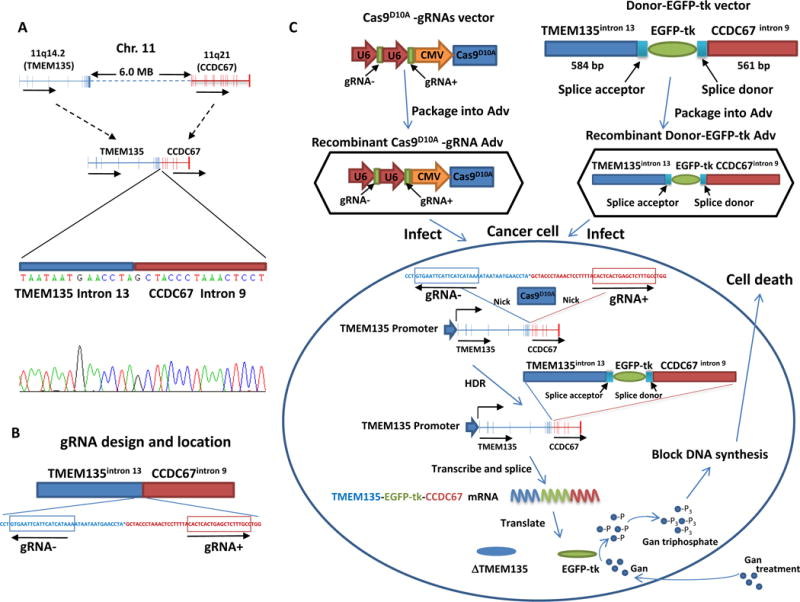
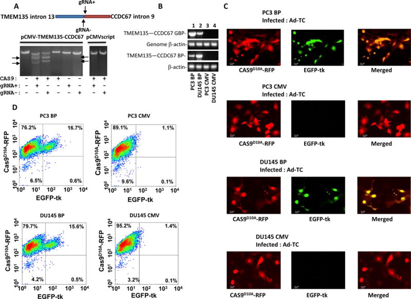
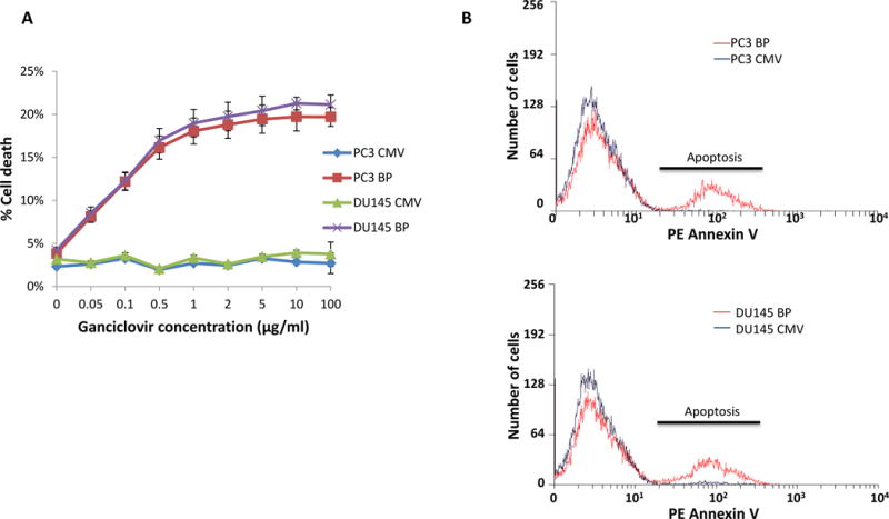
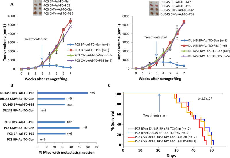
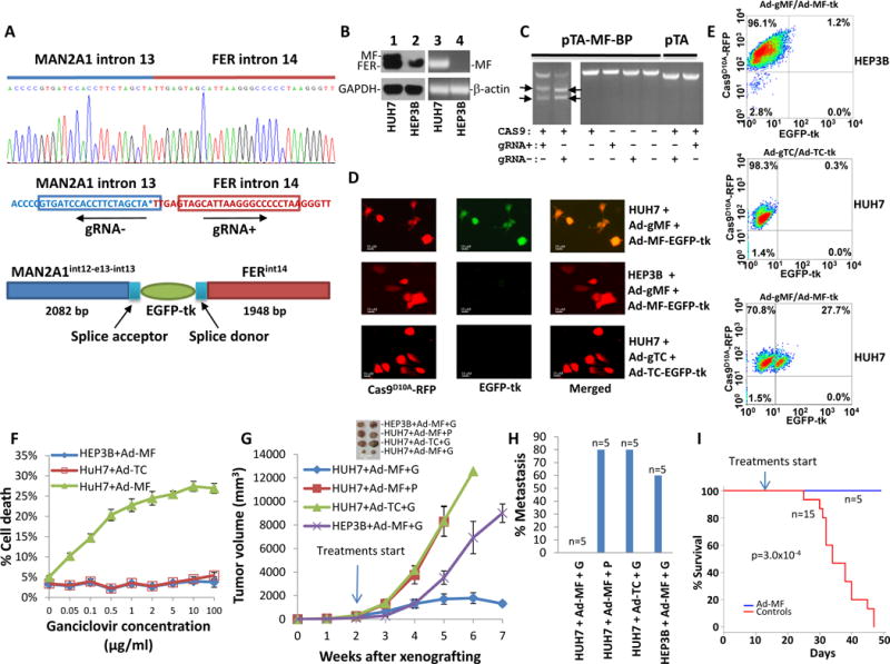
Specifically targeting genomic rearrangements and mutations in tumor cells remains an elusive goal in cancer therapy. Here, we used Cas9-based genome editing to introduce the gene encoding the prodrug-converting enzyme herpes simplex virus type 1 thymidine kinase (HSV1-tk) into the genomes of cancer cells carrying unique sequences resulting from genome rearrangements. Specifically, we targeted the breakpoints of TMEM135-CCDC67 and MAN2A1-FER fusions in human prostate cancer or hepatocellular carcinoma cells in vitro and in mouse xenografts. We designed one adenovirus to deliver the nickase Cas9D10A and guide RNAs targeting the breakpoint sequences, and another to deliver an EGFP-HSV1-tk construct flanked by sequences homologous to those surrounding the breakpoint. Infection with both viruses resulted in breakpoint-dependent expression of EGFP-tk and ganciclovir-mediated apoptosis. When mouse xenografts were treated with adenoviruses and ganciclovir, all animals showed decreased tumor burden and no mortality during the study. Thus, Cas9-mediated suicide-gene insertion may be a viable genotype-specific cancer therapy.


Conflict of interest statement

All authors declare no conflict of financial interest for this study.

Figures

Figure 1
Figure 1. Schema of strategy to introduce EGFP-tk into the breakpoint of TMEM135-CCDC67 fusion gene
(A) Diagram representation of TMEM135 and CCDC67 recombination in Chr.11and Sanger sequencing of TMEM135-CCDC67 chromosome breakpoint. Direction of transcription is indicated by arrow. (B) Diagram of the gRNA design and location. The locations of gRNA− and gRNA+ are indicated by boxes. The directions of gRNA are indicated by arrows. (C) Stepwise schematic diagram of genome therapy strategy to introduce EGFP-tk into the breakpoint of TMEM135-CCDC67. These gRNAs were ligated with Cas9D10A into VQAd5-CMV shuttle vector and recombined into pAd5 virus. Separately, 584 bp of TMEM135 intron 13 sequence and 561 bp of CCDC67 intron 9 sequence are designed to sandwich a promoterless EGFP-tk cDNA, ligated into PAdlox shuttle vector and recombined into adenovirus. A splice acceptor and a splice donor from exon 14 of TMEM135 are inserted between TMEM135 intron 13 and EGFP-tk, and between EGFP-tk and CCDC67 intron 9, respectively, to allow proper EGFP-tk RNA splicing to occur. Cells containing TMEM135-CCDC67 chromosome breakpoint are infected with these recombinant viruses. After nicking at the sites corresponding to gRNA sequences by Cas9D10A, donor DNA containing EGFP-tk is integrated into the breakpoint region of TMEM135-CCDC67 through homology directed recombination (HDR). The integrated EGFP-tk is transcribed by the fusion head gene (TMEM135) promoter in these cells, spliced and translated into protein product of EGFP-tk, which in turn blocks DNA synthesis by converting ganciclovir (Gan) to ganciclovir triphosphate.
Figure 2
Figure 2. EGFP-tk integration and expression in cells expressing TMEM135-CCDC67 fusion breakpoint transcript
(A) gRNA mediated cleavage of pCMV-TMEM135int13-CCDC67int9. In vitro cleavage assays were performed on PVUI linearized pCMV-TMEM135int13-CCDC67int9 vector using recombinant Cas9, S. pyogenes and in vitro transcribed gRNA− or gRNA+ as indicated. The cleavage generated 4317 and 3206 bp fragments of pCMV-TMEM135int13-CCDC67int9 vector for gRNA−, and 4414 and 3109 bp for gRNA+, indicated by arrows. (B) Genome integration and expression of TMEM135int13-CCDC67int9 breakpoint in PC3 and DU145 cells. Top panel: PCR products of TMEM135int13-CCDC67int9 breakpoint from the genome of indicated cells; Second from the top: PCR products of genomic β-actin from the genome of indicated cells. Third from the top: RT-PCR products of TMEM135int13-CCDC67int9 breakpoint from the mRNA of the indicated cells. Bottom panel: RT-PCR products of β-actin from the mRNA of the indicated cells. PC3 BP denotes PC3 cells transfected with pCMV-TMEM135int13-CCDC67int9; DU145 BP denotes DU145 cells transfected with pCMV-TMEM135int13-CCDC67int9; PC3 CMV denotes PC3 transfected with pCMVscript; DU145 CMV denotes DU145 cells transfected with pCMVscript. Primer sequences are listed in Supplementary table 2. (C) Infection of PC3 or DU145 cells containing TMEM135-CCDC67 breakpoint led to expression of EGFP-tk. PC3 or DU145 cells transformed with pCMV-TMEM135int13-CCDC67int9 were infected with pAD5-Cas9D10A-gRNATMEM135int13-gRNACCDC67int9 and pAD-TMEM135int13-EGFP-tk-CCDC67int9 (Ad-TC). Expression of Cas9D10A-RFP is indicated by red fluorescence, while expression EGFP-tk is indicated by green. PC3 or DU145 cells transformed with pCMVscript were used as controls. Selected images were shown. (D) Quantification of EGFP-tk integration/expression by flow cytometry as of (C).
Figure 3
Figure 3. Treatment with nucleotide analogue ganciclovir kills cancer cells expressing EGFP-tk
(A) PC3 or DU145 cells containing TMEM135-CCDC67 breakpoint were infected with pAD5-Cas9D10A-gRNATMEM135int13-gRNACCDC67int9/pAD-TMEM135int13-EGFP-tk-CCDC67int9 (Ad-TC). These cells were then incubated with various concentrations of ganciclovir for 24 hours. Cell deaths were then quantified with phycoerythrin labeled Annexin V through flow cytometer. PC3 or DU145 cells harboring no TMEM135-CCDC67 breakpoint were used as controls. PC3 BP denotes PC3 cells transfected with pCMV-TMEM135int13-CCDC67int9; DU145 BP denotes DU145 cells transfected with pCMV-TMEM135int13-CCDC67int9; PC3 CMV denotes PC3 transfected with pCMVscript; DU145 CMV denotes DU145 cells transfected with pCMVscript. (B) Representative sample of cell death induced by ganciclovir on cells infected with Ad-TC, and treated with 5 μg/ml ganciclovir. PC3 or DU145 cells harboring no TMEM135-CCDC67 breakpoint were used as controls. Apoptosis was indicated by Annexin V staining. PC3 BP denotes PC3 cells transfected with pCMV-TMEM135int13-CCDC67int9; DU145 BP denotes DU145 cells transfected with pCMV-TMEM135int13-CCDC67int9; PC3 CMV denotes PC3 transfected with pCMVscript; DU145 CMV denotes DU145 cells transfected with pCMVscript.
Figure 4
Figure 4. Treatment of ganciclovir induced partial remission of xenografted prostate cancers in SCID mice
(A) PC3 cells harboring TMEM135-CCDC67 breakpoint were xenografted into the subcutaneous regions of SCID mice. These tumors were allowed to grow for 3 week before the treatment. The indicated drugs were applied through intraperitoneal injections 3 times a week until all the mice from control treatments died off. The tumor volumes were measured weekly. PC3 BP denotes PC3 cells transformed with pCMV-TMEM135int13-CCDC67int9; PC3 CMV denotes PC3 cells transformed with pCMVscript; Ad-TC denotes treatment of pAD5-Cas9D10A-gRNATMEM135int13-gRNACCDC67int9 and pAD-TMEM135int13-EGFP-tk-CCDC67int9; Gan denotes Ganciclovir; PBS denotes phosphate buffer saline. (B) DU145 cells harboring TMEM135-CCDC67 breakpoint were xenografted into the subcutaneous regions of SCID mice. These tumors were allowed to grow for 3 week before the treatment. The indicated drugs were applied through intraperitoneal injections 3 times a week until all the mice from control treatments died off. The tumor volumes were measured weekly. DU145 BP denotes DU145 cells transformed with pCMV-TMEM135int13-CCDC67int9; DU145 CMV denotes DU145 cells transformed with pCMVscript; Ad-TC denotes treatment of pAD5-Cas9D10A-gRNATMEM135int13-gRNACCDC67int9 and pAD-TMEM135int13-EGFP-tk-CCDC67int9; Gan denotes Ganciclovir; PBS denotes phosphate buffer saline. (C) Mice treated with TMEM135-CCDC67 breakpoint therapy are free of cancer metastasis. (D) Mice treated TMEM135-CCDC67 breakpoint therapy had no mortality.
Figure 5
Figure 5. Genome therapy targeting at MAN2A1-FER breakpoint
(A) Design of gRNA and recombination donor adenoviruses for MAN2A1-FER fusion gene. Upper panel: Sanger sequencing diagram of MAN2A1-FER chromosome breakpoint of HUH7 cells; Middle panel: Design of gRNA for pAD5-Cas9D10A-gRNAMAN2A1int13-gRNAFERint14; Lower panel: Design of homologous DNA sequences and EGFP-tk for pAD-MAN2A1int13-EGFP-tk-FERint14. The splicing acceptor and donor sequences correspond to the juncture sequences of intron13-exon 14 of MAN2A1and exon15-intron 15 of FER. (B) Expression of MAN2A1-FER in HUH7 cells. Lanes 1 and 2: immunoblots of protein extracts from HUH7 and HEP3B cells with antibodies specific for FER or GAPDH. MAN2A1-FER (MF) and FER protein are indicated. Lanes 3 and 4: RT-PCR of RNA from HUH7 and HEP3B cells with primers specific for MAN2A1-FER (MF) or β-actin. (C) In vitro cleavage assays were performed on BamH1 linearized pTA-MAN2A1int13-FERint14 vector using recombinant Cas9, S. pyogenes and in vitro transcribed gRNA− or gRNA+ as indicated. The cleavage generated 2446 and 1944 bp fragments of pTA-MAN2A1int13-FERint14 vector for gRNA−, and 2484 and 1906 bp for gRNA+. (D) Infection of HUH7 or HEP3B cells led to expression of EGFP-tk in HUH7 but not HEP3B cells. HUH7 and HEP3B cells were infected with pAD5-Cas9D10A-gRNAMAN2A1int13-gRNAFERint14 (ad-gMF) and pAD-MAN2A1int13-EGFP-tk-FERint14 (Ad-MF-EGFP-tk). Expression of Cas9D10A-RFP is indicated by red fluorescence, while expression EGFP-tk is indicated by green. HUH7 cells infected with pAD5-Cas9D10A-gRNATMEM135int13-gRNACCDC67int9 (Ad-gTC) and pAD-TMEM135int13-EGFP-tk-CCDC67int9 (Ad-TC-EGFP-tk) were used as specificity control. Selected images were shown. (E) Quantification of EGFP-tk integration/expression by flow cytometry as of (D). (F) Killing of HUH7 cells with ganciclovir. HUH7 or HEP3B cells were infected with pAD5-Cas9D10A-gRNAMAN2A1int13-gRNAFERint14/pAD-MAN2A1int13-EGFP-tk-FERint14 (Ad-MF). These cells were then incubated with various concentrations of ganciclovir for 24 hours. Cell deaths were then quantified with phycoerythrin labeled Annexin V through flow cytometer. HUH7 cells infected with pAD5-Cas9D10A-gRNATMEM135int13-gRNACCDC67int9/pAD-TMEM135int13-EGFP-tk-CCDC67int9 (Ad-TC) were used as specificity controls. (G) HUH7 and HEP3B cells were xenografted into the subcutaneous regions of SCID mice. These tumors were allowed to grow for 2 weeks before the treatment. These mice were treated with the indicated viruses plus ganciclovir (G, 80mg/kg) or PBS (P). The indicated drugs were applied through intraperitoneal injections 3 times a week until all the mice from control treatments died off. The tumor volumes were measured weekly. (H) Mice treated with MAN2A1-FER breakpoint therapy are free of cancer metastasis. (I) Mice treated MAN2A1-FER breakpoint therapy had no mortality.

Similar articles

Cited by

References

    1. Yu YP, et al. Novel fusion transcripts associate with progressive prostate cancer. The American journal of pathology. 2014;184:2840–2849. - PMC - PubMed
    1. Mojica FJ, Diez-Villasenor C, Garcia-Martinez J, Soria E. Intervening sequences of regularly spaced prokaryotic repeats derive from foreign genetic elements. Journal of molecular evolution. 2005;60:174–182. - PubMed
    1. Esvelt KM, Smidler AL, Catteruccia F, Church GM. Concerning RNA-guided gene drives for the alteration of wild populations. eLife. 2014:e03401. - PMC - PubMed
    1. Jinek M, et al. A programmable dual-RNA-guided DNA endonuclease in adaptive bacterial immunity. Science (New York, NY) 2012;337:816–821. - PMC - PubMed
    1. Ran FA, et al. Double nicking by RNA-guided CRISPR Cas9 for enhanced genome editing specificity. Cell. 2013;154:1380–1389. - PMC - PubMed

Substances