. 2017 Jun;49(6):925-934.
doi: 10.1038/ng.3844. Epub 2017 May 1.

Conserved roles of mouse DUX and human DUX4 in activating cleavage-stage genes and MERVL/HERVL retrotransposons

Affiliations

Conserved roles of mouse DUX and human DUX4 in activating cleavage-stage genes and MERVL/HERVL retrotransposons

Peter G Hendrickson et al. Nat Genet. 2017 Jun.

Abstract

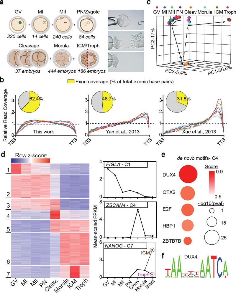
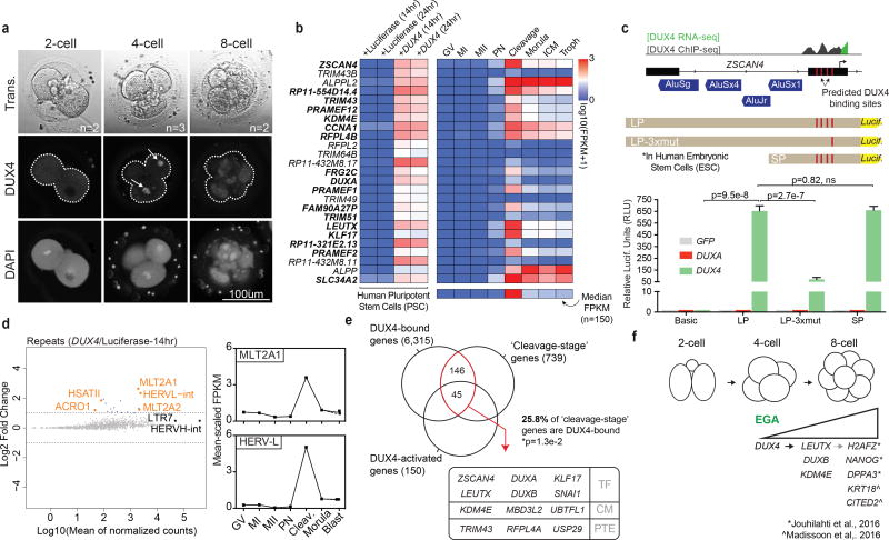
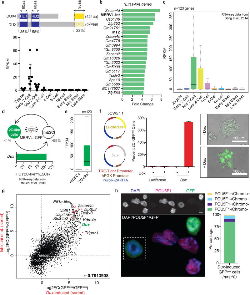
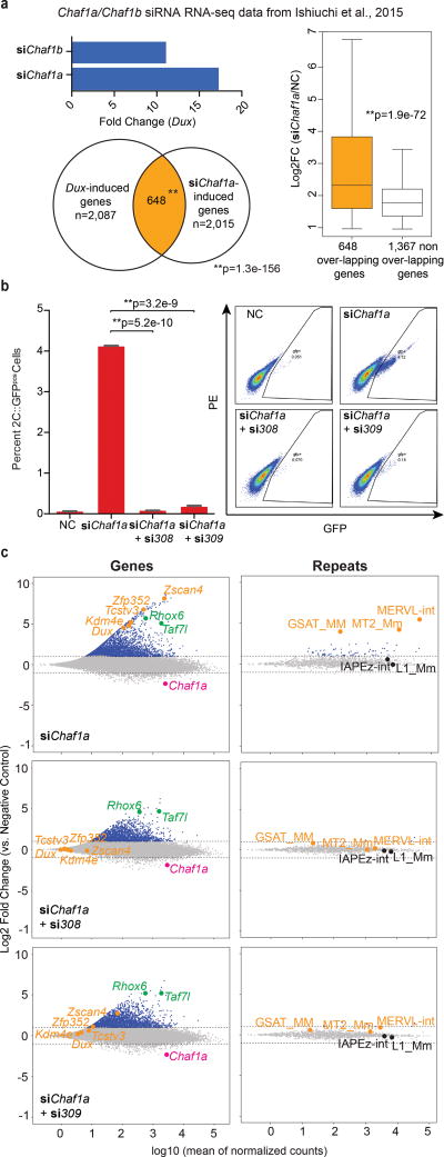
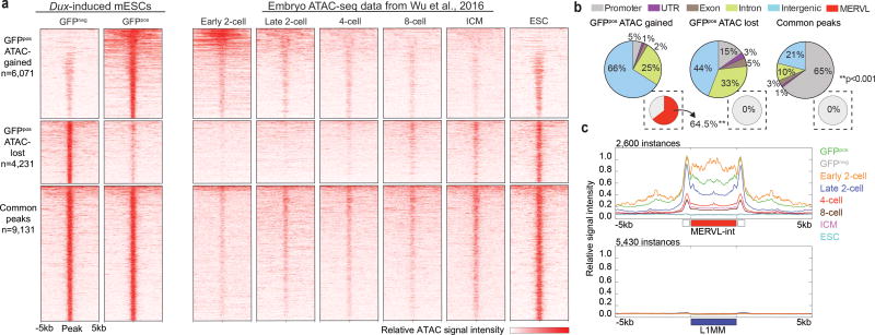
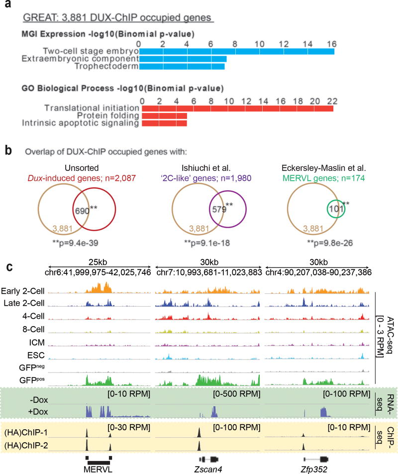
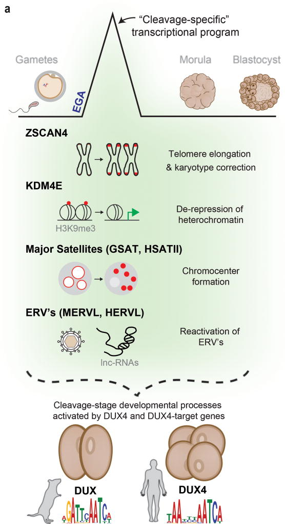
To better understand transcriptional regulation during human oogenesis and preimplantation development, we defined stage-specific transcription, which highlighted the cleavage stage as being highly distinctive. Here, we present multiple lines of evidence that a eutherian-specific multicopy retrogene, DUX4, encodes a transcription factor that activates hundreds of endogenous genes (for example, ZSCAN4, KDM4E and PRAMEF-family genes) and retroviral elements (MERVL/HERVL family) that define the cleavage-specific transcriptional programs in humans and mice. Remarkably, mouse Dux expression is both necessary and sufficient to convert mouse embryonic stem cells (mESCs) into 2-cell-embryo-like ('2C-like') cells, measured here by the reactivation of '2C' genes and repeat elements, the loss of POU5F1 (also known as OCT4) protein and chromocenters, and the conversion of the chromatin landscape (as assessed by transposase-accessible chromatin using sequencing (ATAC-seq)) to a state strongly resembling that of mouse 2C embryos. Thus, we propose mouse DUX and human DUX4 as major drivers of the cleavage or 2C state.


Conflict of interest statement

Competing Financial Interests

The authors have no competing financial interests.

Figures

Figure 1
Figure 1. Improved RNA-sequencing methods reveal new novel transcription, dynamic splice isoform expression, and stage-specific gene expression in human oocytes and pre-implantation development
(a) Summary of the human oocyte and embryonic stages (and cell numbers) collected (left panel), and depiction of the laser mechanical separation of day 5–6 blastocysts into ICM and mural trophectoderm (right panel). (b) Metagene comparison of relative read coverage (from TSS to TTS) in this work and prior studies; each line represents a single developmental stage. Inset pie charts display the corresponding fraction of total exon bases covered by RNA-seq reads. (c) Principal component analysis (PCA) of all egg and embryonic stages based on the highest 50% of all expressed genes (>1 mean FPKM). (d) Statistically determined k-means clusters based on the highest 50% all expressed genes (left panel). Clusters 1, 4, and 7 exhibit stage-specific gene expression and contain prominent developmentally important genes, FIGLA, ZSCAN4, and NANOG, respectively (right panel). (e) The top five de novo motifs enriched in cluster 4 (C4) gene promoters (pre-filtered for ‘best match’ score >0.70). Score- depicted here by color- indicates how strongly the discovered motif matches a known TF binding site. (f) The predicted binding site for DUX4.
Figure 2
Figure 2. DUX4 directly activates the genes and repeat elements that are transiently expressed during human cleavage stage
(a) Immunofluorescence of DUX4 protein in human 2-cell, 4-cell and 8-cell embryos (n=7). (Note: though only one plane is shown, expression was restricted to nuclei of the 4-cell stage, indicated with arrows). (b) Heatmap depicting the top 25 DUX4-activated genes in human iPSCs and their expression in the embryo [two replicates per condition]. Bold font indicates genes belonging to cluster 4 (see Fig. 1d). The bottom row of the heatmap depicts the median embryonic expression of all 150 genes upregulated following DUX4 expression. (c) A diagram of the ZSCAN4 promoter/TSS and the position of the DUX4 ChIP occupancy in DUX4-expressing myoblasts (top panel). ZSCAN4 activation is dependent on DUX4 binding (bottom panel) [four biological replicates per condition. Statistics determined using a two-tailed unpaired t-test. Error bars, s.d.]. (d) MA-plot showing DUX4-mediated induction of specific repeat elements, by subfamily (left panel). Mean-scaled expression of top activated repeats, HERVL and MLT2A1 in human oocytes and embryos (right panel). (e) The overlap of DUX4-ChIP occupied genes [two replicates] with genes enriched in the cleavage-stage embryo and activated by DUX4-overexpression in iPSCs [Overlap statistic calculated by hypergeometric test. Note - only 477 of 739 ‘cleavage genes’ were annotated in GREAT]. In the box, genes encoding notable transcription factors (TF), chromatin modifiers (CM), and post-translational modifying enzymes (PTE) in the overlapping population are listed. (f) Diagram summarizing the timing of DUX4 expression and its effects on embryonic gene expression.
Figure 3
Figure 3. Mouse Dux, a functional ortholog of human DUX4, activates a ‘2C’ transcriptional program and converts mESCs to a ‘2C-like’ state
(a) Sequence level comparison of DUX4 and DUX (top panel) and the normalized expression of Dux in pre-implantation mouse embryos (RNA-seq data from Deng et al., 2014) (bottom panel). (b) Bar graph displaying the top 15 differentially-expressed genes and repeat elements (bold) following ectopic Dux expression in mouse embryonic stem cells (mESCs) [two replicates per condition]. (c) Relative expression of Dux-induced genes (n=123) in the pre-implantation mouse embryo. (d) Diagram of mESC metastability (top panel) and the enrichment of Dux in ‘2C-like’ cells relative to conventional mESCs (bottom panel). (e) Expression of Dux-induced genes (n=123) in ‘2C-like’ cells compared to conventional mESCs. (f) Diagram of doxycycline-inducible lentiviral constructs stably integrated into mESCs (left panel) and their effect (after 24hrs of dox administration) on MERVL::GFP reporter expression evaluated by flow cytometry (middle panel) and live imaging microscopy (right panel) [four biological replicates per condition. Error bars, s.d]. (g) Dot plot showing per gene differential expression in Dux-induced MERVL::GFPpos cells (over MERVL::GFPneg cells), x-axis; compared with per gene differential expression observed in spontaneously converting ‘2C-like’ cells, y-axis. (h) Immunofluorescence quantifying the loss of pluripotency (e.g. POU5F1 protein) and chromocenters in mESCs following ectopic Dux expression (n =110 cells). Scale bar, 10um.
Figure 4
Figure 4. Dux is necessary for spontaneous and CAF-1 mediated conversion of mESCs to a ‘2C-like’ state
(a) Dux is highly upregulated in CAF-1 depleted mESCs (top). Venn diagram displays large overlap of Dux-induced genes with genes activated in Chaf1a-depleted mESCs (bottom) [Overlap statistic calculated by hypergeometric test]. DUX target genes display significantly higher induction than non-targets in Chaf1a-depleted mESCs (right) [Statistics determined using a one-tailed unpaired t-test.] (c) Flow cytometry quantifies the percentage of GFPpos cells following Chaf1a knockdown alone (siChaf1a) and in combination with Dux knockdown (si308 or si309) [three biological replicates per condition. Statistics determined using a two-tailed unpaired t-test. Error bars, s.d]. (c) MA-plots show changes in gene and repeat element expression (by subfamily) in mESCs following knockdown of Chaf1a alone (top panel) and in combination with Dux (si308-middle panel; si309-bottom panel).
Figure 5
Figure 5. Dux-induced ‘2C-like’ cells acquire an open chromatin landscape that resembles an early 2-cell stage embryo
(a) Heatmaps display regions of ATAC-seq signal gain, loss, and found in common between Dux-induced GFPpos and GFPneg cell populations [Two replicates per condition]. Dux-induced GFPpos cells acquire an open/closed chromatin landscape that resembles the early 2-cell stage embryo (Embryo ATAC-seq data from Wu et al., 2016). (b) Pie charts depicting the distribution of ATAC-seq gained, lost and common peaks at basic genomic features. Inset pie charts indicate the percentage of peaks that overlap with MERVL elements (MT2_Mm and MERVL-int) [Enrichment statistic determined empirically]. (c) Metagene analysis of ATAC-seq signal across all MERVL-int instances (top panel) and L1 instances (bottom panel) in Dux-induced GFPpos and GFPneg cells and the early embryo.
Figure 6
Figure 6. DUX binds directly to ‘2C’ gene promoters and retrotransposons
(a) Top enriched ‘MGI expression’ and ‘Gene Ontology (GO)’ terms identified in the 3,881 genes bound by DUX [two replicates]. (b) Overlap of DUX-ChIP occupied genes with genes: upregulated in unsorted mESCs post Dux overexpression (left panel); enriched in ‘2C-like’ cells (middle panel); and driven by MERVL elements (right panel) [Statistics determined by hypergeometric test]. Screenshots demonstrating the overlap of DUX-ChIP occupancy (yellow box) with the acquisition of 2-Cell embryo-like open chromatin and gene/MERVL expression (green box).
Figure 7
Figure 7. A model of DUX4 function during cleavage
(a) A cleavage-specific transcriptional program is activated at EGA in mouse and human cells by DUX or DUX4, respectively. The genes and repetitive elements activated by these DUX4-family genes mediate important molecular events associated with embryonic genome activation (EGA) and reprogramming in the mouse embryo (shaded in green). In human embryos, although activation of these genes and repetitive elements has been shown, their impact on these processes remains to be studied.

References

    1. Liu L, et al. Telomere lengthening early in development. Nat Cell Biol. 2007;9:1436–1441. - PubMed
    1. Matoba S, et al. Embryonic development following somatic cell nuclear transfer impeded by persisting histone methylation. Cell. 2014;159:884–895. - PMC - PubMed
    1. Chung YG, et al. Histone Demethylase Expression Enhances Human Somatic Cell Nuclear Transfer Efficiency and Promotes Derivation of Pluripotent Stem Cells. Cell Stem Cell. 2015;17:758–766. - PubMed
    1. Zalzman M, et al. Zscan4 regulates telomere elongation and genomic stability in ES cells. Nature. 2010;464:858–863. - PMC - PubMed
    1. Kalmbach K, Robinson LG, Wang F, Liu L, Keefe D. Telomere Length Reprogramming in Embryos and Stem Cells. Biomed Res Int. 2014;2014:925121. - PMC - PubMed