. 2017 May 1;12(5):e0175807.
doi: 10.1371/journal.pone.0175807. eCollection 2017.

Transplantation of HGF gene-engineered skeletal myoblasts improve infarction recovery in a rat myocardial ischemia model

Affiliations

Transplantation of HGF gene-engineered skeletal myoblasts improve infarction recovery in a rat myocardial ischemia model

Shu-Ling Rong et al. PLoS One. .

Erratum in

Retraction in

Abstract

Background: Skeletal myoblast transplantation seems a promising approach for the repair of myocardial infarction (MI). However, the low engraftment efficacy and impaired angiogenic ability limit the clinical efficiency of the myoblasts. Gene engineering with angiogenic growth factors promotes angiogenesis and enhances engraftment of transplanted skeletal myoblasts, leading to improved infarction recovery in myocardial ischemia. The present study evaluated the therapeutic effects of hepatocyte growth factor (HGF) gene-engineered skeletal myoblasts on tissue regeneration and restoration of heart function in a rat MI model.

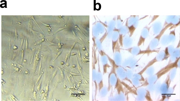
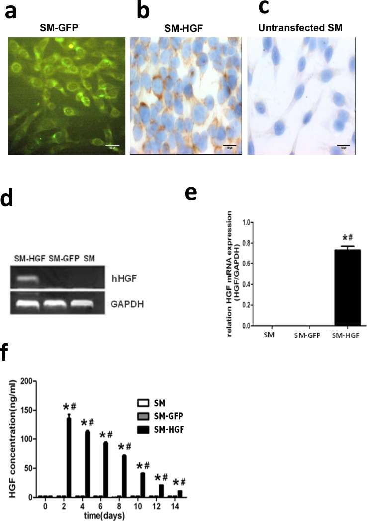
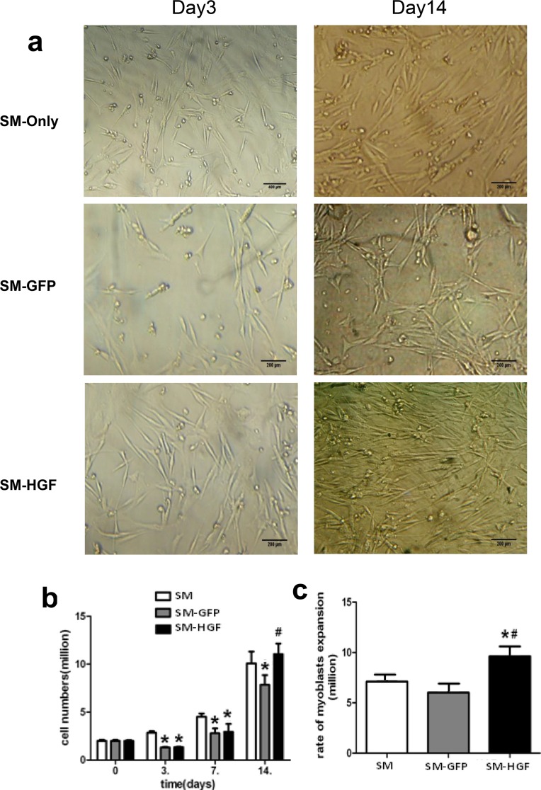
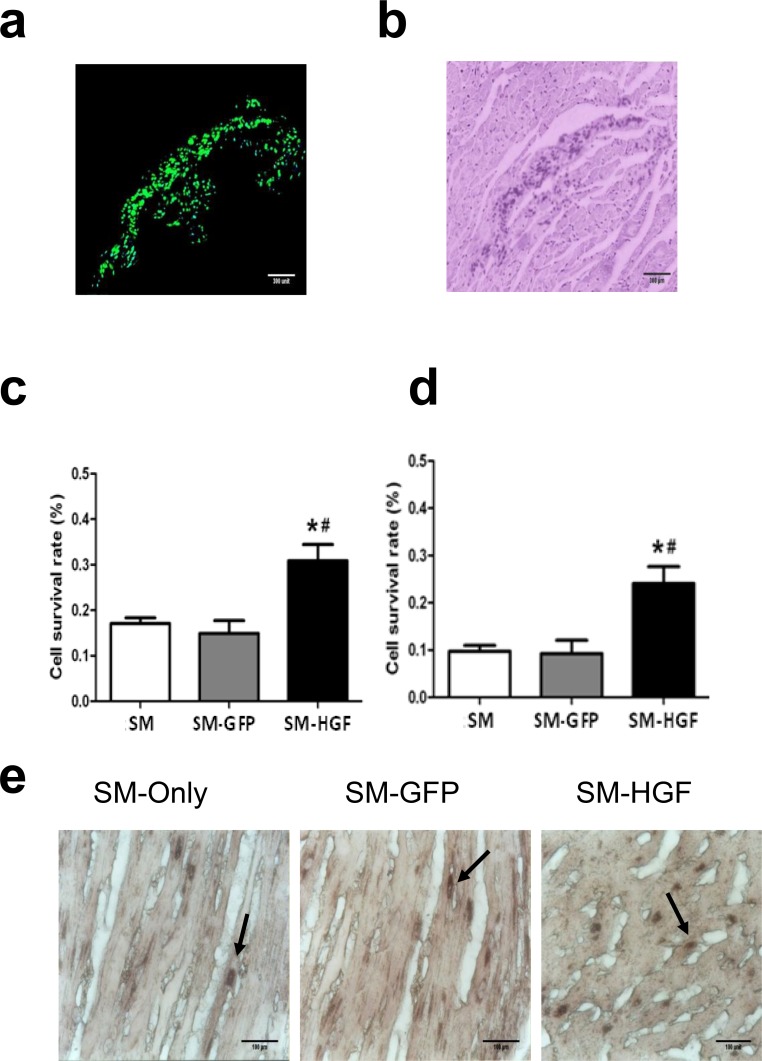
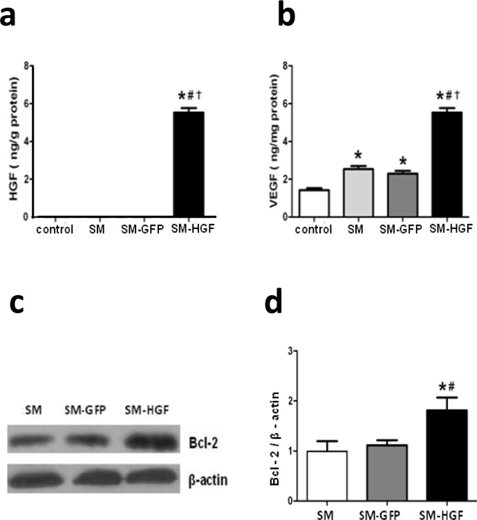
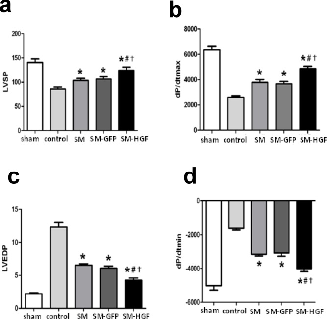
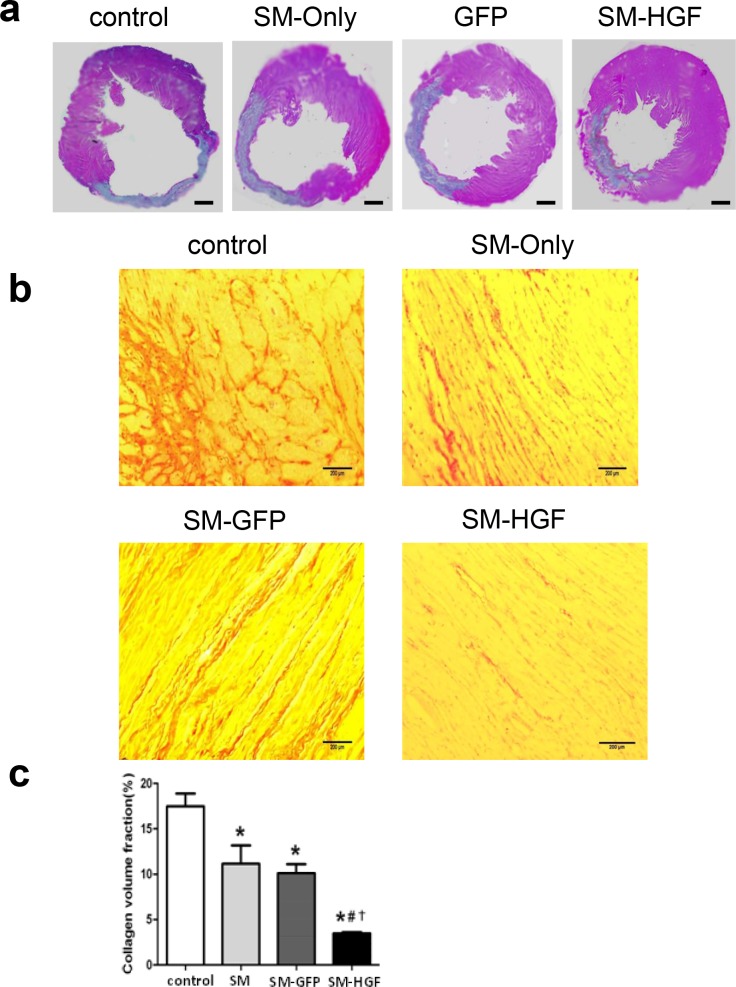
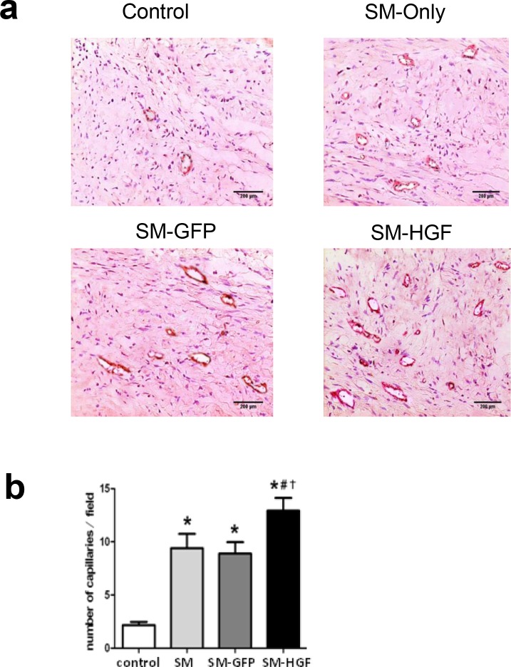
Methods and results: The skeletal myoblasts were isolated, expanded, and transduced with adenovirus carrying the HGF gene (Ad-HGF). Male SD rats underwent ligation of the left anterior descending coronary artery. After 2 weeks, the surviving rats were randomized into four groups and treated with skeletal myoblasts by direct injection into the myocardium. The survival and engraftment of skeletal myoblasts were determined by real-time PCR and in situ hybridization. The cardiac function with hemodynamic index and left ventricular architecture were monitored; The adenovirus-mediated-HGF gene transfection increases the HGF expression and promotes the proliferation of skeletal myoblasts in vitro. Transplantation of HGF-engineered skeletal myoblasts results in reduced infarct size and collagen deposition, increased vessel density, and improved cardiac function in a rat MI model. HGF gene modification also increases the myocardial levels of HGF, VEGF, and Bcl-2 and enhances the survival and engraftment of skeletal myoblasts.

Conclusions: HGF engineering improves the regenerative effect of skeletal myoblasts on MI by enhancing their survival and engraftment ability.


Conflict of interest statement

Competing Interests: The authors have declared that no competing interests exist.

Figures

Fig 1
Fig 1. Identification of myoblasts by immunocytochemistry.
(a) Representative images of myoblasts at day 4 after the primary culture (original magnification ×200). (b) Immunocytochemistry staining of myoblasts by Desmin antibody. Desmin is observed in the cytoplasm (SP, original magnification ×400).
Fig 2
Fig 2. Adenovirus-mediated HGF expression in skeletal myoblasts.
(a) GFP expression in myoblasts transfected with Ad-GFP by fluorescence microscopy (original magnification ×400); (b) Immunocytochemistry staining for human HGF in myoblasts transfected with Ad-HGF (immunocytochemistry staining, original magnification ×400). (c) Immunocytochemistry staining in untransfected control myoblasts (immunocytochemistry staining, original magnification ×400). (d) RT-PCR-based assessment of human HGF gene in myoblasts transfected with Ad-HGF. (e) The RT-PCR analysis of HGF mRNA in SM infected with Ad-HGF, Ad-GFP, and SM only. (f) The Ad-HGF-transduced myoblasts were cultured for 14 days. The conditioned medium was collected at different points in time, and the HGF protein was determined by ELISA. Significant differences were found in myoblasts infected with Ad-HGF (SM-HGF group) as compared to myoblasts infected with Ad-GFP (SM-GFP group) and non-infected myoblasts (SM group) (n = 6, P<0.05, *P<0.05 vs. SM group control cell; #P<0.05 vs. SM-GFP group).
Fig 3
Fig 3. Ad-HGF transfection enhances the proliferation of skeletal myoblasts.
(a) The morphology of skeletal myoblasts transduced with Ad-HGF cultured for 3 (left) and 14 (right) days. (b) The myoblasts were transduced with Ad-HGF and control vector, respectively. 2x10⁶ cells were seeded and cultured for 14 days. The cell numbers were counted at different points in time. (c) The expansion rate of myoblasts in the culture at day 14 post-transfection (n = 6, P<0.05, *P<0.05 vs. SM group control cell; #P<0.05 vs. SM-GFP group).
Fig 4
Fig 4. Assessment of engraftment and cell survival after cell transplantation.
One week after cell transplantation, the GFP+ cells in frozen LV samples (SM-Ad-GFP group) were observed by fluorescence microscope (a). The left myocardium was stained with HE and observed in the bright-field (b). The number of surviving cells (detected using Y chromosome real-time PCR) in the myocardial at week 1 (c) and week 4 (d) after cell transplantation (n = 6, P<0.05, *P<0.05 vs. SM group control cell; #P<0.05 vs. SM-GFP group). The Sry gene-positive cells were identified within the transplanted area in all groups with the use of in situ hybridization 4 weeks after cell transplantation (Fig 4E). The arrows showed the Sry-positive cells.
Fig 5
Fig 5. Implantation of HGF-modified skeletal myoblasts increases the HGF and VEGF expression in the myocardium.
The Ad-GFP- or Ad-HGF- transduced myoblasts were implanted into the myocardium in an MI model for 7 days. The protein levels of HGF (a) and VEGF (b) in the transplanted area were determined by ELISA. (c) Western blot was performed for Bcl-2 expression in SM transduced with Ad-HGF, vector control, and cell control group. (d) The ratio of Bcl-2 to β-actin in the transplanted area was evaluated by Western blot (n = 6, P<0.05, *P<0.05 vs. SM group; #P<0.05 vs. SM-GFP group, P<0.05 vs. control group).
Fig 6
Fig 6. Implantation of HGF-modified skeletal myoblasts improves the heart function.
The Ad-GFP or Ad-HGF transduced myoblasts were implanted into the myocardium in an MI model for 28 days. The cardiac functional parameters including LVSP (a) and +dp/dtmax (b), LVEDP (c), and dp/dtmin (d) were detected (n = 6, P<0.05, *P<0.05 vs. SM group; #P<0.05 vs. SM-GFP group; P<0.05 vs. control group). (d) Western blot for Bcl-2 expression in SM transduced with Ad-HGF, vector control, and cell control group. (n = 6, P<0.05, *P<0.05 vs. SM group; #P<0.05 vs. SM-GFP group; P<0.05 vs. control group).
Fig 7
Fig 7. Implantation of HGF-modified skeletal myoblasts reduces infarct area and reduces collagen deposition.
The Ad-GFP- or Ad-HGF-transduced myoblasts were implanted into the myocardium in an MI model for 28 days. (a) Representative images of histological sections in left ventricular stained with HE. Scale bars, 1 mm. (b) Images show that Sirius Red stained the cardiac sections from SM-GFP, SM only, SM-HGF group, and sham group, respectively. (c) The collagen volume fraction of SM-HGF group compared to the control group and SM group (n = 6, P<0.05, *P<0.05 vs. SM group; #P<0.05 vs. SM-GFP group; P<0.05 vs. control group).
Fig 8
Fig 8. Implantation of HGF-modified skeletal myoblasts increase the vessel density.
The Ad-GFP- or Ad-HGF-transduced myoblasts were implanted into the myocardium in an MI model for 28 days. (a) Sections were stained with antibodies against Factor VIII to facilitate the counting of vessels. The representative images of the capillary density in transplanted area were shown. (b) The numbers of vascular densities of groups indicated (n = 6, P<0.05, *P<0.05 vs. SM group; #P<0.05 vs. SM-GFP group; P<0.05 vs. control group).

Similar articles

Cited by

References

    1. Mozaffarian D, Benjamin EJ, Go AS, Arnett DK, Blaha MJ, Cushman M, et al. Heart disease and stroke statistics—2015 update: a report from the American Heart Association. Circulation, 2015. 131(4): p. e29–322. doi: 10.1161/CIR.0000000000000152 - DOI - PubMed
    1. Taylor DA, Zenovich AG. Cell therapy for left ventricular remodeling. Curr Heart Fail Rep, 2007. 4(1): p. 3–10. - PubMed
    1. Schachinger V, Erbs S, Elsässer A, Haberbosch W, Hambrecht R, Hölschermann H, et al. Intracoronary bone marrow-derived progenitor cells in acute myocardial infarction. N Engl J Med, 2006. 355(12): p. 1210–21. doi: 10.1056/NEJMoa060186 - DOI - PubMed
    1. Henning RJ, Stem cells in cardiac repair. Future Cardiol, 2011. 7(1): p. 99–117. doi: 10.2217/fca.10.109 - DOI - PubMed
    1. Guarita-Souza LC, Carvalho KA, Woitowicz V, Rebelatto C, Senegaglia A, Hansen P, et al. Simultaneous autologous transplantation of cocultured mesenchymal stem cells and skeletal myoblasts improves ventricular function in a murine model of Chagas disease. Circulation, 2006. 114(1 Suppl): p. I120–4. doi: 10.1161/CIRCULATIONAHA.105.000646 - DOI - PubMed

Publication types

MeSH terms