. 2017 Jun 1;140(6):1561-1578.
doi: 10.1093/brain/awx095.

Hypomorphic mutations in POLR3A are a frequent cause of sporadic and recessive spastic ataxia

Martina Minnerop  1   2 Delia Kurzwelly  2   3 Holger Wagner  4 Anne S Soehn  5 Jennifer Reichbauer  6   7 Feifei Tao  8 Tim W Rattay  6   7 Michael Peitz  3   9 Kristina Rehbach  3   9 Alejandro Giorgetti  10   11 Angela Pyle  12 Holger Thiele  13 Janine Altmüller  13   14 Dagmar Timmann  15 Ilker Karaca  4 Martina Lennarz  4 Jonathan Baets  16   17   18 Holger Hengel  6   7 Matthis Synofzik  6   7 Burcu Atasu  7   19 Shawna Feely  20 Marina Kennerson  21   22   23 Claudia Stendel  24   25 Tobias Lindig  26 Michael A Gonzalez  8 Rüdiger Stirnberg  3 Marc Sturm  5 Sandra Roeske  3 Johanna Jung  3 Peter Bauer  5 Ebba Lohmann  7   17   27 Stefan Herms  28   29   30 Stefanie Heilmann-Heimbach  28   29 Garth Nicholson  21   22   23 Muhammad Mahanjah  31   32 Rajech Sharkia  33   34 Paolo Carloni  10 Oliver Brüstle  3   9 Thomas Klopstock  24   25   35 Katherine D Mathews  36 Michael E Shy  20 Peter de Jonghe  16   17   18 Patrick F Chinnery  12   37 Rita Horvath  38 Jürgen Kohlhase  39 Ina Schmitt  2 Michael Wolf  40 Susanne Greschus  41 Katrin Amunts  1   42 Wolfgang Maier  3   4 Ludger Schöls  6   7 Peter Nürnberg  1   3   43   44 Stephan Zuchner  8 Thomas Klockgether  2   3 Alfredo Ramirez  4   28   45 Rebecca Schüle  6   7   8
Affiliations

Hypomorphic mutations in POLR3A are a frequent cause of sporadic and recessive spastic ataxia

Martina Minnerop et al. Brain. .

Erratum in

  • Erratum.
    [No authors listed] [No authors listed] Brain. 2018 Mar 1;141(3):e21. doi: 10.1093/brain/awx329. Brain. 2018. PMID: 29236964 Free PMC article. No abstract available.

Abstract

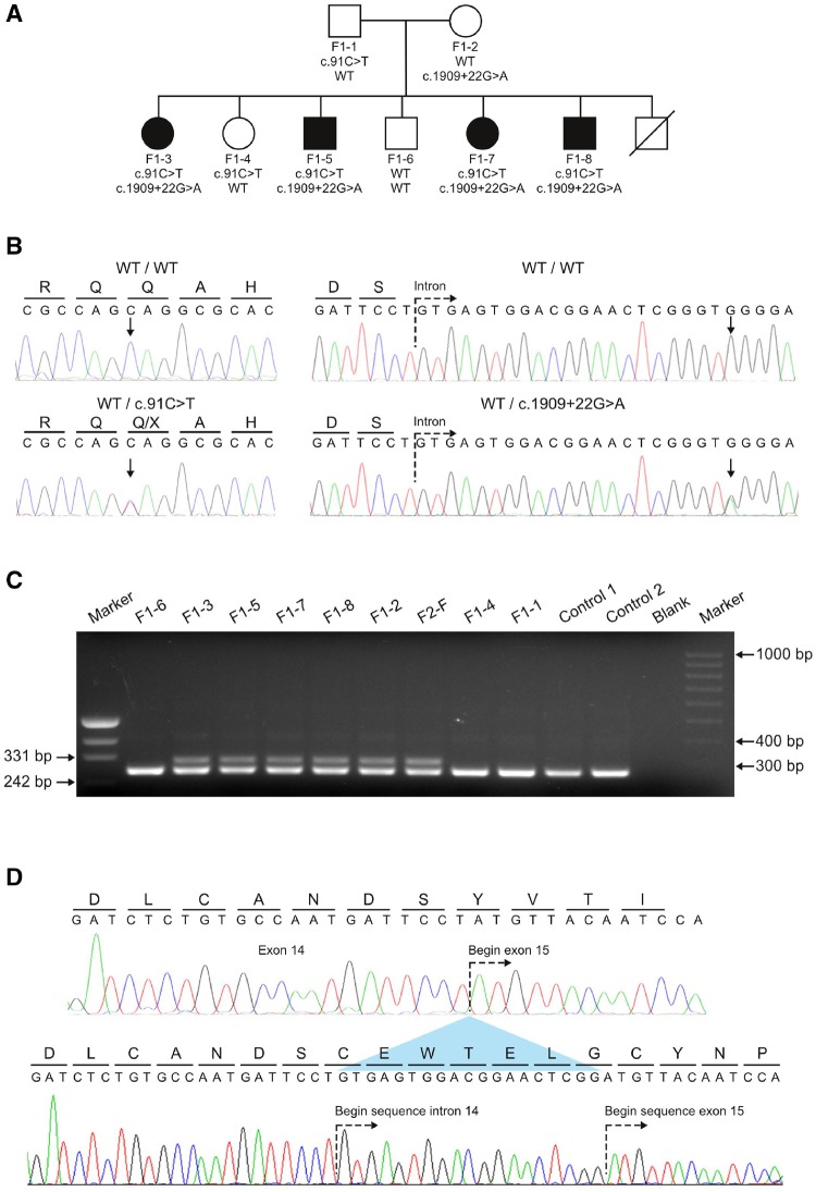
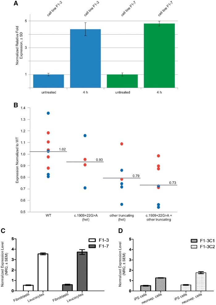
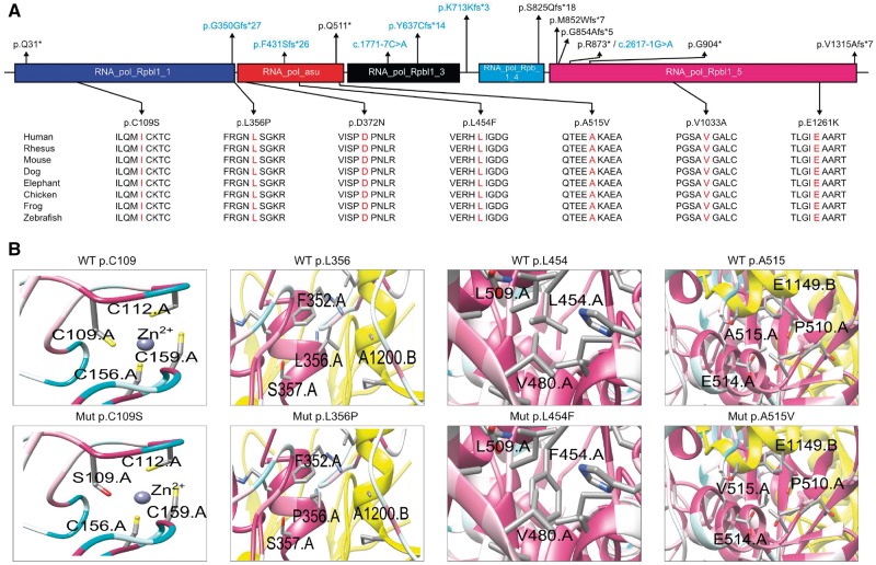
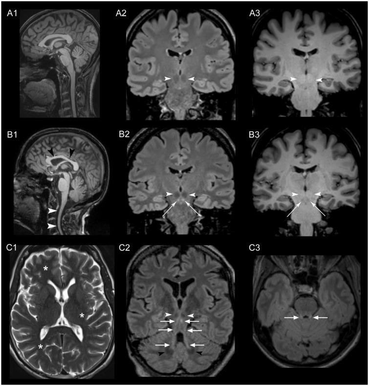
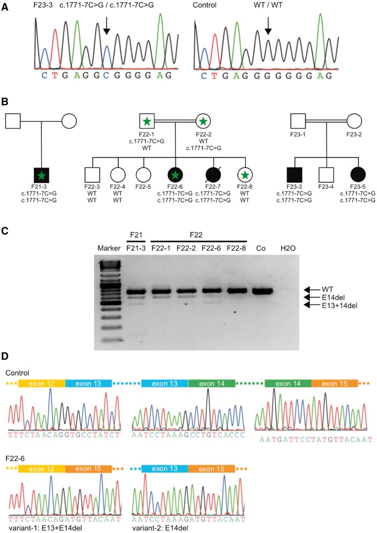
Despite extensive efforts, half of patients with rare movement disorders such as hereditary spastic paraplegias and cerebellar ataxias remain genetically unexplained, implicating novel genes and unrecognized mutations in known genes. Non-coding DNA variants are suspected to account for a substantial part of undiscovered causes of rare diseases. Here we identified mutations located deep in introns of POLR3A to be a frequent cause of hereditary spastic paraplegia and cerebellar ataxia. First, whole-exome sequencing findings in a recessive spastic ataxia family turned our attention to intronic variants in POLR3A, a gene previously associated with hypomyelinating leukodystrophy type 7. Next, we screened a cohort of hereditary spastic paraplegia and cerebellar ataxia cases (n = 618) for mutations in POLR3A and identified compound heterozygous POLR3A mutations in ∼3.1% of index cases. Interestingly, >80% of POLR3A mutation carriers presented the same deep-intronic mutation (c.1909+22G>A), which activates a cryptic splice site in a tissue and stage of development-specific manner and leads to a novel distinct and uniform phenotype. The phenotype is characterized by adolescent-onset progressive spastic ataxia with frequent occurrence of tremor, involvement of the central sensory tracts and dental problems (hypodontia, early onset of severe and aggressive periodontal disease). Instead of the typical hypomyelination magnetic resonance imaging pattern associated with classical POLR3A mutations, cases carrying c.1909+22G>A demonstrated hyperintensities along the superior cerebellar peduncles. These hyperintensities may represent the structural correlate to the cerebellar symptoms observed in these patients. The associated c.1909+22G>A variant was significantly enriched in 1139 cases with spastic ataxia-related phenotypes as compared to unrelated neurological and non-neurological phenotypes and healthy controls (P = 1.3 × 10⁻⁴). In this study we demonstrate that (i) autosomal-recessive mutations in POLR3A are a frequent cause of hereditary spastic ataxias, accounting for about 3% of hitherto genetically unclassified autosomal recessive and sporadic cases; and (ii) hypomyelination is frequently absent in POLR3A-related syndromes, especially when intronic mutations are present, and thus can no longer be considered as the unifying feature of POLR3A disease. Furthermore, our results demonstrate that substantial progress in revealing the causes of Mendelian diseases can be made by exploring the non-coding sequences of the human genome.

Keywords: POLR3A; cerebellar ataxia; hereditary spastic paraplegia; leukodystrophy; spastic ataxia.


Figures

Figure 1
Figure 1
Genetic screening of a non-consanguineous German family. (A) Pedigree of Family F1 affected by the novel spastic ataxia syndrome. Pedigree symbols: circle, female; square, male; filled, affected; unfilled, unaffected; strike through, deceased. The compound heterozygous mutations found in POLR3A are shown below each symbol. Both mutations segregated with the disease phenotype in the family. (B) Sequencing traces showing the wild-type alleles and the mutant alleles c.91C>T and c.1909+22G>A. Arrows denote the nucleotides undergoing a change for the respective mutations. All affected members in Family F1 are compound heterozygous carriers for both mutations. (C) Agarose gel showing PCR product obtained from the amplification of cDNA template derived from patients, healthy family members, and two controls to show the effect of c.1909+22G>A on the splicing of exon 14. A primer pair flanking exon 14 was used to amplify leucocyte-derived cDNA template from six carriers of the c.1909+22G>A variant and three additional family members of Family F1 that carry the wild-type allele (Patients F1-1, F1-4 and F1-6). An additional aberrant larger fragment (298 bp) in carriers of c.1909+22G>A is not present in the five non-carriers of the splice site mutation. (D) Sequencing analysis of the aberrant amplicon. The upper sequence trace shows the wild-type cDNA sequence obtained for exon 14 and 15. The predicted amino acid translation of this sequence is shown above the nucleotides. The lower sequence trace represents direct sequencing of the aberrant fragment obtained in carriers of the c.1909+22G>A allele. An out-of-frame inclusion of the first 19 nucleotides of intron 14 (caused by the activation of a cryptic splice site) causes a shift of the reading frame.
Figure 2
Figure 2
Expression analysis of POLR3A. (A) Specific quantitative-PCR amplification of aberrant splice variant in fibroblast cells from two affected members of Family F1. Expression of POLR3A is normalized with YWHAZ and SDHA as reference genes. Expression levels are shown as relative fold expression levels of cells treated with cycloheximide (100 ng/ml) for 4 h compared to untreated cells. Inhibition of nonsense-mediated mRNA decay led to almost a 5-fold increase of the amount of aberrantly spliced transcript compared to untreated cells. (B) Figure demonstrating interindividual variability in POLR3A expression in controls (wild-type, WT), healthy heterozygous carriers of c.1909+22G>A, healthy heterozygous carriers of other truncating mutations, and affected individuals with compound heterozygous POLR3A mutations (i.e. c.1909+22G>A plus an additional truncating POLR3A mutation). Blue circles are Light Cycler (LC) data from Tübingen (normalized to controls that were available in Tübingen), red circles are TaqMan® data from Bonn (normalized to controls available in Tübingen). Group-wise comparison showed the strongest reduction of total POLR3A levels in the group of affected individuals with compound heterozygous POLR3A mutations of ∼39% of that observed in wild-type. (C) Specific expression levels of the aberrant transcript of POLR3A in adult fibroblasts and leukocytes from Patients F1‐3 and F1‐7 shown relative to average expression level. (D) Specific expression levels of the aberrant transcript of POLR3A in different developmental stages. Fibroblasts from Patients F1‐3 and F1‐7 were reprogrammed to establish iPSC lines. Two iPSC clones from Patient F1‐3 (F1‐3C1 and F1‐3C2), were differentiated to neuroepithelial cells (neuroep. cells). The plot shows expression of cDNA template derived from iPSC lines and iPSC-derived neuroepithelial cells. The level of cryptic splice site activation is lowest in the iPSCs reprogrammed from patient fibroblasts.
Figure 3
Figure 3
Schematic representation of RPC1 and 3D structure of amino acid exchanges. (A) Schematic representation of the RPC1 protein. Exonic and intronic mutations that result in missense, nonsense, or frameshift mutations are mapped to the respective domains. Intronic variants that affect splicing are labelled in blue. The amino acid conservation across species was determined for all missense variants from the UCSC Genome Browser (genome.ucsc.edu). (B) Protein 3D representations of POLR3A missense mutations p.C109S, p.L356P, p.L454F and p.A515V. 3D representations of POLR3A missense mutations p.D372N, p.E1261K and p.V1033A are shown in Supplementary Fig. 8A. In the panels, the colour of the main chain of the protein indicates highly conserved (pink) to poorly conserved (white) amino acid residues (based on the entire phylogenetic tree, see ‘Materials and methods’ section). The interacting subunits (chain B: RPB2, chain E: RPABC1 and chain K: RPABC5) are indicated in yellow, green and red, respectively. For a model of the full-length RPC1 and the localization of each of the mutants in relation with the accessory proteins with the same colour code, see Supplementary Fig. 8B.
Figure 4
Figure 4
Brain MRI. MRI images of a healthy control (A13) and Patients F1‐5 (B1), F13-1 (B2, B3), F1‐5 (C1), F1‐7 (C2) and F5-1 (C3), obtained using a 3 T Scanner (MAGNETOM Skyra, Siemens Healthcare). White arrowheads in B1 indicate the atrophy of the cervical spinal cord and the comparatively hypoplastic corpus callosum (black arrowheads) in a sagittal T1-weighted image compared to a healthy control in A1. In B2, C2 and C3 a standard coronal (B2), standard axial (C3) and a paracoronal view (C2)—the latter angulated along the course of the superior cerebellar peduncle—were reformatted from the 3D FLAIR sequence dataset. Striking bilateral hyperintensity (bright signal) along the entire superior cerebellar peduncles (arrows in B2, C2 and C3) ranging from the cerebellar dentate nucleus (black arrowheads in C2) to the midbrain just below the red nucleus (white arrowheads in B2 and C2) was found. The hyperintensity in the FLAIR images had a hypointense correlate in T1-weighted images (B3) indicating secondary myelin degradation rather than hypomyelination (Schiffmann and van der Knaap, 2009). No signs of generalized hypomyelination in T2 images as described in hypomyelinating leukodystrophy were observed (asterisks in C1, axial T2-weighted image indicating a normal myelination in subcortical white matter and internal capsule exemplarily shown in Patient F1‐5).
Figure 5
Figure 5
Functional consequences and co-segregation of c.1771-7C>G. (A) Sequencing of genomic DNA of the proband Patient F23-3 and a healthy control. The proband carries a homozygous intronic variant (c.1771-7C>G). (B) The same intronic variant was found in three different unrelated families. Segregation of the variant with the disease status was demonstrated for two of the families (Families F22 and F23). Genotypes are depicted below the pedigree symbols. Family members with available leucocyte RNA for analysis are marked by a green asterisk. (C) PCR products obtained from the amplification of patient and control cDNA leucocyte samples showing the effect of the c.1771-7C>G variant on the splicing of exon 14. The sequence traces for controls showing the exon–intron boundaries of exons 12 to 15 are depicted. In the probands carrying the c.1771-7C>G variant (the sequence shown is for proband Patient F22-6) two additional splice variants occur (E13+E14del and E14del). (D) Sequence analysis of proband Patient F22-6. In variant-1 exons 13 (128 bp) and 14 (139 bp) are skipped, leading to a product missing 267 bp. The combined loss of exons 13 and 14 does not lead to a shift of the reading frame. However, a total of 90 amino acids are predicted to be missing from the final protein product (aa 548–637) including a part of the Rpb1_3 domain, which likely represents the pore through which nucleotides enter the active site (Severinov et al., 1996). In variant-2, only exon 14 (139 bp) is skipped, resulting in a shift of the reading frame and the introduction of a pre terminal stop codon nine codons after the mutated codon (p.P591Mfs*9).

Comment in

References

    1. Altschul SF, Madden TL, Schaffer AA, Zhang J, Zhang Z, Miller W. et al. Gapped BLAST and PSI-BLAST: a new generation of protein database search programs. Nucleic Acids Res 1997; 25: 3389–402. - PMC - PubMed
    1. Azmanov DN, Siira SJ, Chamova T, Kaprelyan A, Guergueltcheva V, Shearwood AJ. et al. Transcriptome-wide effects of a POLR3A gene mutation in patients with an unusual phenotype of striatal involvement. Hum Mol Genet 2016; 25: 4302–14. - PubMed
    1. Beers J, Gulbranson DR, George N, Siniscalchi LI, Jones J, Thomson JA. et al. Passaging and colony expansion of human pluripotent stem cells by enzyme-free dissociation in chemically defined culture conditions. Nat Protoc 2012; 7: 2029–40. - PMC - PubMed
    1. Bernard G, Chouery E, Putorti ML, Tetreault M, Takanohashi A, Carosso G. et al. Mutations of POLR3A encoding a catalytic subunit of RNA polymerase Pol III cause a recessive hypomyelinating leukodystrophy. Am J Hum Genet 2011; 89: 415–23. - PMC - PubMed
    1. Boespflug-Tanguy O, Aubourg P, Dorboz I, Begou M, Giraud G, Sarret C. et al. Neurodegenerative disorder related to AIMP1/p43 mutation is not a PMLD. Am J Hum Genet 2011; 88: 392–3; author reply 3–5. - PMC - PubMed

MeSH terms

Supplementary concepts