. 2017 Apr 18;8(16):26471-26491.
doi: 10.18632/oncotarget.15594.

Histamine therapeutic efficacy in metastatic melanoma: Role of histamine H4 receptor agonists and opportunity for combination with radiation

Affiliations

Histamine therapeutic efficacy in metastatic melanoma: Role of histamine H4 receptor agonists and opportunity for combination with radiation

Noelia A Massari et al. Oncotarget. .

Abstract

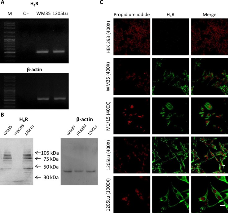
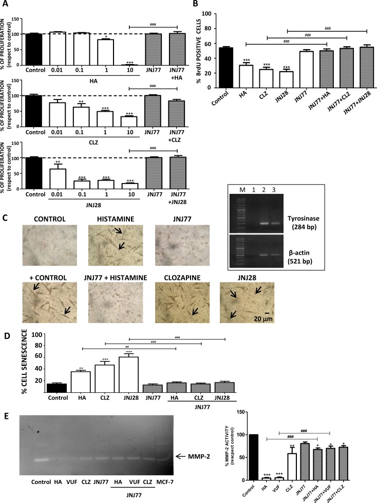
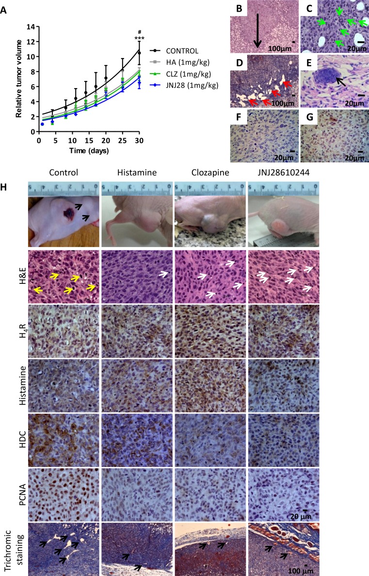
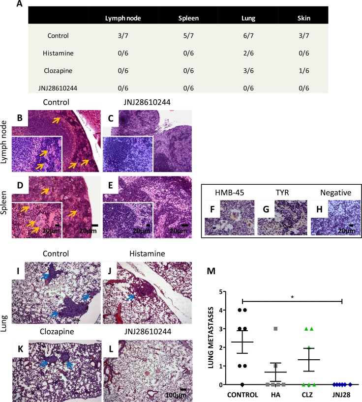
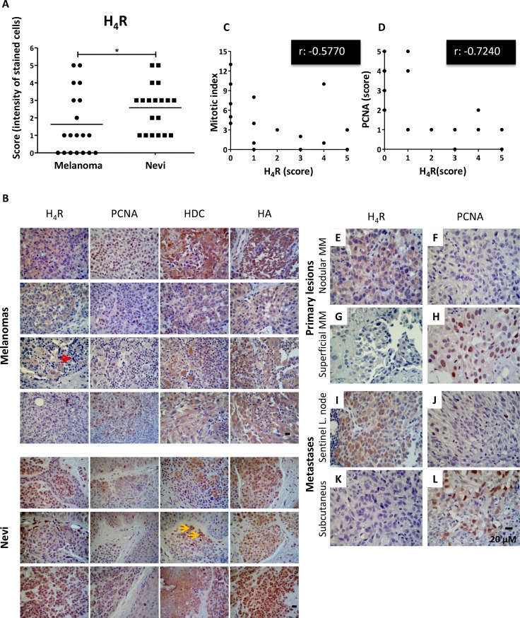
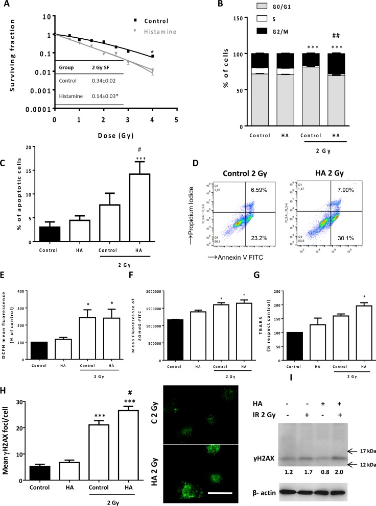
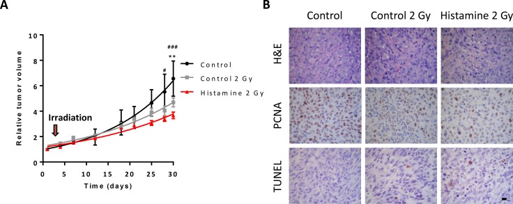
The aims of the work were to improve our knowledge of the role of H4R in melanoma proliferation and assess in vivo the therapeutic efficacy of histamine, clozapine and JNJ28610244, an H4R agonist, in a preclinical metastatic model of melanoma. Additionally, we aimed to investigate the combinatorial effect of histamine and gamma radiation on the radiobiological response of melanoma cells.Results indicate that 1205Lu metastatic melanoma cells express H4R and that histamine inhibits proliferation, in part through the stimulation of the H4R, and induces cell senescence and melanogenesis. Daily treatment with H4R agonists (1 mg/kg, sc) exhibited a significant in vivo antitumor effect and importantly, compounds reduced metastatic potential, particularly in the group treated with JNJ28610244, the H4R agonist with higher specificity. H4R is expressed in benign and malignant lesions of melanocytic lineage, highlighting the potential clinical use of histamine and H4R agonists. In addition, histamine increased radiosensitivity of melanoma cells in vitro and in vivo. We conclude that stimulation of H4R by specific ligands may represent a novel therapeutic strategy in those tumors that express this receptor. Furthermore, through increasing radiation-induced response, histamine could improve cancer radiotherapy for the treatment of melanoma.

Keywords: H4R; JNJ28610244; experimental melanoma; ionizing radiation; tumor growth.


Conflict of interest statement

CONFLICTS OF INTEREST

The authors have no conflicts of interest to declare.

Figures

Figure 1
Figure 1. H4R expression in 1205Lu cells
H4R receptor expression was determined by RT-PCR, Western blot and Immunofluorescence. (A) RT-PCR of H4R. Lanes: M, DNA ladder molecular size marker; WM35, human primary melanoma cells were used as positive control; 1205Lu, human metastatic melanoma cells. β-actin (521 bp) was used as load control. (B) Western blot of H4R. WM35 cells were used as positive control. HEK293 cells were used as negative control. β-actin (42 kDa) was used as load control. (C) Immunofluorescence (green) of H4R in 1205Lu cells evaluated by confocal microscopy. Nuclei were counterstained with ethidium bromide (red). Pictures were taken at 400X-fold and 1000X-fold magnification. Scale bar = 20 μm. Representative results of three independent experiments. WM35 and M1/15 cells were used as positive control. HEK293 cells were used as negative control.
Figure 2
Figure 2. H4R-induced biological responses in 1205Lu cells
Cells were left untreated (control) or treated with histamine (HA), clozapine (CLZ), JNJ28610244 (JNJ28) or VUF8430 (VUF) and/or 10 μM JNJ7777120 (JNJ77) (A) Clonogenic assay. (B) Incorporation of BrdU-positive cells assessed by immunocytochemistry 48 h after treatment. (C) L-dopa staining with melanin precursor (10 mM L-dopa). evaluated 10 days after treatment. Localization of dopa-oxidase was indicated by the presence of an insoluble brown/black precipitate. Pictures were taken at 200X-fold magnification. Scale bar = 20 μm. Arrows indicate cell prolongations. Positive control: 16 nM TPA + L-dopa. Inset: Tyrosinase expression in 1205Lu was evaluated by RT-PCR (284 bp). Lanes: M, DNA ladder molecular size marker; 1, Negative control (without cDNA); 2: Positive control (M1/15 human melanoma cells), 3: 1205Lu cells. β-actin (521 bp) was used as load control. (D) Senescence-associated to β-galactosidase staining evaluated 48 h after treatment. (E) MMP-2 gelatinololytic activity determined 24 h after treatment, MCF-7 cells were used as positive control. Error bars represent the means ± SEM of three independent experiments (ANOVA and Dunnett's Multiple Comparison Test, *P < 0.05, **P < 0.01, ***P < 0.001 vs. control; ANOVA and Newman–Keuls Multiple Comparison Test, ##P < 0.01, ###P < 0.001 vs. JNJ77+HA or H4R agonist). Results are representative of three independent experiments.
Figure 3
Figure 3. Antitumoral effect of H4R agonists in 1205Lu xenografted tumor induced in nude mice
(A) Evaluation of relative tumor volume. Daily sc. treatment 1 mg/kg of histamine (HA), clozapine (CLZ) or JNJ28610244 (JNJ28) significantly diminished the tumor volume, evidencing this effect at the end of the experiment. Non-linear regression fit was performed to evaluate the exponential growth, (Repeated Measures ANOVA, ***P < 0.001 HA, CLZ and JNJ28 vs. control; Two-way ANOVA and Bonferroni post-test #P < 0.05 JNJ28 vs. control). (B) 1205Lu tumors showed vertical growth (arrow), with invasion of reticular dermis (H&E staining, 200X-fold magnification). (C) 1205Lu tumors demonstrated intratumoral neutrophils (green arrow). (D) Dilated lymphatic vessels (red arrows, Masson´s trichromic staining, 200X-fold magnification). (E) Lymphatic emboli (arrow, H&E staining, 400X-fold magnification). Presence of tumor cells in mitosis. (F) HMB-45 positive and (G) Tyrosinase positive immunostaining. Scale bars = 100 and 20 μm. (H) Tumors of the control group showed significant anisocytosis and anisokaryosis, nuclear pleomorphism and atypical mitosis (yellow arrows). Tumors of mice treated with histamine, clozapine or JNJ28610244 presented cell homogeneity, with rounded and uniform nuclei with typical mitosis, and presence of inflammatory infiltrates (white arrows), (H&E staining, 400X-fold magnification). Immunohistochemistry of 1205Lu xenografted mice. Formalin-fixed paraffin embedded tissue sections of control, histamine, clozapine, and JNJ28610244 mice were stained to evaluate intracellular levels of histamine, HDC, H4R expression, proliferation and vascular and connective tissue morphology. Pictures were taken at a 400X-fold magnification for immunostaining (Scale bar = 20 μm) and 50X-fold magnification for trichromic stain (Scale bar = 100 μm).
Figure 4
Figure 4. Analysis of the metastatic potential of 1205Lu xenografted tumor in mice
(A) Number of animals with metastases of the total number of animals in the experimental group. (B) Giant multinucleated cells (yellow arrows) in lymph nodes and (D) in spleen of control mice. (C) Lymph nodes and (E) spleen of 1 mg/kg JNJ28610244 treated mice showing no particularities (H&E staining, 100X-fold magnification, inset: 400X-fold magnification). Scale bar = 20 μm. (F) Representative pictures of HMB-45 and (G) tyrosinase (TYR) positive immunostaining of giant multinucleated cells of spleen, confirming linage specificity. (H) Spleen of treated animals did not give signal for these antigens (negative immunostaining), confirming the absence of metastases. Pictures were taken at 400X-fold magnification. Scale bar = 20 μm. Representative images of lungs from (I) control, (J) histamine, (K) clozapine and (L) JNJ28610244 groups. Blue arrows indicate presence of lung metastases (H&E, 50X-fold magnification, scale bar = 100 μm). (M) The number of lung metastases was evaluated in mice treated with saline solution (control), 1 mg/kg histamine (HA), clozapine (CLZ,) or JNJ28610244 (JNJ28). Each dot represents the number of metastasis per mouse. The middle line represents the average number (Kruskal-Wallis non-parametric Test and Dunn's Multiple Comparison test, *P = 0.0214).
Figure 5
Figure 5. Immunohistochemical detection of H4R, histamine, HDC and PCNA in benign and malignant lesions derived from human melanocytic tissues
(A) H4R expression level in nevi and melanoma tissues. Each dot represents a score (intensity of stained cells). The middle line represents the average number (Mann-Whitney non-parametric two tail Comparison Test, *P = 0.0452). (B) Representative pictures show examples of immunostaining for H4R, histamine, HDC and PCNA in superficial spreading melanoma, nodular melanoma, acral lentiginous amelanotic melanoma, subcutaneous melanoma metastasis, intradermal nevus, and junctional nevus. Red arrow indicates lymphoid cells positively stained. Yellow arrows indicate melanin content. Pictures were taken at 630X-fold magnification. Scale bar = 20 μm. (C) Spearman's inverse correlation between H4R and mitotic index (correlation coefficient rho, r: −0.5770, **P = 0.0097); (D) Spearman's inverse correlation between H4R and PCNA expression (correlation coefficient rho, r: −0.7240, ***P = 0.0005). Primary melanoma: (E, F) Nodular melanoma with high H4R expression levels and low PCNA; (G, H) Superficial amelanotic melanoma with low expression of H4R and high PCNA levels. Metastases: (I, J) Sentinel lymph node with melanoma partial metastases with high H4R levels and low PCNA expression, (K, L) Subcutaneous melanoma metastasis with low H4R expression and high PCNA levels. Pictures were taken at 630X-fold magnification. Scale bar = 20 mm.
Figure 6
Figure 6. Effect of histamine on the radiosensitivity of melanoma cells
1205Lu cells were cultured in presence or absence of histamine (HA) and were irradiated 24 h after treatment. (A) Clonogenic survival was determined. Radiobiological parameters of 1205Lu cells were obtained from the survival curves adjusted to the linear quadratic model [SF= e−(αD+βD2)]. Values are means ± SEM of 3 independent experiments performed in triplicates. Inset: 2 Gy SF of untreated and histamine-treated cells. (B) The percentage of cells in different phases of the cell cycle was monitored 24 h after irradiation using flow cytometry. Results represent the mean value of 3 independent experiment (***P < 0.001 vs. % S phase of Control; ##P < 0.01 vs. % G2/M phase of 2 Gy Control. ANOVA and Newman-Keuls post test). (C) Percentage of apoptotic cells was determined 24 h after irradiation by the TUNEL assay (***P < 0.001 vs. Control; #P < 0.05 vs. 2 Gy Control. ANOVA and Newman-Keuls post test), and by (D) Annexin-V staining and flow cytometry. Annexin-V positive cells are shown in both right quadrants of dot plot. (E) Measurement of intracellular ROS by flow cytometry (*P < 0.05, ANOVA and Newman-Keuls post test). (F) Mean fluorescence analysis of 8-OHdG determined by flow cytometry (*P < 0.05 vs. Control, ANOVA and Newman-Keuls post test). (G) Percentage of thiobarbituric acid reactive species (TBARS) with respect to untreated cells (control), (*P < 0.05 vs. Control, ANOVA and Newman-Keuls post test). (H) DNA double strand breaks were evidenced by γH2AX foci formation. The average number of foci per cell was determined 20 min after irradiation. Representative pictures were taken at a 400X-fold magnification (Scale bar = 20 μm). (I). γH2AX (15 kDa) was assayed by Western blot. β-actin (42 kDa) was used as loading control. Semiquantitative analyses of band intensities are shown (n = 3).
Figure 7
Figure 7. Combined effect of radiation and histamine on melanoma tumors induced in nude mice
(A) Evaluation of relative tumor volume in untreated (control), untreated and 2 Gy irradiated (control 2 Gy) and histamine-treated and 2 Gy irradiated animals (Histamine 2 Gy). Tumor volumes were measured by day and non-linear regression fit was performed to evaluate the exponential growth, (Repeated Measures ANOVA and Dunnet post-test, *P < 0.01 Histamine 2 Gy vs. control; Two-way ANOVA and Bonferroni post-test #P < 0.05, ###P < 0.001 Histamine 2 Gy vs. control). (B) Histological and immunohistochemical analysis. Formalin-fixed paraffin embedded tissue sections of the different groups were stained to evaluate histopathological characteristics (H&E), proliferation (PCNA) and apoptosis (TUNEL). 630X original magnification. Scale bar = 20 μm.

Similar articles

Cited by

References

    1. Block KI, Gyllenhaal C, Lowe L, Amedei A, Amin AR, Amin A, Aquilano K, Arbiser J, Arreola A, Arzumanyan A, Ashraf SS, Azmi AS, Benencia F, et al. Designing a broad-spectrum integrative approach for cancer prevention and treatment. Semin Cancer Biol. 2015;35:S276–304. - PMC - PubMed
    1. Marzuka A, Huang L, Theodosakis N, Bosenberg M. Melanoma Treatments: Advances and Mechanisms. J Cell Physiol. 2015;230:2626–2633. - PubMed
    1. Bernatchez C, Cooper ZA, Wargo JA, Hwu P, Lizée G. Novel Treatments in Development for Melanoma. Cancer Treat Res. 2016;167:371–416. - PubMed
    1. Prieto PA1, Reuben A, Cooper ZA, Wargo JA. Targeted therapies combined with immune checkpoint therapy. Cancer J. 2016;22:138–146. - PMC - PubMed
    1. Medina VA, Rivera ES. Histamine receptors and cancer pharmacology. Br J Pharmacol. 2010;161:755–767. - PMC - PubMed

MeSH terms