. 2017 Apr 18;8(16):26687-26701.
doi: 10.18632/oncotarget.15803.

The vitamin D receptor is involved in the regulation of human breast cancer cell growth via a ligand-independent function in cytoplasm

Affiliations

The vitamin D receptor is involved in the regulation of human breast cancer cell growth via a ligand-independent function in cytoplasm

Trupti Trivedi et al. Oncotarget. .

Abstract

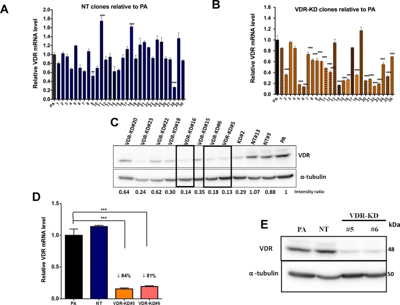
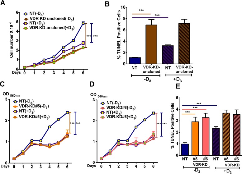
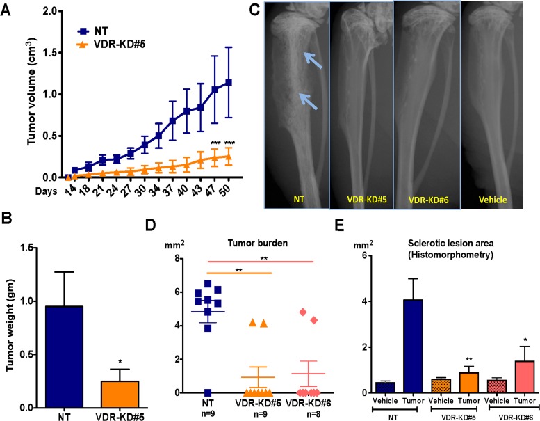
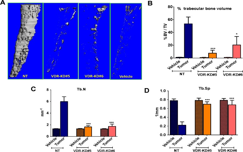
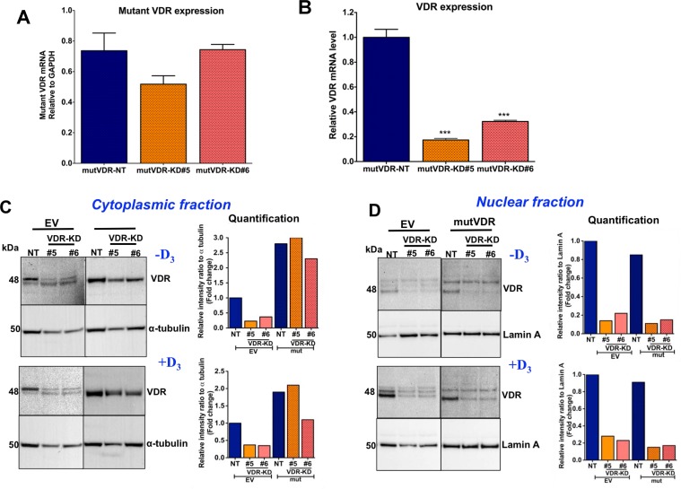
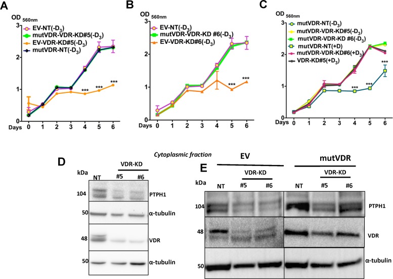
Vitamin D has pleiotropic effects on multiple tissues, including malignant tumors. Vitamin D inhibits breast cancer growth through activation of the vitamin D receptor (VDR) and via classical nuclear signaling pathways. Here, we demonstrate that the VDR can also function in the absence of its ligand to control behaviour of human breast cancer cells both outside and within the bone microenvironment. Stable shRNA expression was used to knock down VDR expression in MCF-7 cells, generating two VDR knockdown clonal lines. In ligand-free culture, knockdown of VDR in MCF-7 cells significantly reduced proliferation and increased apoptosis, suggesting that the VDR plays a ligand-independent role in cancer cell growth. Implantation of these VDR knockdown cells into the mammary fat pad of nude mice resulted in reduced tumor growth in vivo compared with controls. In the intra-tibial xenograft model, VDR knockdown greatly reduced the ability of the cells to form tumors in the bone microenvironment. The in vitro growth of VDR knockdown cells was rescued by the expression of a mutant form of VDR which is unable to translocate to the nucleus and hence accumulates in the cytoplasm. Thus, our data indicate that in the absence of ligand, the VDR promotes breast cancer growth both in vitro and in vivo and that cytoplasmic accumulation of VDR is sufficient to produce this effect in vitro. This new mechanism of VDR action in breast cancer cells contrasts the known anti-proliferative nuclear actions of the VDR-vitamin D ligand complex.

Keywords: bone metastasis; breast cancer; ligand independent; vitamin D; vitamin D receptor.


Conflict of interest statement

CONFLICTS OF INTEREST

The authors declare that they have no competing interests.

Figures

Figure 1
Figure 1. Stable knockdown of VDR in MCF-7 cells
MCF-7 cells were transduced with a lentivirus expressing either a non-target shRNA (NT) or shRNA against VDR (VDR-KD) and single cell clones were selected. (AB) Level of expression of VDR mRNA was measured using quantitative RT-PCR in (A) MCF-7 NT clones and (B) MCF-7 VDR-KD clones and compared to parental MCF-7 cells (PA). (C) Level of VDR protein in cell lysates of PA, NT and VDR-KD clones was assessed using Western blotting. (DE) MCF-7 PA cells and NT and VDR-KD clones were grown for 8 weeks in absence of antibiotic and VDR mRNA and protein levels were reassessed to ensure stable knockdown. Results are expressed as the mean ± SEM (n = 3). ***p < 0.001 using one-way ANOVA with Tukey's post-test.
Figure 2
Figure 2. VDR knockdown abrogates vitamin D signaling in MCF-7 cells
(A) In response treatment with 1,25D3 for 24 hours, VDR mRNA was increased by 2-fold in NT cells as compared to vehicle treated cells. In contrast, VDR-KD clones showed marginal response to the 1,25D3 treatment. (B) After 48-hour treatment with 1,25D3, VDR protein levels were significantly increased in NT treated cells. The VDR-KD clones show marginally increased VDR protein levels following treatment. (C) After 24-hour treatment of NT cells, a significant induction of CYP24 mRNA was observed compared to vehicle treated cells. In contrast, CYP24 mRNA induction was attenuated in VDR-KD clones with 1,25D3 treatment. Results are expressed as the mean ± SEM (n = 3). ***p < 0.001 NT(−D3) compared to NT (+D3) using one-way ANOVA with Tukey's post test.
Figure 3
Figure 3. VDR knockdown reduces MCF-7 cell growth and induces apoptosis in a ligand-independent manner
(A) In ligand-free culture, MCF7-VDR-KD non-clonal cells showed reduced growth by 53% compared to NT cells. Treatment of NT cells with 1,25D3 reduced cell growth by 44% compared to untreated cells. (B) In ligand free culture, MCF7-VDR-KD uncloned cells showed 6.8-fold increased apoptosis compared to NT cells, as measured by TUNEL assay. In response to 1,25D3, NT uncloned cells showed 3-fold increased apoptosis as compared to untreated cells.(C) Similar to non-clonal cells, VDR-KD#5 showed 41% growth reduction compared to MCF7-NT cells. Treatment of MCF7-NT cells with 10−8 M 1,25D3 reduced cell growth by ∼50% compared to untreated MCF7-NT cells. In contrast, the same treatment has no effect on growth of VDR-KD#5 cells. (D) Similarly, on day 6, VDR-KD#6 had a decreased growth by 51%, compared to NT in ligand-free culture. The magnitude of growth inhibition in VDR knockdown clones is comparable to the ligand mediated growth inhibition in control cell lines. Treatment of VDR-KD#6 cells with 1,25D3 does not reduce cell growth. (E) Both VDR-KD clones exhibited a 3-fold increased rate of apoptosis compared to NT cells in ligand-free culture. Upon 1,25D3 treatment, NT cell apoptosis increased 1.3-fold while VDR knockdown cells showed slightly (but insignificant) increased apoptosis compared to vehicle treated cells. Results are expressed as the mean ± SEM. For growth assay (n = 6) ***p < 0.001 compared to NT (−D3) using 2-way ANOVA with Bonferroni post-test. For TUNEL using 1-way ANOVA with Tukey's post-test.
Figure 4
Figure 4. VDR knockdown in MCF-7 cells reduces orthotopic tumor growth and also reduces its capability to form tumor in the bone environments
(A) Orthotopic tumor growth: Tumors derived from VDR-KD#5 cells grew significantly slower than NT cell-derived tumors. (B) Orthotopic tumor weight: At the experiment endpoint (day 50), the tumor mass of mice that received the VDR-KD clone was reduced by 72% compared to MCF7-NT. (C) Representative radiographs: Representative images showing osteoblastic lesions as indicated by arrows in tibiae injected with MCF7-NT cells 21 weeks p.i. In contrast, no bone changes can be seen in bones inoculated with VDR-KD clonal cells. (D) Quantitative histomorphometry: Of the mice implanted with MCF7-NT, 88% developed tumors as confirmed by the presence of cancer cells in tibiae. In contrast, only 25% of mice implanted with VDR-KD cells developed tumors. (E) Bone histomorphometry: Sclerotic lesion area was significantly greater in MCF7-NT injected bones than in tibiae inoculated with VDR-KD cells. Results are expressed as the mean ± SEM: Figure A and B (n = 5/group). ***p < 0.001 compared to MCF-N7 NT tumors, using 2-way ANOVA with Bonferroni post-test. Figure C to E (n = 8–9/group)., *p < 0.05, **p < 0.01 compared to NT tumor using 1-way ANOVA with Tukey's post-test.
Figure 5
Figure 5. VDR knockdown in MCF-7 cells decreased osteosclerotic bone formation in bones of nude mice
Bone micro CT analysis was performed only for trabecular bone by excluding cortical bone (A) Bone microCT: Representative images showing tumor-mediated trabecular bone formation in tibiae injected with MCF7-NT cells 21 weeks p.i. In contrast, negligible amounts of trabecular bone formation seen in bones inoculated with VDR-KD clonal cells. (BD) Quantitative analysis of micro-CT scans: Increased trabecular bone volume (BV/TV %) was observed in tibiae injected with NT cells compared to contralateral tibiae and VDR-KD inoculated tibiae. Trabecular number in the tibiae inoculated with NT cells was significantly higher compared to vehicle control. Tibiae injected with VDR knockdown cells showed significantly higher trabecular number compared to vehicle. Similarly, trabecular separation was decreased by 72% in tibiae with NT cells, compared to vehicle inoculated tibiae. Trabecular separation in VDR-KD inoculated tibia was reduced up to 13% compared to vehicle-inoculated tibiae. Results are expressed as the mean ± SEM. *p < 0.05, ***p < 0.001 compared to NT tumor using 1-way ANOVA with Tukey's post-test.
Figure 6
Figure 6. Stable expression of nuclear localization mutant VDR (mutVDR) in VDR knockdown clones
(A) After stable transfection of mutVDR into VDR knockdown cells the level of mutVDR mRNA in VDR knockdown clones was comparable to MCF7-NT cells using mutant VDR specific primers. (B) Expression of mutVDR in VDR-KD#5 and VDR-KD#6 does not change its shRNA knockdown effects. (C) Western blot of cytoplasmic fractions and its quantification. After stable transfection of the mutVDR and empty vector (EV) into VDR knockdown cells, Western blots demonstrated a ∼3-fold increase in mutVDR protein in the cytoplasmic fraction of VDR-KD clones compared to EV-transfected NT cells in the absence of vitamin D. (D) Western blot of nuclear fractions and its quantification. Despite stable transfection of mutVDR and EV into VDR-KD cells, the mutVDR marginally translocated into the nucleus following treatment with 10−8M 1,25D3 for 48 hours. In contrast, the intact VDR considerably translocated to the nucleus in EV transfected control cells in response to treatment with 1,25D3. Results are expressed as the mean ± SEM. ***p < 0.001 compared to mutVDR-NT using 1-way ANOVA with Tukey's post-test.
Figure 7
Figure 7. Expression of mutVDR into the VDR knockdown cells restores their growth
(AB) Similar to VDR-KD cells, in ligand-free culture the EV-VDR-KD clones were slow in growth compared to EV-NT. Stable expression of the mutVDR in VDR-KD clones rescued the growth phenotype of VDR knockdown cells. mutVDR expression in both knockdown clones resulted in similar increases in growth rate. This was comparable to the growth rate of EV-NT cells. (C) When mutVDR -VDR-KD clones were treated with 1,25D3, they were unresponsive to treatment, thus cell growth remained the same until day 5. A slight reduction in growth was observed on day 6. (D) Western blots of the cytoplasmic fraction showed that compared to NT, PTPH1 protein levels were decreased by 50% in VDR-KD#5 and 40% in VDR-KD#6 (E) After mutVDR expression, PTPH1 protein was increased by 2 fold in cytoplasm of mutVDR -VDR-KD clones compared to their respective EV controls. Results are expressed as the mean ± SEM (n = 6). ***p < 0.001 compared to EV-NT (−D3), mutVDR-VDR-KD#5 (−D3), mutVDR-VDR-KD#6 (−D3), mutVDR-NT(−D3), mutVDR-VDR-KD#5 (+D3), mutVDR-VDR-KD#6 (+D3) using 2-way ANOVA with Bonferroni post-test.

Similar articles

Cited by

References

    1. Bray F, Ren JS, Masuyer E, Ferlay J. Global estimates of cancer prevalence for 27 sites in the adult population in 2008. Int J Cancer. 2013;132:1133–1145. - PubMed
    1. Siegel R, Naishadham D, Jemal A. Cancer statistics, 2013. CA Cancer J Clin. 2013;63:11–30. - PubMed
    1. Yip CH, Smith RA, Anderson BO, Miller AB, Thomas DB, Ang ES, Caffarella RS, Corbex M, Kreps GL, McTiernan A. Guideline implementation for breast healthcare in low- and middle-income countries: early detection resource allocation. Cancer. 2008;113:2244–2256. - PubMed
    1. Coleman RE, Rubens RD. The clinical course of bone metastases from breast cancer. Br J Cancer. 1987;55:61–66. - PMC - PubMed
    1. Smith HS, Barkin RL. Painful Boney Metastases. Am J Ther. 2014;21:106–30. - PubMed

MeSH terms