

A novel oncolytic adenovirus based on simian adenovirus serotype 24
- PMID: 28460470
- PMCID: PMC5432303
- DOI: 10.18632/oncotarget.15845
A novel oncolytic adenovirus based on simian adenovirus serotype 24
Abstract
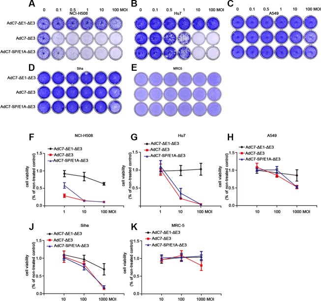
Among the oncolytic virotherapy, an emerging treatment for tumor, adenoviruses are widely used at present in preclinical and clinical trials. Traditionally, oncolytic adenoviruses were developed based on the human adenovirus serotype 5 (AdHu5). However, AdHu5 has the drawbacks of preexisting anti-AdHu5 immunity in most populations, and extensive sequestration of Adhu5 by the liver through hexon, blood coagulation factor X (FX), and FX receptor interactions. To tackle these problems, we explored a novel oncolytic adenovirus AdC7-SP/E1A-ΔE3 for cancer treatment. AdC7-SP/E1A-ΔE3 was constructed by replacing the E1A promoter with tumor specific promoter survivin promoter and deleting E3 region using direct cloning methods based on simian adenovirus serotype 24 (namely AdC7). We showed that AdC7-SP/E1A-ΔE3 significantly killed tumor cell lines NCI-H508 and Huh7, and inhibited tumor growth in both NCI-H508 and Huh7 xenograft tumor models. Importantly, AdC7-SP/E1A-ΔE3 exhibited the antitumor efficacy via systemic administration. Mechanistically, infected cells were killed by AdC7-SP/E1A-ΔE3 via the p53-independent mitochondrial apoptosis pathway in which phosphorylation of BAD markedly declined and the expresses of Bik significantly went up. Therefore, AdC7-SP/E1A-ΔE3 is a promising candidate for liver and colon tumor treatment.
Keywords: AdC7; chimpanzee adenoviruses; oncolytic adenoviruses; p53-independent mitochondrial apoptosis; tumor treatment.
Conflict of interest statement
The Authors do not have any conflicts of interest.
Figures


Similar articles
-
[Anti-Tumor Effect of a Novel Oncolytic Virus Based on Chimpanzee Adenovirus Type 6].Sichuan Da Xue Xue Bao Yi Xue Ban. 2022 Jan;53(1):71-76. doi: 10.12182/20220160504. Sichuan Da Xue Xue Bao Yi Xue Ban. 2022. PMID: 35048603 Free PMC article. Chinese.
-
A survivin-mediated oncolytic adenovirus induces non-apoptotic cell death in lung cancer cells and shows antitumoral potential in vivo.J Gene Med. 2006 Oct;8(10):1232-42. doi: 10.1002/jgm.953. J Gene Med. 2006. PMID: 16900558
-
Overexpression of tumor suppressor TSLC1 by a survivin-regulated oncolytic adenovirus significantly inhibits hepatocellular carcinoma growth.J Cancer Res Clin Oncol. 2012 Apr;138(4):657-70. doi: 10.1007/s00432-011-1138-2. Epub 2012 Jan 12. J Cancer Res Clin Oncol. 2012. PMID: 22237452 Free PMC article.
-
Genetically modified adenoviruses against gliomas: from bench to bedside.Neurology. 2004 Aug 10;63(3):418-26. doi: 10.1212/01.wnl.0000133302.15022.7f. Neurology. 2004. PMID: 15304571 Review.
-
Recent advances in genetic modification of adenovirus vectors for cancer treatment.Cancer Sci. 2017 May;108(5):831-837. doi: 10.1111/cas.13228. Epub 2017 May 7. Cancer Sci. 2017. PMID: 28266780 Free PMC article. Review.
Cited by
-
Adenovirus-Neutralizing and Infection-Promoting Activities Measured in Serum of Human Brain Cancer Patients Treated with Oncolytic Adenovirus Ad5-∆24.RGD.Int J Mol Sci. 2025 Jan 20;26(2):854. doi: 10.3390/ijms26020854. Int J Mol Sci. 2025. PMID: 39859568 Free PMC article.
-
Non-Human Primate-Derived Adenoviruses for Future Use as Oncolytic Agents?Int J Mol Sci. 2020 Jul 8;21(14):4821. doi: 10.3390/ijms21144821. Int J Mol Sci. 2020. PMID: 32650405 Free PMC article. Review.
-
Nonhuman Primate Adenoviruses of the Human Adenovirus B Species Are Potent and Broadly Acting Oncolytic Vector Candidates.Hum Gene Ther. 2022 Mar;33(5-6):275-289. doi: 10.1089/hum.2021.216. Hum Gene Ther. 2022. PMID: 34861769 Free PMC article.
-
Chimpanzee adenoviral vector prime-boost regimen elicits potent immune responses against Ebola virus in mice and rhesus macaques.Emerg Microbes Infect. 2019;8(1):1086-1097. doi: 10.1080/22221751.2019.1644968. Emerg Microbes Infect. 2019. PMID: 31339465 Free PMC article.
-
Development of novel vaccine vectors: Chimpanzee adenoviral vectors.Hum Vaccin Immunother. 2018 Jul 3;14(7):1679-1685. doi: 10.1080/21645515.2017.1419108. Epub 2018 Jan 23. Hum Vaccin Immunother. 2018. PMID: 29300685 Free PMC article. Review.
References
-
- Rosa DD, Ismael G, Lago LD, Awada A. Molecular-targeted therapies: lessons from years of clinical development. Cancer treatment reviews. 2008;34:61–80. - PubMed
-
- Bischoff JR, Kim DH, Williams A, Heise C, Horn S, Muna M, Ng L, Nye JA, SampsonJohannes A, Fattaey A, McCormick F. An adenovirus mutant that replicates selectively in p53-deficient human tumor cells. Science. 1996;274:373–376. - PubMed
-
- Fueyo J, Gomez-Manzano C, Alemany R, Lee PS, McDonnell TJ, Mitlianga P, Shi YX, Levin VA, Yung WK, Kyritsis AP. A mutant oncolytic adenovirus targeting the Rb pathway produces anti-glioma effect in vivo. Oncogene. 2000;19:2–12. - PubMed
MeSH terms
Substances
LinkOut - more resources
Full Text Sources
Other Literature Sources
Research Materials
Miscellaneous
