. 2017 Apr 18;8(16):26927-26940.
doi: 10.18632/oncotarget.15879.

Triptolide suppresses the in vitro and in vivo growth of lung cancer cells by targeting hyaluronan-CD44/RHAMM signaling

Affiliations

Triptolide suppresses the in vitro and in vivo growth of lung cancer cells by targeting hyaluronan-CD44/RHAMM signaling

Jung Min Song et al. Oncotarget. .

Abstract

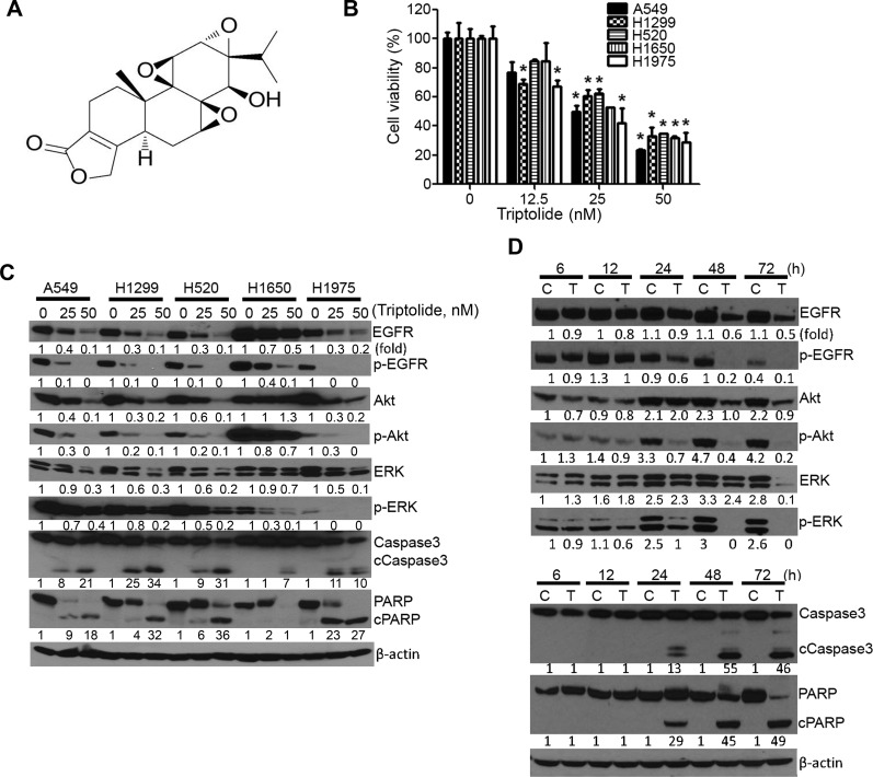
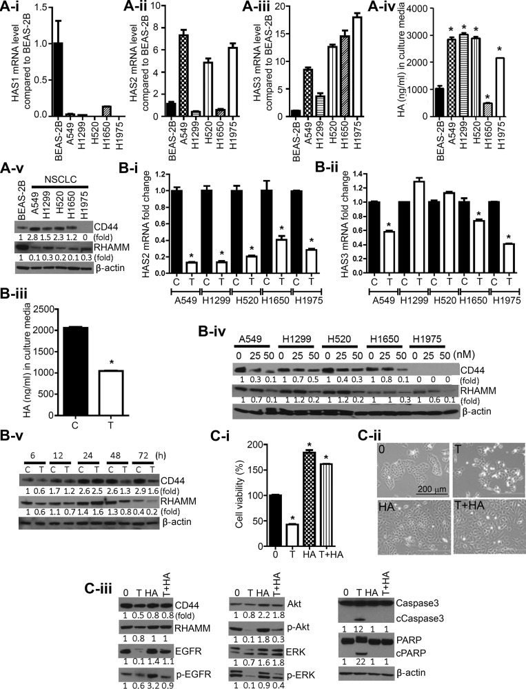
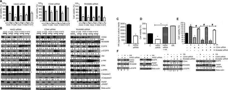
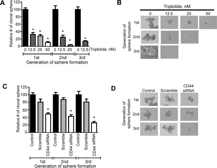
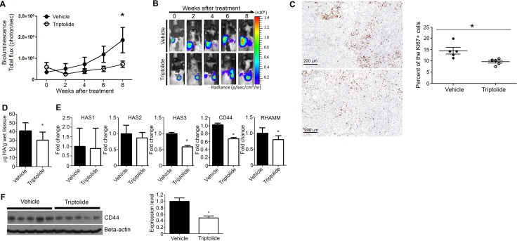
Higher levels of hyaluronan (HA) and its receptors CD44 and RHAMM have been associated with poor prognosis and metastasis in NSCLC. In the current study, our goal was to define, using cellular and orthotopic lung tumor models, the role of HA-CD44/RHAMM signaling in lung carcinogenesis and to assess the potential of triptolide to block HA-CD44/RHAMM signaling and thereby suppress the development and progression of lung cancer. Triptolide reduced the viability of five non-small cell lung cancer (NSCLC) cells, the proliferation and self-renewal of pulmospheres, and levels of HA synthase 2 (HAS2), HAS3, HA, CD44, RHAMM, EGFR, Akt and ERK, but increased the cleavage of caspase 3 and PARP. Silencing of HAS2, CD44 or RHAMM induced similar effects. Addition of excess HA to the culture media completely abrogated the effects of triptolide and siRNAs targeting HAS2, CD44, or RHAMM. In an orthotopic lung cancer model in nude rats, intranasal administration of liposomal triptolide (400 μg/kg) for 8 weeks significantly reduced lung tumor growth as determined by bioluminescence imaging, lung weight measurements and gross and histopathological analysis of tumor burden. Also, triptolide suppressed expressions of Ki-67, a marker for cell proliferation, HAS2, HAS3, HA, CD44, and RHAMM in lung tumors. Overall, our results provide a strong rationale for mitigating lung cancer by targeting the HA-CD44/RHAMM signaling axis.

Keywords: NSCLC; hyaluronan; hyaluronan receptor; orthotopic; triptolide.


Conflict of interest statement

CONFLICTS OF INTEREST

We confirm that there are no conflicts of interest associated with this publication.

Figures

Figure 1
Figure 1. Triptolide modulated the viability of NSCLC cells and levels of cell proliferation- and apoptosis-related proteins
(A) Chemical structure of triptolide. (B) Dose-dependent anti-proliferative effects of triptolide in NSCLC cells. MTT assays were performed in five NSCLC cell lines treated with DMSO or triptolide (12.5, 25 and 50 nM) for 72 h and the data were presented as percentage mean ± SD of cell viability compared to DMSO-treated cells. (C, D) Representative western immunoblotting results showing dose-dependent (C) and time-dependent (D) effects of triptolide on the expression of cell proliferation- and apoptosis-related proteins in NSCLC cells. Cells were treated with different concentrations of triptolide (0, 25 and 50 nM) for 72 h or A549 cells were treated with 50 nM of triptolide for different time periods (6, 12, 24, 48 and 72 h). Three independent assays were performed from different samples as described in materials and methods section. *P < 0.05, compared with the control group. Assays were performed in triplicate and repeated three times on different days. C, Control; T, triptolide.
Figure 2
Figure 2. Triptolide suppressed levels of HASs, CD44, and RHAMM, and HA in NSCLC cells and exogenous HA conferred protection against the anti-proliferative and pro-apoptotic effects of triptolide
(A) Constitutive levels of HAS1 (2A-i), HAS2 (2A-ii), HAS3 (2A-iii), HA (2A-iv) and CD44 and RHAMM (2A-v) in immortalized BEAS-2B bronchial cells and NSCLC cell lines. (B) Modulation of levels of HAS2 (2B-i), HAS3 (2B-ii), HA (2B-iii), CD44 and RHAMM (2B-iv; 2B-v) in NSCLC cells treated with triptolide (25 nM) or DMSO. Cells were treated with 25 nM of triptolide for 72 h with the exception of the results shown in Figure 2B-iv in which cell were exposed to 0, 25 or 50 nM of triptolide. (C) Exogenous HA attenuated triptolide-induced cytotoxicity and modulation of cell proliferation and survival-related proteins. C-i, A549 cells grown in RPMI media supplemented with 2.5% FBS were treated with DMSO, triptolide (25 nM), HA (2.5 mg/mL), or triptolide + HA for 72 h and cell viability determined by MTT assay. C-ii, images of A549 cells exposed to DMSO, triptolide (25 nM), HA, or triptolide + HA for 72 h. C-iii, Western immunoblotting assays showing attenuation by HA of triptolide-induced modulation in the expression of cell proliferation and survival-related proteins. *P < 0.05. Assays were performed in triplicate and repeated three times on different days. C, Control; T, triptolide.
Figure 3
Figure 3. siRNA-mediated silencing of HAS2, CD44 or RHAMM reduced NSCLC cell viability and the level of cell proliferation- and apoptosis-related proteins and these effects were modulated by exogenous HA
(A) Effects of HAS2, CD44 and RHAMM siRNA on the viability of NSCLC cells. Each cell line was transfected with the individual siRNAs and cell viability was determined by MTT assay as described in the Materials and Methods section. (B) HAS2, CD44 and RHAMM siRNAs modulated the expression of cell proliferation-and survival-related proteins. NSCLC cells were transfected with the siRNAs and Western immunoblotting was performed as described in the Materials and Methods section. (C) Effect of HAS2 siRNA on HA synthesis. A549 cells were transfected with HAS2 siRNA and accumulation of HA in the culture media was determined as described in the Materials and Methods section. (D, E) Exogenous HA (2.5 mg/mL) rescued cells from the cytotoxic effects of siRNAs targeting HAS2 (D), CD44, RHAMM or CD44 + RHAMM (E). A549 cells were treated with HAS2, CD44, RHAMM or CD44 + RHAMM siRNAs or HA alone or siRNA + HA and cell viability determined by MTT assay. (F) Exogenous HA attenuated the effects of triptolide on cell proliferation-and apoptosis-related proteins. A549 cells grown in RPMI media supplemented with 2.5% FBS were treated with HAS2, CD44, RHAMM or CD44 + RHAMM siRNAs or HA alone or siRNA + HA and expression of the proteins determined by Western immunoblotting. For all experiments, at least three independent assays were carried out. *P < 0.05, compared to treatment with DMSO-treated cells; Δ, compared to DMSO-treated cells; ♯, compared to treatment with siRNA only.
Figure 4
Figure 4. Triptolide and CD44 siRNA reduced the proliferation and self-renewal of putative lung cancer stem cells expressing CD44
(A, C) Effects of triptolide (A) or CD44 siRNA (C) on the number of primary-, secondary and tertiary-pulmospheres generated from A549 cells. Cells were treated with triptolide (12.5, 25 and 50 nM) or CD44 siRNA and the frequency of pulmospheres determined as described in the Materials and Methods section. (B, D) Images of primary, secondary and tertiary pulmospheres generated from triptolide (B)- or CD44 siRNA (D)- treated A549 cells. At least three independent assays were carried for these assays. *P < 0.05.
Figure 5
Figure 5. Liposomal triptolide significantly reduced the growth of orthotopic lung tumor in nude rats
(A) Effects of triptolide on the growth of orthotopically transplanted A549 cells as determined by bioluminescence imaging. Rats in which luciferase expressing A549 cells were implanted in the lung were given liposome-encapsulated triptolide (400 μg/kg) and the growth of lung tumors monitored by bioluminescence imaging as described in the Materials and Methods section. (B) Representative results of bioluminescence imaging studies in the vehicle and triptolide groups. (C) Representative images of Ki-67 expression in the lung tissues of vehicle- (top panel) or triptolide-treated (bottom panel) rats. Right panel: Bar graph showing the percent of Ki-67-positive lung tumor cells (mean ± SD) in vehicle- or triptolide-treated rats. (D) Levels of HA in lung tumor tissues of vehicle- and triptolide-treated rats. HA levels were measured by HA ELISA-like assay kit as described in the Materials and Methods section. (E) Levels of HAS1, HAS2, HAS3, CD44, and RHAMM mRNA transcripts in lung tumor tissues of vehicle- and triptolide-treated rats. (F) Left panel: Representative results showing Western immunoblotting analyses of CD44 expression in lung tumor tissues of vehicle- and triptolide-treated rats. Right panel: Quantification of CD44 expression in lung tumor tissues. The results from Figure 5D–5F were obtained from at least three independent assays performed on different days. *P < 0.05.

Similar articles

Cited by

References

    1. Tammi RH, Passi AG, Rilla K, Karousou E, Vigetti D, Makkonen K, Tammi MI. Transcriptional and post-translational regulation of hyaluronan synthesis. FEBS J. 2011;278:1419–1428. doi: 10.1111/j.1742-4658.2011.08070.x. - DOI - PubMed
    1. Toole BP. Hyaluronan: from extracellular glue to pericellular cue. Nature Rev Cancer. 2004;4:528–539. doi: 10.1038/nrc1391. - DOI - PubMed
    1. Tammi RH, Kultti A, Kosma VM, Pirinen R, Auvinen P, Tammi MI. Hyaluronan in human tumors: pathobiological and prognostic messages from cell-associated and stromal hyaluronan. Semin Cancer Biol. 2008;18:288–295. doi: 10.1016/j.semcancer.2008.03.005. - DOI - PubMed
    1. Itano N, Kimata K. Altered hyaluronan biosynthesis in cancer progression. Semin Cancer Biol. 2008;18:268–274. doi: 10.1016/j.semcancer.2008.03.006. - DOI - PubMed
    1. Toole BP, Slomiany MG. Hyaluronan: a constitutive regulator of chemoresistance and malignancy in cancer cells. Semin Cancer Biol. 2008;18:244–250. doi: 10.1016/j.semcancer.2008.03.009. - DOI - PMC - PubMed

MeSH terms