

doi: 10.1016/j.ophtha.2017.03.058.
Epub 2017 Apr 28.
Aqueous Angiography: Aqueous Humor Outflow Imaging in Live Human Subjects
Affiliations
- PMID: 28461013
- PMCID: PMC5522757
- DOI: 10.1016/j.ophtha.2017.03.058
Item in Clipboard
Aqueous Angiography: Aqueous Humor Outflow Imaging in Live Human Subjects
Ophthalmology.
2017 Aug.
No abstract available
Figures
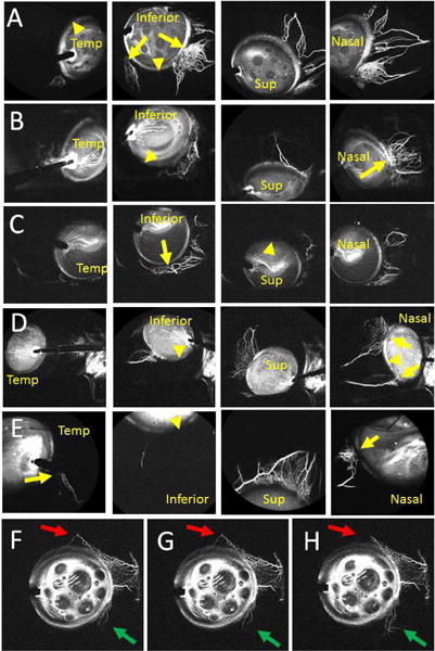
Aqueous Angiography Shows Segmental Outflow Patterns in Living Human Subjects Aqueous angiography in different positions for subject 1 (Row A; right eye), subject 2 (Row B; right eye), subject 3 (Row C; right eye), subject 4 (Row D; left eye), and subject 5 (Row E; left eye). Arrows identify areas with segmental angiographic signal and arrowheads point out regions without angiographic signal. F-H, Subject 6 showed a simultaneous dynamic inferonasal increase (green arrows) and superior decrease (red arrows) in angiographic signal. Sup = superior; Temp = temporal.
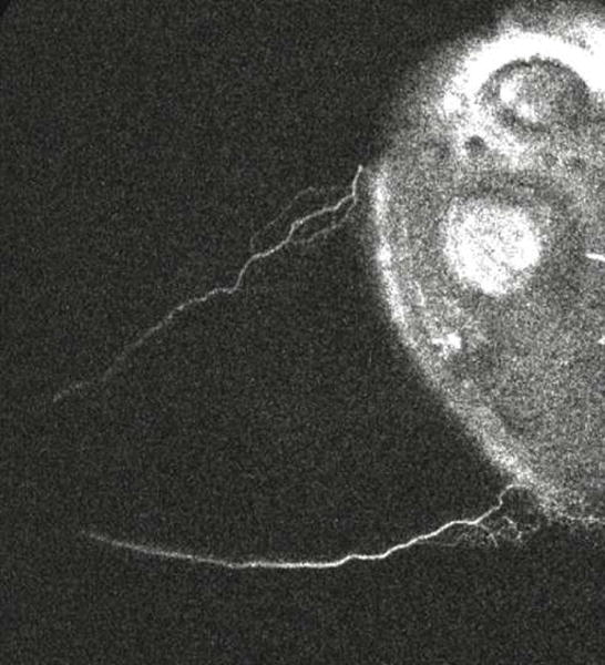
A clip was taken from subject 4. The left/bottom/top of the image are nasal/inferior/superior quadrants respectively in this left eye. The video starts 50 seconds after introduction of tracer and lasts for ~ 46 seconds. Angiographic signal appeared almost immediately inferonasally (bottom left) with pulsatile flow in this eye within the first 5 seconds. At 10 seconds, the angiographic signal extended more inferior and continued to grow at 16 seconds. At 22 seconds, this inferior extension started to fade. At 26 seconds, superonasal (top left) signal arose with a pulsatile quality. At 35 seconds, the inferior signal that first appeared at 10 seconds and then disappeared at 22 seconds started to re-appear.
References
-
- Jacobs DS, Cox TA, Wagoner MD, et al. Capsule staining as an adjunct to cataract surgery: a report from the American Academy of Ophthalmology. Ophthalmology. 2006;113(4):707–13. - PubMed
Publication types
MeSH terms
Substances
Grants and funding
LinkOut - more resources
Full Text Sources
Other Literature Sources
