. 2017 Aug:36:98-107.
doi: 10.1016/j.cellsig.2017.04.021. Epub 2017 Apr 28.

Differential manipulation of arrestin-3 binding to basal and agonist-activated G protein-coupled receptors

Affiliations

Differential manipulation of arrestin-3 binding to basal and agonist-activated G protein-coupled receptors

Susanne Prokop et al. Cell Signal. 2017 Aug.

Abstract

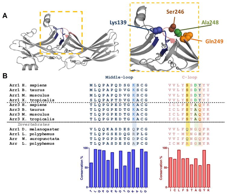
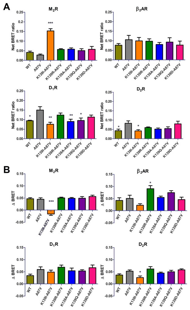
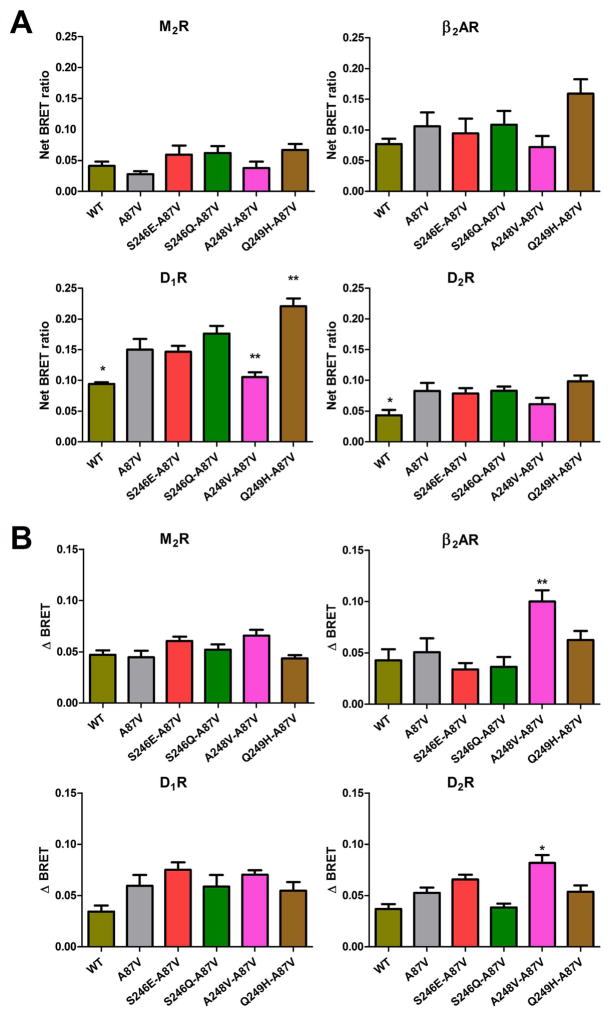
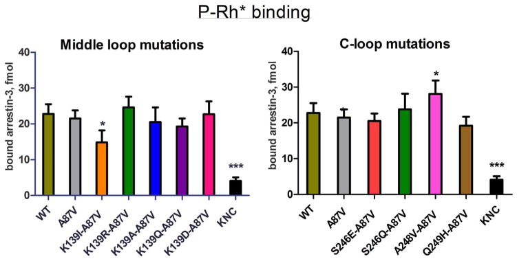
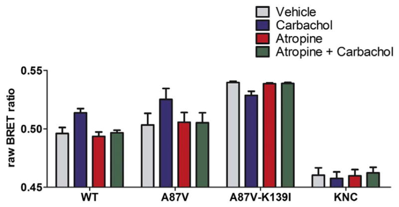
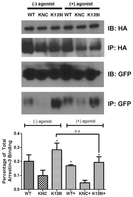
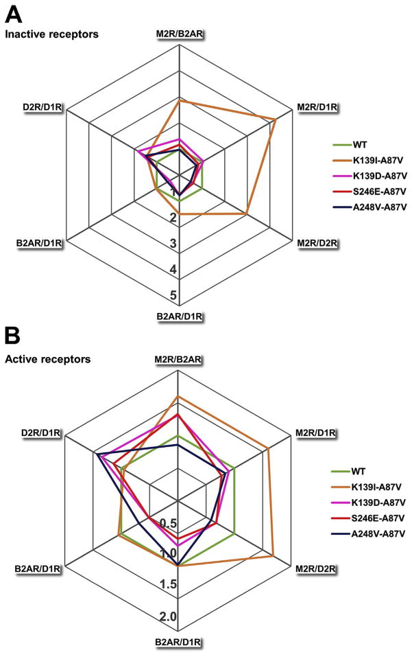
Non-visual arrestins interact with hundreds of different G protein-coupled receptors (GPCRs). Here we show that by introducing mutations into elements that directly bind receptors, the specificity of arrestin-3 can be altered. Several mutations in the two parts of the central "crest" of the arrestin molecule, middle-loop and C-loop, enhanced or reduced arrestin-3 interactions with several GPCRs in receptor subtype and functional state-specific manner. For example, the Lys139Ile substitution in the middle-loop dramatically enhanced the binding to inactive M2 muscarinic receptor, so that agonist activation of the M2 did not further increase arrestin-3 binding. Thus, the Lys139Ile mutation made arrestin-3 essentially an activation-independent binding partner of M2, whereas its interactions with other receptors, including the β2-adrenergic receptor and the D1 and D2 dopamine receptors, retained normal activation dependence. In contrast, the Ala248Val mutation enhanced agonist-induced arrestin-3 binding to the β2-adrenergic and D2 dopamine receptors, while reducing its interaction with the D1 dopamine receptor. These mutations represent the first example of altering arrestin specificity via enhancement of the arrestin-receptor interactions rather than selective reduction of the binding to certain subtypes.

Keywords: Arrestin; GPCRs; Protein engineering; Protein-protein interactions; Receptor specificity.


Figures

Fig. 1
Fig. 1
Structure and sequence of the middle and C-loops of arrestins. A. Crystal structure of rhodopsin-bound arrestin-1 (Protein Data Bank entry 4ZWJ [27] ). The middle and C-loops are shown in blue and pink, respectively. Residues mutated in this study are shown as CPK models. B. Multiple sequence alignment of arrestin-1 (Arr1), arrestin-3 (Arr3) and arrestin homologs (Arr) from invertebrate species. Residues mutated in this study are highlighted. Bar graph under the alignment shows the extent of residue conservation at each position. (For interpretation of the references to color in this figure legend, the reader is referred to the web version of this article.)
Fig. 2
Fig. 2
Basal and agonist-induced binding of Lys139 mutants to GPCRs. A. Basal (agonist-independent) association of the indicated luciferase-tagged receptor and wild type or mutant Venus-Arrestin-3 in COS-7 cells at 10 min of vehicle treatment. Nonspecific (bystander) BRET was measured using non-receptor-binding arrestin-3 mutant (Arrestin3-KNC [23,30] ). Net BRET ratio was calculated by subtracting non-specific BRET from the raw BRET data in each experiment. B. The agonist-induced arrestin-3 recruitment to indicated receptors. The BRET change (ΔBRET) was determined by the difference between the BRET ratio between ligand and vehicle treated cells (nonspecific BRET was subtracted from both). Means ± S.E. of at least three independent experiments are shown. Each experiment was performed in quadruplicate. Statistical significance was determined by one-way ANOVA, followed by Dunnett’s post-hoc test: *, p < 0.05; **, p < 0.01; ***, p < 0.001, as compared with A87V base mutant binding to each receptor.
Fig. 3
Fig. 3
The effect of C-loop mutations on basal and agonist-induced receptor binding. A. Basal BRET (net BRET ratio) and B. agonist-induced BRET change (delta BRET) between indicated luciferase-tagged receptors and Venus-tagged arrestin-3C-loop mutants was calculated, as described in the legend to Fig. 2. The cells were stimulated for 10 min with the appropriate agonist. BRET ratio obtained with negative control (arrestin-3 KNC [,83] ) was subtracted. Means ± S.E. of at least three independent experiments are shown. Each experiment was performed in quadruplicate. Statistical significance was determined by one-way ANOVA: *, p < 0.05; **, p < 0.01; ***, p < 0.001, as compared with A87V base mutant binding to each receptor.
Fig. 4
Fig. 4
Direct binding of arrestin-3 mutants to light-activated phosphorylated rhodopsin. WT and mutant forms of arrestin-3 produced in cell-free translation in the presence of radiolabeled leucine (2 nM) were incubated with 0.3 μg of light-activated phosphorylated rhodopsin (P-Rh*) for 5 min at 37 °C. Free and rhodopsin-bound arrestin was separated by gel filtration on 2-ml Sepharose 2B–CL columns. The amount of bound arrestin eluting with rhodopsin-containing membranes was quantified by scintillation counting. Non-specific binding (in the absence of rhodopsin) was subtracted. Means ± S.D. of three independent experiments performed in duplicate are shown. The data were analyzed by one-way ANOVA with arrestin type as the main factor, followed by Dunnett’s post-hoc test.
Fig. 5
Fig. 5
Lys139Ile mutant binds inactive M2R. COS-7 cells co-expressing M2R-RLuc8 and indicated form of Venus-arrestin-3. Cells were pretreated for 5 min with vehicle or 10 μM inverse agonist atropine [50], and stimulated with carbamylcholine as in Fig. 2. Raw BRET ratios are shown. Basal arrestin-3 binding was not altered by atropine treatment. *, p < 0.05 (vs. vehicle control), analyzed with one-way repeated measures ANOVA, followed by Dunnett’s post-hoc test. Atropine prevented the effects of carbachol. #, p < 0.05 (significant interaction between the two treatments), analyzed with two-way repeated measures ANOVA, followed by Bonferroni post-hoc test.
Fig. 6
Fig. 6
Co-immunoprecipitation of Venus-arrestin constructs with M2R. HEK arrestin-2/3 KO cells [31] were co-transfected with Venus-arrestin constructs and HA-M2R-RLuc8. After 48 h, the cells were incubated in either serum-free media alone, or serum-free media with 10 μM carbamylcholine for 15 min. The cells were lysed in IP buffer (50 mM Tris-HCl pH 7.5, 2 mM EDTA, 250 mM NaCl, 10% glycerol, 0.5% NP-40, 20 mM NaF, and 1 mM NaVO3) and the supernatant was cleared by centrifugation at max speed for 15 min, then pre-cleared with protein G agarose. The supernatant was immunoprecipitated using a rat HA antibody against HA-M2R-Rluc8. The input (IB) and immunoprecipitated material (IP) were subjected to Western blotting to detect HA and GFP, as indicated. The GFP blot of the immunoprecipitated samples was analyzed using Versadoc. The results were statistically analyzed using one-way ANOVA followed by Dunnett’s post-hoc test. The difference with KNC is shown (*, p < 0.05, n = 3). The effect of agonist treatment on Venus-arrestin-3 (K139I) was not statistically significant (n.s.).
Fig. 7
Fig. 7
Receptor selectivity of arrestin-3 mutants. Each receptor pair is provided as radially arranged axis that starts from the centre. The relative binding of each arrestin-3 mutant was plotted along all axes, and the connected values create a polygon. The binding ratio of WT arrestin-3 was set at 1 for each receptor pair. If the relative binding is> 1, the indicated arrestin has a preference for the first member of the receptor pair (receptor1/receptor2). Panel A shows the basal arrestin interactions. The green regular hexagon indicates WT arrestin. Only those mutants are highlighted that have at least a 1.5-fold bias against one receptor. Panel B shows the selectivity of the arrestin3 mutants after ligand treatment. (For interpretation of the references to color in this figure legend, the reader is referred to the web version of this article.)

Similar articles

Cited by

References

    1. Schoneberg T, Schulz A, Biebermann H, Hermsdorf T, Rompler H, Sangkuhl K. Pharmacol Ther. 2004;104:173–206. - PubMed
    1. Stoy H, Gurevich VV. Genes Dis. 2015;2:108–132. - PMC - PubMed
    1. Russo D, Arturi F, Schlumberger M, Caillou B, Monier R, Filetti S, Suarez HG. Oncogene. 1995;11:1907–1911. - PubMed
    1. Hebrant A, van Staveren WC, Maenhaut C, Dumont JE, Leclere J. Eur J Endocrinol. 2011;164:1–9. - PubMed
    1. Carman CV, Benovic JL. Curr Opin Neurobiol. 1998;8:335–344. - PubMed