

A non-contrast CMR index for assessing myocardial fibrosis
- PMID: 28461132
- PMCID: PMC6294294
- DOI: 10.1016/j.mri.2017.04.012
A non-contrast CMR index for assessing myocardial fibrosis
Abstract
Purpose: Safe, sensitive, and non-invasive imaging methods to assess the presence, extent, and turnover of myocardial fibrosis are needed for early stratification of risk in patients who might develop heart failure after myocardial infarction. We describe a non-contrast cardiac magnetic resonance (CMR) approach for sensitive detection of myocardial fibrosis using a canine model of myocardial infarction and reperfusion.
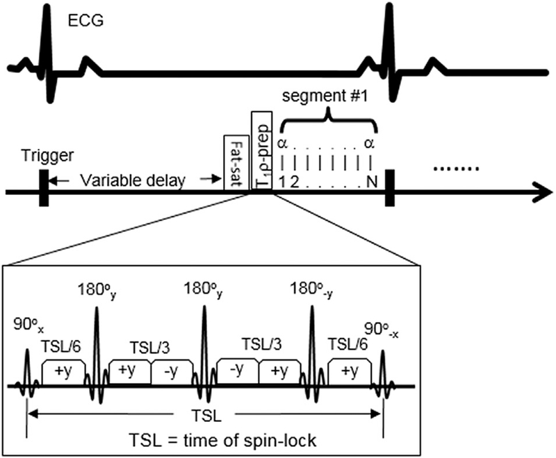
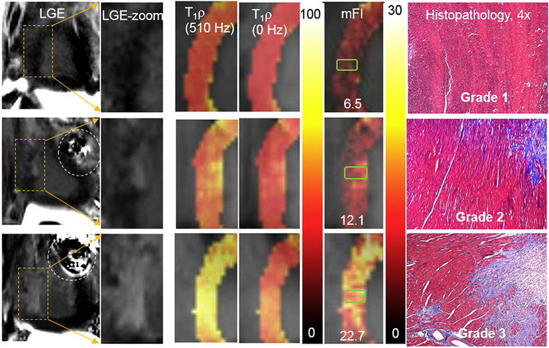
Methods: Seven dogs had coronary thrombotic occlusion of the left anterior descending coronary arteries followed by fibrinolytic reperfusion. CMR studies were performed at 7days after reperfusion. A CMR spin-locking T1ρ mapping sequence was used to acquire T1ρ dispersion data with spin-lock frequencies of 0 and 511Hz. A fibrosis index map was derived on a pixel-by-pixel basis. CMR native T1 mapping, first-pass myocardial perfusion imaging, and post-contrast late gadolinium enhancement imaging were also performed for assessing myocardial ischemia and fibrosis. Hearts were dissected after CMR for histopathological staining and two myocardial tissue segments from the septal regions of adjacent left ventricular slices were qualitatively assessed to grade the extent of myocardial fibrosis.
Results: Histopathology of 14 myocardial tissue segments from septal regions was graded as grade 1 (fibrosis area, <20% of a low power field, n=9), grade 2 (fibrosis area, 20-50% of field, n=4), or grade 3 (fibrosis area, >50% of field, n=1). A dramatic difference in fibrosis index (183%, P<0.001) was observed by CMR from grade 1 to 2, whereas differences were much smaller for T1ρ (9%, P=0.14), native T1 (5.5%, P=0.12), and perfusion (-21%, P=0.05).
Conclusion: A non-contrast CMR index based on T1ρ dispersion contrast was shown in preliminary studies to detect and correlate with the extent of myocardial fibrosis identified histopathologically. A non-contrast approach may have important implications for managing cardiac patients with heart failure, particularly in the presence of impaired renal function.
Keywords: CMR; Myocardial diffuse fibrosis; Myocardial infarction; Spin-lock T(1)ρ.
Copyright © 2017 Elsevier Inc. All rights reserved.
Figures
Similar articles
-
In vivo chronic myocardial infarction characterization by spin locked cardiovascular magnetic resonance.J Cardiovasc Magn Reson. 2012 Jun 15;14(1):37. doi: 10.1186/1532-429X-14-37. J Cardiovasc Magn Reson. 2012. PMID: 22704222 Free PMC article.
-
First-pass perfusion CMR two days after infarction predicts severity of functional impairment six weeks later in the rat heart.J Cardiovasc Magn Reson. 2011 Aug 3;13(1):38. doi: 10.1186/1532-429X-13-38. J Cardiovasc Magn Reson. 2011. PMID: 21812990 Free PMC article.
-
Single Breath-Hold T1ρ-Mapping of the Heart for Endogenous Assessment of Myocardial Fibrosis.Invest Radiol. 2016 Aug;51(8):505-12. doi: 10.1097/RLI.0000000000000261. Invest Radiol. 2016. PMID: 26895195
-
Cardiovascular magnetic resonance imaging to assess myocardial fibrosis in valvular heart disease.Int J Cardiovasc Imaging. 2018 Jan;34(1):97-112. doi: 10.1007/s10554-017-1195-y. Epub 2017 Jun 22. Int J Cardiovasc Imaging. 2018. PMID: 28642994 Free PMC article. Review.
-
T1 and T2 Mapping in Cardiology: "Mapping the Obscure Object of Desire".Cardiology. 2017;138(4):207-217. doi: 10.1159/000478901. Epub 2017 Aug 17. Cardiology. 2017. PMID: 28813699 Review.
Cited by
-
Role of endogenous T1ρ and its dispersion imaging in differential diagnosis of cardiac amyloidosis.J Cardiovasc Magn Reson. 2024 Winter;26(2):101080. doi: 10.1016/j.jocmr.2024.101080. Epub 2024 Aug 8. J Cardiovasc Magn Reson. 2024. PMID: 39127261 Free PMC article.
-
Endogenous T1ρ cardiovascular magnetic resonance in hypertrophic cardiomyopathy.J Cardiovasc Magn Reson. 2021 Oct 25;23(1):120. doi: 10.1186/s12968-021-00813-5. J Cardiovasc Magn Reson. 2021. PMID: 34689798 Free PMC article.
-
Clinical Heart Failure Stratification Through Native T1 Mapping: Experience of a Referral Service.Arq Bras Cardiol. 2021 May;116(5):919-925. doi: 10.36660/abc.20190782. Arq Bras Cardiol. 2021. PMID: 34008815 Free PMC article. English, Portuguese.
-
Fast myocardial T1ρ mapping in mice using k-space weighted image contrast and a Bloch simulation-optimized radial sampling pattern.MAGMA. 2022 Apr;35(2):325-340. doi: 10.1007/s10334-021-00951-y. Epub 2021 Sep 7. MAGMA. 2022. PMID: 34491466 Free PMC article.
-
Free-breathing simultaneous native myocardial T1, T2 and T1ρ mapping with Cartesian acquisition and dictionary matching.J Cardiovasc Magn Reson. 2023 Nov 9;25(1):63. doi: 10.1186/s12968-023-00973-6. J Cardiovasc Magn Reson. 2023. PMID: 37946191 Free PMC article.
References
-
- Brown RD, Ambler SK, Mitchell MD, Long CS. The cardiac fibroblast: therapeutic target in myocardial remodeling and failure. Annu Rev Pharmacol Toxicol 2005;45:657–87. - PubMed
-
- Kosmala W, Przewlocka-Kosmala M, Wojnalowicz A, Mysiak A, Marwick TH. Integrated backscatter as a fibrosis marker in the metabolic syndrome: association with biochemical evidence of fibrosis and left ventricular dysfunction. Eur Heart J Cardiovasc Imaging 2012;13:459–67. - PubMed
-
- Wickline SA, Thomas LJ, Miller JG, Sobel BE, Perez JE. Sensitive detection of the effects of reperfusion on myocardium by ultrasonic tissue characterization with integrated backscatter. Circulation 1986;74:389–400. - PubMed
-
- Di Bello V, Cucco C, Giannini C, et al. Myocardial tissue characterization and aortic stenosis. J Am Soc Echocardiogr 2010;23:1067–70. - PubMed
MeSH terms
Grants and funding
LinkOut - more resources
Full Text Sources
Other Literature Sources
Medical
