. 2017 Jun 5;10(6):846-865.
doi: 10.1016/j.molp.2017.04.008. Epub 2017 Apr 28.

Mass Spectrometric Analyses Reveal a Central Role for Ubiquitylation in Remodeling the Arabidopsis Proteome during Photomorphogenesis

Affiliations

Mass Spectrometric Analyses Reveal a Central Role for Ubiquitylation in Remodeling the Arabidopsis Proteome during Photomorphogenesis

Victor Aguilar-Hernández et al. Mol Plant. .

Abstract

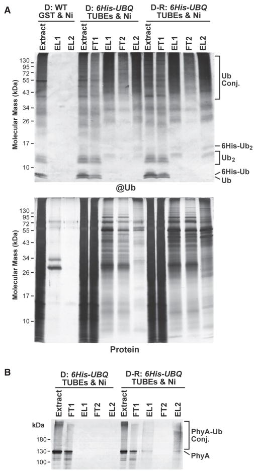
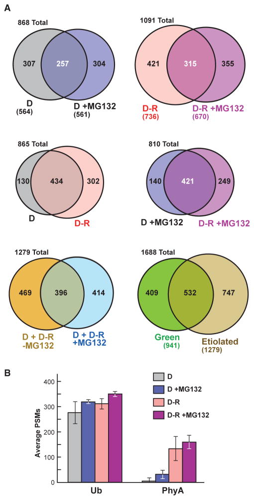
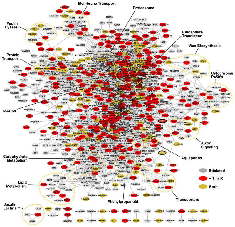
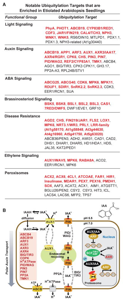
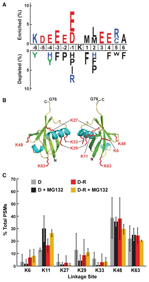
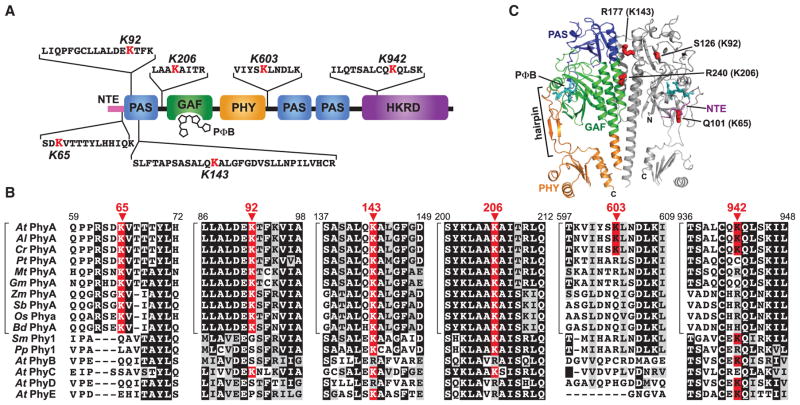
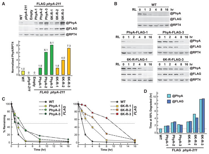
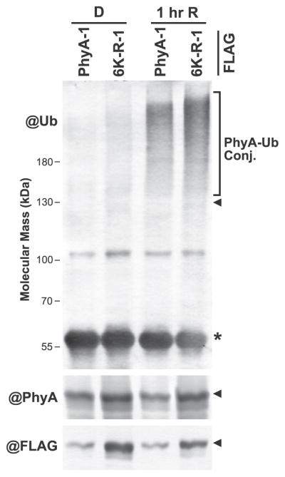
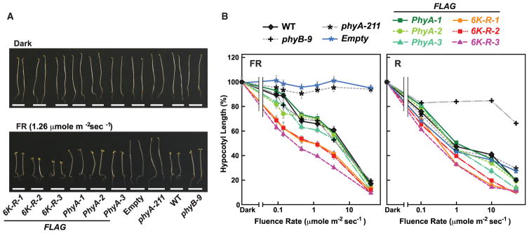
The switch from skotomorphogenesis to photomorphogenesis is a key developmental transition in the life of seed plants. While much of the underpinning proteome remodeling is driven by light-induced changes in gene expression, the proteolytic removal of specific proteins by the ubiquitin-26S proteasome system is also likely paramount. Through mass spectrometric analysis of ubiquitylated proteins affinity-purified from etiolated Arabidopsis seedlings before and after red-light irradiation, we identified a number of influential proteins whose ubiquitylation status is modified during this switch. We observed a substantial enrichment for proteins involved in auxin, abscisic acid, ethylene, and brassinosteroid signaling, peroxisome function, disease resistance, protein phosphorylation and light perception, including the phytochrome (Phy) A and phototropin photoreceptors. Soon after red-light treatment, PhyA becomes the dominant ubiquitylated species, with ubiquitin attachment sites mapped to six lysines. A PhyA mutant protected from ubiquitin addition at these sites is substantially more stable in planta upon photoconversion to Pfr and is hyperactive in driving photomorphogenesis. However, light still stimulates ubiquitylation and degradation of this mutant, implying that other attachment sites and/or proteolytic pathways exist. Collectively, we expand the catalog of ubiquitylation targets in Arabidopsis and show that this post-translational modification is central to the rewiring of plants for photoautotrophic growth.

Keywords: Arabidopsis; mass spectrometry; photomorphogenesis; phytochrome degradation; ubiquitin.


Figures

Figure 1
Figure 1. Two-Step Affinity Purification of Ubiquitylated Proteins from Etiolated Arabidopsis Seedlings Before and After Red-Light Irradiation
Ub conjugates were affinity purified by sequential TUBEs (1) and Ni-NTA (Ni) chromatography (2) steps from 7-day-old 6His-UBQ seedlings either kept in the dark (D) or exposed to 1 h of red light (D-R). Samples from dark-grown wild-type (WT) seedlings, which were subjected to the same purification protocol using GST and Ni-NTA beads, were included for comparison. (A) SDS–PAGE analysis of the various fractions during the purification. The gels were either subjected to immunoblotting with anti-Ub antibodies (@Ub) or stained for protein with silver (Protein). FT and EL represent flow through and elution fractions, respectively. Extract represents the initial clarified seedling extract. Ub conjugates and the Ub and 6His-Ub monomers and dimers are indicated. (B) The two-step affinity purification also enriches for PhyA-Ub conjugates that accumulate in etiolated seedlings after R. The samples analyzed in (A) were immunoblotted with the monoclonal antibody 073D against PhyA.
Figure 2
Figure 2. Comparisons of the Ubiquitylated Protein MS Datasets Obtained from Etiolated Arabidopsis Seedlings Before (D) and After Red-Light Irradiation (R) and/or MG132 Exposure
(A) Venn diagrams showing overlap of the ubiquitylated protein data-sets. The number of proteins in each sector is indicated. The numbers in parentheses indicate the total number of proteins in each dataset. The etiolated dataset represents the combined catalog of proteins detected in dark-grown seedlings with or without exposure to 50 μM MG132 and/or red light. The green MS dataset was obtained from 14-day-old, light-grown seedlings and was described previously (Kim et al., 2013). (B) Abundance of Ub and PhyA peptides in the MS datasets as quantified by peptide spectral matches (PSMs). Each bar represents the average of three biological replicates (±SD).
Figure 3
Figure 3. Protein Interaction Network of Ubiquitylated Proteins Identified in Etiolated Arabidopsis Seedlings Before and Soon After Red-Light (R) Irradiation
Protein interaction networks were generated with the complete list of 955 (etiolated only) and 1071 (etiolated and following 1-h R irradiation) ubiquitylated proteins using the STRING database and visualized using the Cytoscape program. The node colors reflect proteins identified only in etiolated (D and D + MG132) (gray), R-irradiated (D-R and D-R + MG132) (red), or in both pairs of datasets. Important functional clusters are surrounded by dashed lines. The red circles locate PhyA and PHOT1. Either the Arabidopsis thaliana locus identifier or the protein name abbreviation (where known) is shown for each substrate.
Figure 4
Figure 4. Ubiquitylation Targets Influential in Etiolated Arabidopsis Seedlings
(A) Notable ubiquitylation targets involved in various aspects of hormone and light signaling, peroxisome biology, and disease resistance. Targets shown in red were only detected in etiolated seedlings but not previously in green seedlings (Kim et al., 2013). See Supplemental Datasets 2 and 3 for the complete list. (B) Schematic diagram of auxin transport and perception highlighting the key proteins that are ubiquitylated in etiolated seedlings. The proteins and their proposed position within the auxin system are indicated. The structure of IAA is shown at the top right.
Figure 5
Figure 5. Analysis of Ub-Binding Sites within Various Targets and within Ub Itself from Etiolated Seedlings
(A) Analysis of the amino acid sequences surrounding Ub attachment sites in the catalog of Ub targets from both etiolated and green seedlings (Kim et al., 2013). The analysis used a window of six residues N- and C-terminal to the modified lysine for 413 peptides with 478 Ub footprints to identify significant position-specific under- or over-representation of amino acids flanking the Ub-attachment lysine. (B) 3D ribbon front and side models of plant Ub highlighting the lysines that could bind Ub covalently. α helices, β strands, and the side chains of lysines are shown in cyan, green, and red, respectively. N, N terminus. C, C terminus. G76, C-terminal active-site glycine. Adapted from PDB: 1UBQ (Vijay-Kumar et al., 1987). (C) Percentage of Ub footprints identified by tandem MS that represents poly-Ub chains linked internally through the various Ub lysines. Ubiq-uitylated proteins were purified from dark-grown (D) seedlings before or after red-light (R) irradiation with or without pretreatment with 50 μM MG132. Each bar represents the average of three independent MS analyses (±SD).
Figure 6
Figure 6. Location and Conservation of the Ubiquitylation Sites in Arabidopsis PhyA
(A) Diagram of the PhyA locating the identified ubiquitylation sites relative to the PAS, GAF, PHY, and HKRD domains. The MS-identified peptide sequence bearing each Ub footprint is shown with the modified lysine colored in red. NTE, N-terminal extension. PΦB, phytochromobilin. (B) Amino acid sequence conservation surrounding the ubiquitylation sites. The alignment windows include sequences from representative PhyA isoforms from A. thaliana (At), A. lyrata (Al), Capsella rubella (Cr), Populus trichocarpa (Pt), Medicago truncatula (Mt), Glycine max (Gm), Zea mays (Zm), Sorghum bicolor (Sb), Oryza sativa (Os) and Brachypodium distachyon (Bd), PhyA proteins from the bryophytes Selaginella moellendorffii (Sm) and Physcomitrella patens (Pp), and the PhyB, PhyC, PhyD, and PhyE isoforms from A. thaliana. Identical and similar amino acids are highlighted in black and grey boxes, respectively. Dashes denote gaps. The modified lysines are highlighted by the red boxes with the residue number indicated above the arrowheads. Brackets locate members of the PhyA subfamily. See Supplemental Figure 4 for the complete sequence alignment and protein identifier numbers. (C) Predicted 3D structure of the PhyA photosensory module showing the expected positions of the four ubiquitylated lysines in this region. The model was generated by SWISS-MODEL (http://swissmodel.expasy.org) using the equivalent region in Arabidopsis PhyB as the template (PDB: 4OUR; Burgie et al., 2014). N, amino terminus. C, carboxyl terminus.
Figure 7
Figure 7. Preventing Ubiquitylation at the Six Known Sites in Arabidopsis PhyA Slows but Does Not Block Degradation as Pfr
Either the normal PhyA sequence (PhyA) or one in which the six ubiquitylated lysines were substituted for arginines (6K-R) were expressed with a FLAG tag in the phyA-211 background. (A) Upper panel: Levels of PhyA in 4-day-old etiolated seedlings before irradiation. Clarified extracts from WT, phyA-211, or phyA-211 seedlings transformed with an empty vector or the PHYA-FLAG and 6K-R-FLAG transgenes were subjected to SDS–PAGE and immunoblotting with either the 073D anti-PhyA or anti-FLAG monoclonal antibody. Three independent transformants were analyzed for PhyA and 6K-R. Near equal protein loading was confirmed by immunoblotting with anti-RPT4 antibodies. Lower panel: Quantification of PhyA levels by densitometric scans of the anti-PhyA antibody immunoblots shown in (A). The values were normalized to the PhyA level in WT. (B) Degradation of PhyA in R. Four-day-old etiolated seedlings were irradiated with red light (R) for the indicated times and harvested. Clarified extracts were subjected to immunoblotting as in (A). (C) Quantification of PhyA degradation rates by densitometric scans of the anti-PhyA antibody immunoblots shown in (B). Each point represents the average of three biological replicates (±SD), which was normalized to the value for PhyA at t = 0. Dashed lines highlight the time when 50% of PhyA was degraded. Each bar represents the average of three independent experiments. (D) Estimated time needed to degrade 50% of PhyA or 6K-R as Pfr based on the degradation kinetics shown in (C) using either the anti-PhyA or FLAG monoclonal antibodies for detection. Dashed line highlights the value for WT PhyA.
Figure 8
Figure 8. Substitution of the Six Known Ubiquitylation Sites in PhyA for Arginines Does Not Block Pfr Ubiquitylation
PhyA-FLAG was immunoprecipitated from PhyA-1 phyA-211 and 6K-R-1 phyA-211 plants (see Figure 7A) before or after a 1-h irradiation with red light (R), and immunoblotted with antibodies against Ub, PhyA, or FLAG. PhyA-Ub conjugates are located by the bracket. The arrowheads and asterisk indicate the position of PhyA-FLAG and the anti-FLAG IgG heavy chain used for immunoprecipitation, respectively.
Figure 9
Figure 9. PhyA Missing the Ubiquitylated Lysines Are Hyperactive in Seedling Photomorphogenesis
Plants expressing FLAG-tagged PhyA without (PhyA) or with the known ubiquitylated lysines converted to arginines (6K-R) were germinated in the dark or under various fluences of continuous far-red (FR) or red light (R). After 7 days, hypocotyl lengths were measured. (A) Images of representative seedlings either kept in the dark or irradiated with FR. The three independent transformants of phyA-211 with the FLAG-tagged versions of either PhyA or 6K-R as described in Figure 7 were compared. WT, phyB-9, and phyA-211 seedlings either alone or transformed with an empty vector were included as controls. (B) Fluence responses of the mutant collection in (A) under continuous R or FR. Each point (±SE) represents the average of three independent experiments, each containing at least 30 seedlings.

References

    1. Abramoff M, Magalhaes P, Ram S. Image processing with ImageJ. Biophotonics Int. 2004;11:36–42.
    1. Al-Sady B, Ni W, Kircher S, Schafer E, Quail PH. Photoactivated phytochrome induces rapid PIF3 phosphorylation prior to proteasome-mediated degradation. Mol Cell. 2006;23:439–446. - PubMed
    1. Bernhardt A, Lechner E, Hano P, Schade V, Dieterle M, Anders M, Dubin MJ, Benvenuto G, Bowler C, Genschik P, et al. CUL4 associates with DDB1 and DET1 and its downregulation affects diverse aspects of development in Arabidopsis thaliana. Plant J. 2006;47:591–603. - PubMed
    1. Boller T, Felix G. A renaissance of elicitors: perception of microbe-associated molecular patterns and danger signals by pattern-recognition receptors. Annu Rev Plant Biol. 2009;60:379–406. - PubMed
    1. Book AJ, Gladman NP, Lee SS, Scalf M, Smith LM, Vierstra RD. Affinity purification of the Arabidopsis 26S proteasome reveals a diverse array of plant proteolytic complexes. J Biol Chem. 2010;285:25554–25569. - PMC - PubMed

Publication types

MeSH terms

LinkOut - more resources