

Metabolic In Vivo Visualization of Pituitary Adenomas: a Systematic Review of Imaging Modalities
- PMID: 28461279
- PMCID: PMC5821230
- DOI: 10.1016/j.wneu.2017.04.128
Metabolic In Vivo Visualization of Pituitary Adenomas: a Systematic Review of Imaging Modalities
Abstract
Objective: Pituitary adenomas (PAs) are the most common intrasellar mass. Functional PAs constitute most of pituitary tumors and can produce symptoms related to hormonal overproduction. Timely and accurate detection is therefore of vital importance to prevent potentially irreversible sequelae. Magnetic resonance imaging is the gold standard for detecting PAs, but is limited by poor sensitivity for microadenomas and an inability to differentiate scar tissue from tumor residual or predict treatment response. Several new modalities that detect PAs have been proposed.
Methods: A systematic review of the PubMed database was performed for imaging studies of PAs since its inception. Data concerning study characteristics, clinical symptoms, imaging modalities, and diagnostic accuracy were collected.
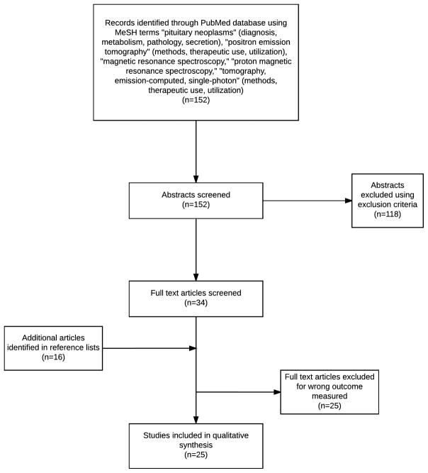
Results: After applying exclusion criteria, 25 studies of imaging PAs using positron emission tomography (PET), magnetic resonance spectroscopy (MRS), and single photon emission computed tomography were reviewed. PET reliably detects PAs, particularly where magnetic resonance imaging is equivocal, although its efficacy is limited by high cost and low availability. Single photon emission computed tomography possesses good sensitivity for neuroendocrine tumors but its use with PAs is poorly documented. MRS consistently detects cellular proliferation and hormonal activity, but warrants further study at higher magnetic field strength.
Conclusions: PET and MRS appear to have the strongest predictive value in detecting PAs. MRS has the advantage of low cost, but the literature is lacking in specific studies of the pituitary. Due to high recurrence rates of functional PAs and low sensitivity of existing diagnostic workups, further investigation of metabolic imaging is necessary.
Keywords: Functional pituitary adenoma; Hormonally active pituitary adenoma; Magnetic resonance spectroscopy; Metabolic imaging; Positron emission tomography; Single photon emission computed tomography.
Copyright © 2017 Elsevier Inc. All rights reserved.
Figures
References
-
- Chanson P, Raverot G, Castinetti F, et al. Management of clinically non-functioning pituitary adenoma. Ann Endocrinol (Paris) 2015;76(3):239–247. - PubMed
-
- Milker-Zabel S, Debus J, Thilmann C, Schlegel W, Wannenmacher M. Fractionated stereotactically guided radiotherapy and radiosurgery in the treatment of functional and nonfunctional adenomas of the pituitary gland. Int J Radiat Oncol Biol Phys. 2001;50(5):1279–1286. - PubMed
-
- Tang BN, Levivier M, Heureux M, et al. 11C-methionine PET for the diagnosis and management of recurrent pituitary adenomas. European journal of nuclear medicine and molecular imaging. 2006;33(2):169–178. - PubMed
-
- Alzahrani AS, Farhat R, Al-Arifi A, Al-Kahtani N, Kanaan I, Abouzied M. The diagnostic value of fused positron emission tomography/computed tomography in the localization of adrenocorticotropin-secreting pituitary adenoma in Cushing’s disease. Pituitary. 2009;12(4):309–314. - PubMed
-
- Chanson P, Salenave S. Diagnosis and treatment of pituitary adenomas. Minerva endocrinologica. 2004;29(4):241–275. - PubMed
Publication types
MeSH terms
Substances
Grants and funding
LinkOut - more resources
Full Text Sources
Other Literature Sources
Medical
