. 2017 Aug;15(8):1106-1116.
doi: 10.1158/1541-7786.MCR-17-0053. Epub 2017 May 1.

RETRACTED: Role of Rac1 Pathway in Epithelial-to-Mesenchymal Transition and Cancer Stem-like Cell Phenotypes in Gastric Adenocarcinoma

Affiliations

RETRACTED: Role of Rac1 Pathway in Epithelial-to-Mesenchymal Transition and Cancer Stem-like Cell Phenotypes in Gastric Adenocarcinoma

Changhwan Yoon et al. Mol Cancer Res. 2017 Aug.

Retraction in

Abstract

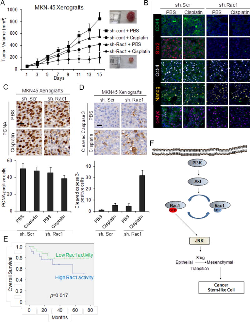
Rac1, a Rho GTPase family member, is dysregulated in a variety of tumor types including gastric adenocarcinoma, but little is known about its role in cancer stem-like cells (CSCs). Therefore, Rac1 activity and inhibition were examined in gastric adenocarcinoma cells and mouse xenograft models for epithelial-to-mesenchymal transition (EMT) and CSC phenotypes. Rac1 activity was significantly higher in spheroid-forming or CD44+ gastric adenocarcinoma CSCs compared with unselected cells. Rac1 inhibition using Rac1 shRNA or a Rac1 inhibitor (NSC23766) decreased expression of the self-renewal transcription factor, Sox-2, decreased spheroid formation by 78%-81%, and prevented tumor initiation in immunodeficient mice. Gastric adenocarcinoma CSCs had increased expression of the EMT transcription factor Slug, 4.4- to 8.3-fold greater migration, and 4.2- to 12.6-fold greater invasion than unselected cells, and these increases could be blocked completely with Rac1 inhibition. Gastric adenocarcinoma spheroid cells were resistant to 5-fluorouracil and cisplatin chemotherapy, and this chemotherapy resistance could be reversed with Rac1 shRNA or NSC23766. The PI3K/Akt pathway may be upstream of Rac1, and JNK may be downstream of Rac1. In the MKN-45 xenograft model, cisplatin inhibited tumor growth by 50%, Rac1 inhibition by 35%, and the combination by 77%. Higher Rac1 activity, in clinical specimens from gastric adenocarcinoma patients who underwent potentially curative surgery, correlated with significantly worse survival (P = 0.017). In conclusion, Rac1 promotes the EMT program in gastric adenocarcinoma and the acquisition of a CSC state. Rac1 inhibition in gastric adenocarcinoma cells blocks EMT and CSC phenotypes, and thus may prevent metastasis and augment chemotherapy.Implications: In gastric adenocarcinoma, therapeutic targeting of the Rac1 pathway may prevent or reverse EMT and CSC phenotypes that drive tumor progression, metastasis, and chemotherapy resistance. Mol Cancer Res; 15(8); 1106-16. ©2017 AACR.


Conflict of interest statement

Conflict of interest: The authors declare no conflicts of interest.

Figures

Figure 1
Figure 1. Rac1 activity in GA CSCs
A. Western blot demonstrating levels of active and total Rac1, RhoA, and Cdc42 in eight gastric adenocarcinoma cell lines. Western blot of active and total Rac1 in three GA cell lines grown as monolayers and as spheroids (B) or following FACS for CD44 (−) and CD44(+) cells (C). D. Western blot for total Rac1 and putative CSC markers in AGS, MKN-45and NCI-N87 spheroid cells following transduction with Rac1 shRNA (sh.Rac1) or scrambled control shRNA (sh.Scr). E. Immunofluorescence of AGS spheroids for DAPI (blue), Rac1 (red) and CD44 (green). F. Photos and graph of AGS, MKN-45, and MNI-N87 cells following transduction with sh.Scr or sh. Rac1 and grown in spheroid formation conditions. Bars represent standard deviation. *p<0.05 compared to control.
Figure 2
Figure 2. Rac1 promotes Sox2 expression and self renewal in GA CSCs
A. Western blot demonstrating levels of self-renewal proteins for Sox2, Oct-4, Nanog, and c-Myc in three GA cell lines grown as monolayers or as spheroids. Western blot for Sox2, Oct-4, Nanog, and c-Myc in GA cell lines grown as spheroids and transduced with Rac1 shRNA (sh.Rac1) or scrambled shRNA (sh.Scr). C. Immunofluorescence of AGS spheroids for DAPI (blue), CD44 (green), and Sox2 (red) following transduction with sh.Rac1 or sh.Scr and treatment with NSC23766 or carrier (DMSO). D. Photos from single cell assay for AGS and MKN-45 spheroid cells following transduction with sh.Scr and sh. Rac1. E. Photos of immunodeficient mice injected with 5000 or 20,000 MKN-45 spheroid cells stably transduced with sh.Scr and sh. Rac1.
Figure 3
Figure 3. PI3K/Akt pathway activates Rac1 and Rac1 activates JNK in GA CSCs
A. Immunofluorescence photos of AGS, MKN-45, and NCI-N87 CD44(+) spheroid cells treated with the Akt inhibitor LY294002, JNK inhibitor SP600125, ERK inhibitor U0126, p38 inhibitor SB203580, or carrier (DMSO). Scale bar 50 μm. B. Western blot of GA spheroid cells for total Rac1 and total and phosphorylated MAPK proteins following stable transduction of cells with Rac1 shRNA (sh.Rac1) or control shRNA (sh.Scr). C. Western blot of GA spheroid cells for total and phosphorylated Akt and JNK along with total and active Rac1 following treatment with the Akt inhibitor LY294002 or carrier (DMSO). Western blot of gastric adenocarcinoma CD44(+) spheroid cells for total and phosphorylated MAPK proteins and total and active Rac1 following treatment with the Akt inhibitor LY294002 or carrier (DMSO) (D) or with the JNK inhibitor SP00125 or carrier (DMSO) (E). Immunohistochemical staining of gastric tumors with low or high Rac1 activity for phosphorylated Rac1, phosphorylated JNK, and Sox2. Scale bar 20 μm.
Figure 4
Figure 4. Rac1 activity promotes EMT, migration, and invasion in GA CSCs
A. Western blot demonstrating levels of N-cadherin, Slug, Snail, and Zeb1 in gastric adenocarcinoma monolayer and spheroid cells. B. Western blot demonstrating levels of N-cadherin, Slug, Snail, and Zeb1 in GA spheroid cells transduced with Rac1 shRNA (sh.Rac1) or scrambled shRNA (sh.Scr). C. Immunofluorescence photos of AGS spheroids transduced with Rac1 shRNA (sh.Rac1) or scrambled shRNA (sh.Scr). Photos and graphs of migration (D) and invasion (E) assays for GA spheroid cells transduced with Rac1 shRNA (sh.Rac1) or scrambled shRNA (sh.Scr). Bars represent standard deviation. *p<0.05 compared to control.
Figure 5
Figure 5. Rac1 activity promotes chemoresistance in GA CSCs
A. Lethal dose 50(LD50) of GA monolayer and spheroid cells for 5-fluorouracil (5-FU) and cisplatin. Proliferation assays for GA monolayer cells (B) and spheroid cells (C) following treatment with Rac1 shRNA (sh.Rac1), scrambled control shRNA (sh.Scr), 5-fluorouracil (5-FU) or cisplatin chemotherapy, and/or carrier (DMSO). D. Western blot of AGS spheroid cells for total Rac1 and cleaved caspase 3 following treatment with Rac1 shRNA (sh.Rac1), scrambled control shRNA (sh.Scr), 5-fluorouracil (5-FU) or cisplatin chemotherapy, and/or carrier (DMSO). Bars represent standard deviation. *p<0.05.
Figure 6
Figure 6
A. Tumor growth curves for MKN-45 xenografts treatment with Rac1 shRNA (sh.Rac1), scrambled control shRNA (sh.Scr), cisplatin, and/or PBS. B. Photos following immunohistochemical analysis of treated tumors for CD44 (green), Sox2 (red), Oct-4 (white), Nanog (yellow), and c-Myc (tomato). Scale bar 20 μm. Photos and graphs of immunohistochemical analysis of treated tumors for proliferation (PCNA) (C) and apoptosis (cleaved caspase 3) (D). Scale bar 20 μm. E. Kaplan-Meier overall survival curves for patients undergoing surgical resection for GA stratified by low versus high Rac1 activity in the primary tumor. F. Diagram of Rac1 pathway in GA CSCs. Bars represent standard deviation. *p<0.05 compared to control.

References

    1. Jemal A, Bray F, Center MM, Ferlay J, Ward E, Forman D. Global cancer statistics. CA Cancer J Clin. 2011 Mar;61(2):69–90. - PubMed
    1. Wagner AD, Grothe W, Haerting J, Kleber G, Grothey A, Fleig WE. Chemotherapy in advanced gastric cancer: a systematic review and meta-analysis based on aggregate data. J Clin Oncol. 2006 Jun 20;24(18):2903–9. - PubMed
    1. Cunningham D, Starling N, Rao S, Iveson T, Nicolson M, Coxon F, et al. Capecitabine and oxaliplatin for advanced esophagogastric cancer. N Engl J Med. 2008 Jan 3;358(1):36–46. - PubMed
    1. Rocco A, Compare D, Nardone G. Cancer stem cell hypothesis and gastric carcinogenesis: Experimental evidence and unsolved questions. World J Gastrointest Oncol. 2012 Mar 15;4(3):54–9. - PMC - PubMed
    1. Alison MR, Lin WR, Lim SM, Nicholson LJ. Cancer stem cells: in the line of fire. Cancer Treat Rev. 2012 Oct;38(6):589–98. - PubMed

Publication types

MeSH terms