. 2017 Jun 16;292(24):9919-9931.
doi: 10.1074/jbc.M116.772566. Epub 2017 May 1.

Sirtuin 2 mutations in human cancers impair its function in genome maintenance

Affiliations

Sirtuin 2 mutations in human cancers impair its function in genome maintenance

PamelaSara E Head et al. J Biol Chem. .

Abstract

Sirtuin 2 (SIRT2) is a sirtuin family deacetylase, which maintains genome integrity and prevents tumorigenesis. Although Sirt2 deficiency in mice leads to tumorigenesis, the functional significance of somatic SIRT2 mutations in human tumors is unclear. Using structural insight combined with bioinformatics and functional analyses, we show that naturally occurring cancer-associated SIRT2 mutations at evolutionarily conserved sites disrupt its deacetylation of DNA-damage response proteins by impairing SIRT2 catalytic activity or protein levels but not its localization or binding with substrate. We observed that these SIRT2 mutant proteins fail to restore the replication stress sensitivity, impairment in recovery from replication stress, and impairment in ATR-interacting protein (ATRIP) focus accumulation of SIRT2 deficiency. Moreover, the SIRT2 mutant proteins failed to rescue the spontaneous induction of DNA damage and micronuclei of SIRT2 deficiency in cancer cells. Our findings support a model for SIRT2's tumor-suppressive function in which somatic mutations in SIRT2 contribute to genomic instability by impairing its deacetylase activity or diminishing its protein levels in the DNA-damage response. In conclusion, our work provides a mechanistic basis for understanding the biological and clinical significance of SIRT2 mutations in genome maintenance and tumor suppression.

Keywords: DNA; DNA damage; DNA damage response; acetylation; cancer; genomic instability; sirtuin; tumor suppressor gene.


Conflict of interest statement

The authors declare that they have no conflicts of interest with the contents of this article

Figures

Figure 1.
Figure 1.
Cancer-associated SIRT2 mutations are evolutionarily conserved and predicted to be functionally significant. A, table listing cBioPortal data for nine naturally occurring SIRT2 mutations in human cancer samples along with occurrence in patient cohort, cancer type, and study type. B, conservation of cancer-associated SIRT2 mutation amino acids among diverse species, including chimpanzee, mouse, chicken, zebrafish, fruit fly, yeast, and nematode, and their location in the protein structure. The NES is in yellow, and the deacetylase domain is highlighted in blue. C, impact prediction of cancer mutations by Grantham, SIFT, and PolyPhen, and their conservation scores as calculated by ConSurf. TCGA, The Cancer Genome Atlas; COSMIC, Catalogue of Somatic Mutations in Cancer.
Figure 2.
Figure 2.
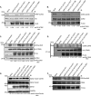
Cancer-associated mutations impair SIRT2 deacetylase activity and protein level but not localization. A, Western blot analysis demonstrating protein levels of SIRT2-FLAG WT and mutants expressed in U2OS cells. B, U2OS cells were transfected with SIRT2-FLAG WT or mutants. 72 h after transfection, cells were incubated with or without 5 nm LMB for 4 h to inhibit SIRT2 nuclear export, fixed, and processed for indirect immunofluorescence using anti-FLAG antibodies (green) and DAPI staining. Representative images are shown. C, in vitro measurements of SIRT2 deacetylase activity using the Fleur de Lys deacetylase assay are shown. SIRT2-FLAG WT and mutants were purified from 293T cells and eluted from FLAG M2 beads. Protein concentration was measured by Coomassie staining with a BSA standard control. 120 ng of SIRT2-FLAG WT and mutants was incubated in a reaction with an acetylated p53 Lys-320 peptide as a substrate. The mean of the relative fluorescence measured from three replicas is shown. Error bars represent S.D. Deacetylase activities of mutants were compared with that of SIRT2-FLAG WT. *, p < 0.05; **, p < 0.01. IB, immunoblotting.
Figure 3.
Figure 3.
Structural analysis of SIRT2 mutations yields insights into their functional significance. A, of the nine mutations selected from cBioPortal, four are found in either the NAD+-binding pocket or the catalytic domain. Mutations P128L, P140H, and F190V create the NAD+-binding pocket. NAD+ can be seen in blue, and the amino acids of interest are in red. Ala-186 lies next to the catalytic amino acid His-187. The protein structure was obtained from the Structural Bioinformatics Protein Data Bank, specifically structure 4RMH (40). B, a close-up of Ala-186 in a non-space-filling model. C, a close-up of Ala-186 in a space-filling model. D, of the nine mutations selected from cBioPortal, four are located in non-catalytic domains of the SIRT2 protein structure. These mutations are highlighted in purple. E, a close-up of two of the four mutations from D reveals they are mainly surface or hydrophobic amino acids. F, hypothetical crystal model of amino acid Arg-42.
Figure 4.
Figure 4.
Cancer-associated mutations impair SIRT2 deacetylation of DDR substrates in vitro and in cells but not interaction with substrate. Shown are representative Western blots of several replicas. A, acetylated ATRIP was isolated from 293T cells transfected with HA-ATRIP and histone acetyltransferases (HATs) pCAF, p300, and CBP and incubated in an in vitro deacetylation assay with SIRT2-FLAG WT or mutants isolated from 293T cells and in the presence of TSA with or without NAD and nicotinamide. The reaction mixtures were separated by SDS-PAGE and immunoblotted (IB) with site-specific anti-acetyl ATRIP Lys-32, HA, and FLAG antibodies. B, acetylated CDK9 was isolated from 293T cells transfected with CDK9-GFP and HATs and incubated in an in vitro deacetylation assay with SIRT2-FLAG WT or mutants isolated from 293T cells and in the presence of TSA with or without NAD and nicotinamide. The reaction mixtures were separated by SDS-PAGE and immunoblotted with site-specific anti-acetyl CDK9 Lys-48, HA, and FLAG antibodies. C, 293T cells were transfected with CDK9-HA and HATs or were left untransfected. After 24 h, cells were subsequently transfected with SIRT2-FLAG WT or mutants and incubated in the presence of TSA for 24 h. Deacetylation of CDK9-HA was assessed by Western blot analysis using site-specific anti-acetyl CDK9 Lys-48, HA, FLAG, and GAPDH antibodies. D, immunoprecipitation of samples from Fig. 2C. 293T cells were transfected with CDK9-HA and HATs or were left untransfected. After 24 h, cells were subsequently transfected with SIRT2-FLAG WT or mutants and incubated in the presence of TSA for 24 h. Lysate were immunoprecipitated with an anti-HA antibody, separated by SDS-PAGE, and immunoblotted with site-specific anti-acetyl CDK9 Lys-48, HA, and FLAG antibodies. E, 293T cells were transfected with HATs. After 24 h, cells were subsequently transfected with SIRT2-FLAG WT or mutants and incubated in the presence of TSA for 24 h. Deacetylation of endogenous α-tubulin was assessed by Western blot analysis using site-specific anti-acetyl-α-tubulin Lys-40, α-tubulin, SIRT2, and GAPDH antibodies. F, acetylated CDK9 was isolated from 293T cells transfected with CDK9-GFP and HATs and incubated in an in vitro deacetylation assay without SIRT2 (Mock), with SIRT2-FLAG WT only, or with equal amounts of both SIRT2-FLAG WT and one of four SIRT2-FLAG mutants isolated from 293T cells (SIRT2-FLAG H187Y, P128L, P140H, or A186V) in the presence of TSA with NAD+. The reaction mixtures were separated by SDS-PAGE and immunoblotted with site-specific anti-acetyl CDK9 Lys-48, CDK9, and FLAG antibodies.
Figure 5.
Figure 5.
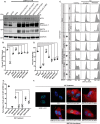
Cancer-associated SIRT2 mutations fail to rescue RSR defects of SIRT2 deficiency. A, Western blot analysis demonstrating efficiency of SIRT2 and ATR knockdown and expression of SIRT2-FLAG WT and mutants in HCT-116 cells. B, quantitation of cells with 4N DNA content following 10-h release from HU treatment in cells transfected with NS, ATR, or SIRT2-10 UTR siRNA with or without complementation with SIRT2-FLAG WT or mutants. The mean from two replicas is shown, and error bars represent S.D. NS indicates p ≥ 0.05; *, p < 0.05; **, p < 0.01. C, HCT116 cells were transfected with NS, ATR, or SIRT2-10 UTR siRNA, treated with 3 mm HU for 24 h (arrested), and released into nocodazole for 10 h (released). SIRT2–10 UTR knockdown was complemented with SIRT2-FLAG WT and mutants. DNA content was analyzed by flow cytometry. Representative cell cycle profiles are shown. D, HU sensitivity as measured by alamarBlue cell viability staining of HCT116 cells transfected with NS, siATR, or SIRT2-10 UTR siRNA with or without complementation with SIRT2-FLAG WT or mutants and treated with 1.6 mm HU for 24 h followed by a 24-h release. The mean of HU-treated to untreated viability relative to NS siRNA in triplicate is shown, and error bars represent S.D. E, quantitation of cells with >5 GFP-ATRIP foci following 24-h 3 mm HU treatment in a GFP-ATRIP stable U2OS cell line transfected with NS or SIRT2-5 siRNA with or without complementation with EV (pcDNA3.1) or SIRT2-FLAG WT or mutants (SIRT2-FLAG WT constructs contained wobble mutations to protect against knockdown). The mean from three replicas of 100 cells counted in each is shown, and error bars represent S.D. NS indicates p ≥ 0.05; *, p < 0.05; **, p < 0.01. F, representative images of conditions quantified and outlined in E with GFP-ATRIP in green, FLAG in red, and DAPI. IB, immunoblotting.
Figure 6.
Figure 6.
Cancer-associated SIRT2 mutations fail to rescue genomic instability of SIRT2 deficiency. A, U2OS SIRT2 KO cells generated by CRISPR/Cas9 were complemented with or without SIRT2-FLAG WT or mutants. Western blot analysis demonstrating efficiency of expression of SIRT2-FLAG WT and mutants in U2OS SIRT2 KO cells or endogenous SIRT2 in U2OS WT cells is shown. The arrow indicates a nonspecific band beneath the SIRT2 protein band. B, U2OS SIRT2 KO cells demonstrate increased spontaneous γH2AX foci and were complemented with or without SIRT2-FLAG WT or mutants. The degree of alleviation of spontaneous γH2AX foci observed was quantified. Quantitation of the percentage of cells with >5 spontaneous γH2AX foci is shown. The mean was calculated from three replicas of 100 cells for each condition, and error bars represent S.D. *, p < 0.05; **, p < 0.01. C, representative images of U2OS SIRT2 KO cells complemented with or without SIRT2-FLAG WT or mutants and stained for γH2AX foci (green), FLAG (red), and DAPI. U2OS SIRT2 KO cells with SIRT2 construct expression stain positive for FLAG (red), and examples of this are highlighted by white arrows, whereas cells in the same population that did not express SIRT2 construct do not exhibit red staining in the cytoplasm. D, U2OS SIRT2 KO cells were transfected with or without SIRT2-FLAG WT or mutants and stained for FLAG in red and DAPI. Induced micronuclei were counted. Quantitation of micronuclei is shown. The mean was calculated from three replicas of 100 cells for each condition, and error bars represent S.D. *, p < 0.05; **, p < 0.01. E, representative images of micronuclei conditions from D. IB, immunoblotting.

Similar articles

Cited by

References

    1. Guarente L. (2011) Franklin H. Epstein Lecture: sirtuins, aging, and medicine. N. Engl. J. Med. 364, 2235–2244 - PubMed
    1. Finkel T., Deng C. X., and Mostoslavsky R. (2009) Recent progress in the biology and physiology of sirtuins. Nature 460, 587–591 - PMC - PubMed
    1. Saunders L. R., and Verdin E. (2007) Sirtuins: critical regulators at the crossroads between cancer and aging. Oncogene 26, 5489–5504 - PubMed
    1. Donmez G., and Outeiro T. F. (2013) SIRT1 and SIRT2: emerging targets in neurodegeneration. EMBO Mol. Med. 5, 344–352 - PMC - PubMed
    1. Zhang H., Head P. E., and Yu D. S. (2016) SIRT2 orchestrates the DNA damage response. Cell Cycle 15, 2089–2090 - PMC - PubMed

Publication types

MeSH terms