

doi: 10.1503/cmaj.160472.
Dual-stent retrieval for mechanical thrombectomy of refractory clot in acute stroke as a rescue technique
Affiliations
- PMID: 28461375
- PMCID: PMC5415391
- DOI: 10.1503/cmaj.160472
Item in Clipboard
Dual-stent retrieval for mechanical thrombectomy of refractory clot in acute stroke as a rescue technique
CMAJ.
.
No abstract available
Conflict of interest statement
Competing interests: None declared.
Figures
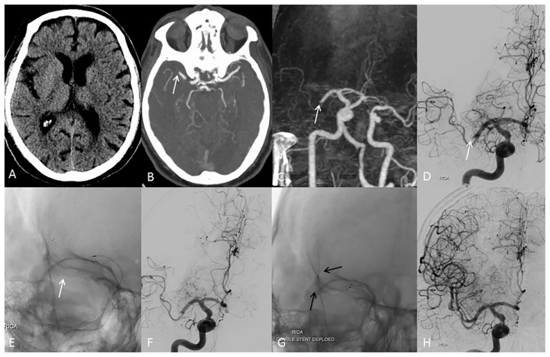
(A) Axial computed tomography (CT) scan of the head showing no obvious infarct. (B) Axial CT angiography and (C) coronal subtraction CT angiography of the head showing occlusion (white arrow) of the right middle cerebral artery (MCA). (D) A frontal angiogram showing an occluded right MCA. (E) Fluoroscopic anteroposterior image showing the deployed stent retriever (white arrow) in the right MCA. (F) Persistent occlusion of the right MCA after four passes with the stent retriever. (G) Fluoroscopic anteroposterior projection showing two stent retrievers deployed in the right MCA (black arrows). (H) Frontal angiogram showing complete recanalization of the right MCA.
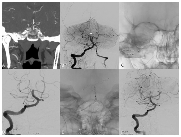
(A) Coronal and (B) frontal computed tomography (CT) angiograms of the head showing an occluded basilar tip (white arrow). (C) Fluoroscopic anteroposterior image showing the stent retriever deployed at the basilar tip. (D) Persistent occlusion of the basilar tip after three passes with a single stent retriever. (E) Fluoroscopic image showing two stent retrievers overlapped at the basilar trunk and their distal ends extended into the right and left posterior cerebral arteries (PCAs) to create the Y configuration. (F) Angiogram after dual-stent retrieval showing complete recanalization of the basilar tip with a residual clot in the distal left PCA.
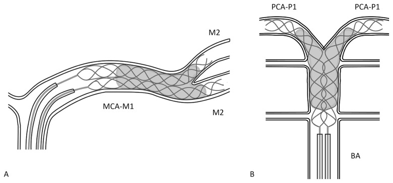
(A) Schematic diagram of the simultaneous deployment of dual stent retrievers within the clot in the middle cerebral artery (MCA) M1 (MCA-M1) segment with their distal ends extending to both limbs of the MCA bifurcation (M2 segments). (B) Schematic diagram showing the overlap of the two stent retrievers at the basilar trunk and their distal ends extending into both posterior cerebral arteries (PCA-P1 segments) to create the Y configuration. BA = basilar artery.
References
-
- Rha JH, Saver JL. The impact of recanalization on ischemic stroke outcome: a meta-analysis. Stroke 2007;38:967–73. - PubMed
-
- Berkhemer OA, Fransen PS, Beumer D, et al. A randomized trial of intraarterial treatment for acute ischemic stroke. N Engl J Med 2015;372:11–20. - PubMed
-
- Campbell BC, Mitchell PJ, Kleinig TJ, et al. Endovascular therapy for ischemic stroke with perfusion-imaging selection. N Engl J Med 2015;372:1009–18. - PubMed
-
- Goyal M, Demchuk AM, Menon BK, et al. Randomized assessment of rapid endovascular treatment of ischemic stroke. N Engl J Med 2015;372:1019–30. - PubMed
MeSH terms
LinkOut - more resources
Full Text Sources
Other Literature Sources
Medical