. 2017 Jun 29;37(14):e00048-17.
doi: 10.1128/MCB.00048-17. Print 2017 Jul 15.

CREBH Maintains Circadian Glucose Homeostasis by Regulating Hepatic Glycogenolysis and Gluconeogenesis

Affiliations

CREBH Maintains Circadian Glucose Homeostasis by Regulating Hepatic Glycogenolysis and Gluconeogenesis

Hyunbae Kim et al. Mol Cell Biol. .

Abstract

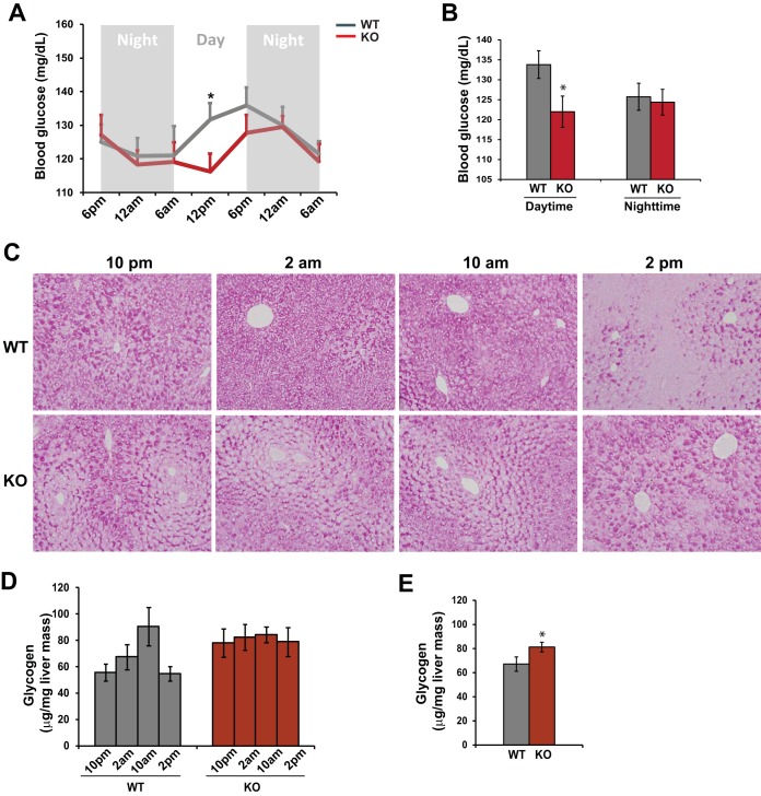
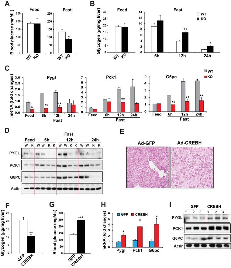
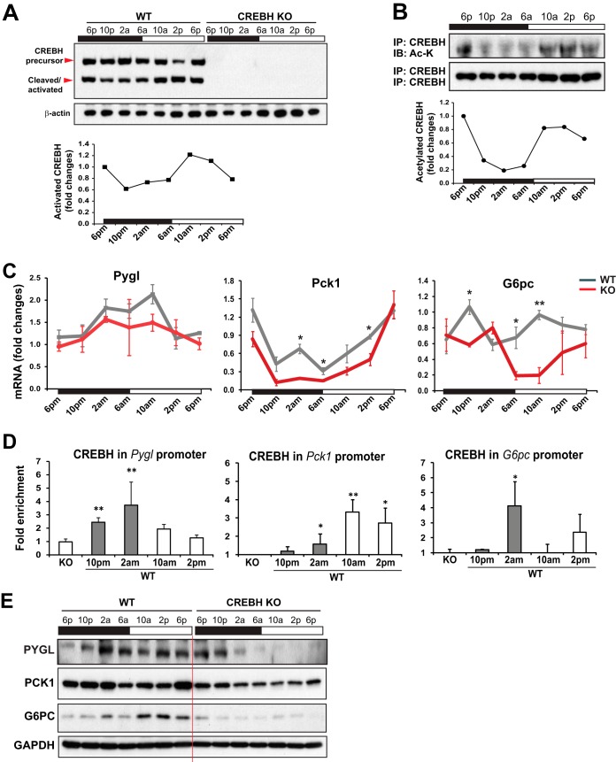
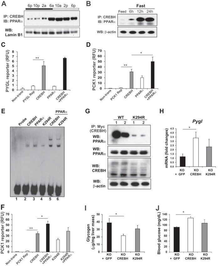
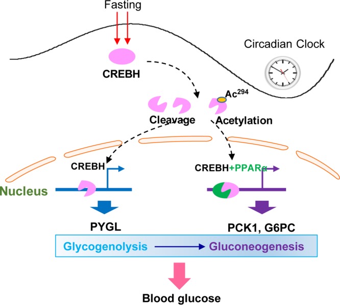
Cyclic AMP-responsive element binding protein, hepatocyte specific (CREBH), is a liver-enriched, endoplasmic reticulum-tethered transcription factor known to regulate the hepatic acute-phase response and lipid homeostasis. In this study, we demonstrate that CREBH functions as a circadian transcriptional regulator that plays major roles in maintaining glucose homeostasis. The proteolytic cleavage and posttranslational acetylation modification of CREBH are regulated by the circadian clock. Functionally, CREBH is required in order to maintain circadian homeostasis of hepatic glycogen storage and blood glucose levels. CREBH regulates the rhythmic expression of the genes encoding the rate-limiting enzymes for glycogenolysis and gluconeogenesis, including liver glycogen phosphorylase (PYGL), phosphoenolpyruvate carboxykinase 1 (PCK1), and the glucose-6-phosphatase catalytic subunit (G6PC). CREBH interacts with peroxisome proliferator-activated receptor α (PPARα) to synergize its transcriptional activities in hepatic gluconeogenesis. The acetylation of CREBH at lysine residue 294 controls CREBH-PPARα interaction and synergy in regulating hepatic glucose metabolism in mice. CREBH deficiency leads to reduced blood glucose levels but increases hepatic glycogen levels during the daytime or upon fasting. In summary, our studies revealed that CREBH functions as a key metabolic regulator that controls glucose homeostasis across the circadian cycle or under metabolic stress.

Keywords: CREBH; circadian metabolism; gluconeogenesis; glucose metabolism; glycogenolysis; liver metabolism; nuclear receptor; transcriptional regulation.


Figures

FIG 1
FIG 1
CREBH regulates rhythmic levels of blood glucose and hepatic glycogen storage in mice under the control of the circadian clock. (A) Levels of blood glucose in CREBH-null and WT control mice across the circadian cycle. Blood glucose levels were measured every 6 h for 36 h in constant darkness. Data are presented as means ± standard errors of the means (n, 8 mice per time point) at each time point. *, P ≤ 0.05. (B) Average blood glucose levels in CREBH-null and WT control mice during the daytime or nighttime. Bars, means; error bars, standard errors of the means (n, 16 mice per group). (C) PAS staining reveals hepatic glycogen levels in CREBH-null and WT control mice at 10 p.m., 2 a.m., 10 a.m., and 2 p.m. (magnification, ×200). (D) Quantitative enzymatic analysis of hepatic glycogen levels in CREBH-null and WT control mice at 10 p.m., 2 a.m., 10 a.m., and 2 p.m. Bars, means; error bars, standard errors of the means (n, 3 mice per group per time point). (E) Quantitative enzymatic analysis of glycogen levels in pooled liver tissue samples collected from 12 CREBH-null or 12 WT control mice across the circadian cycle. Bars, means; error bars, standard errors of the means (n, 12 mice per group per time point).
FIG 2
FIG 2
CREBH promotes hepatic glycogenolysis and gluconeogenesis upon fasting or CREBH overexpression. (A) Levels of blood glucose in CREBH-null and WT control mice under feeding conditions or after a 12-h fast. Bars, means; error bars, standard errors of the means (n, 3 mice per time point). *, P < 0.05. (B) Hepatic glycogen levels in CREBH-null and WT control mice under feeding conditions or after fasting for 6, 12, or 24 h. Bars, means; error bars, standard errors of the means (n, 3 mice per time point). **, P < 0.01. (C) Expression levels of Pygl, Pck1, and G6pc mRNAs in the livers of CREBH-null and WT control mice under feeding conditions or after fasting for 6, 12, or 24 h. mRNA expression levels were determined by qRT-PCR. Fold changes in mRNA levels were determined by comparison to the mRNA levels in one of the wild-type control mice under the feeding condition. Bars, means; error bars, standard errors of the means (n, 3 or 4 mice per time point). (D) Western blot analysis of PYGL, PCK1, and G6PC proteins in mouse livers under feeding conditions or after a 6-, 12-, or 24-h fast. (E) PAS staining of glycogen in the livers of mice overexpressing GFP or activated CREBH (magnification, ×200). WT mice were injected through the tail vein with a recombinant adenovirus (Ad) expressing activated CREBH or GFP as a control. At 3 days after the injection, the animals were euthanized for the examination of hepatic glycogen and blood glucose. (F and G) Levels of hepatic glycogen (F) and blood glucose (G) in mice overexpressing GFP or activated CREBH in the liver. Bars, means; error bars, standard errors of the means (n, 3 mice). ***, P < 0.001. (H) Expression levels of Pygl, Pck1, and G6pc mRNAs in the livers of mice overexpressing GFP or activated CREBH. mRNA expression levels were determined by qRT-PCR. Fold changes in mRNA levels were determined by comparison to the mRNA levels in one of the mice overexpressing GFP. Bars, means; error bars, standard errors of the means (n, 3 mice). (I) Western blot analysis of PYGL, PCK1, and G6PC proteins in the livers of mice overexpressing GFP or activated CREBH. Levels of β-actin were determined as loading controls.
FIG 3
FIG 3
CREBH regulates rhythmic expression of the key genes involved in hepatic glycogenolysis and gluconeogenesis in mice under the control of the circadian clock. (A) (Top) Western blot analysis of levels of CREBH precursor and activated/cleaved forms in mouse livers over the circadian cycle. Liver tissues were collected from WT or KO mice every 4 h over a 24-h circadian cycle (n, 3 mice/genotype/time point). Tissues from each group at each time point were pooled, and cellular proteins were prepared from the pooled tissues. Levels of β-actin protein were determined as controls. (Bottom) Graph showing the quantification of activated/cleaved CREBH protein in mouse livers over the circadian cycle. The intensity of the CREBH protein signal, determined by Western blot densitometry, was normalized to that of β-actin. Fold changes in protein levels were determined by comparison to the protein level at 6 p.m. (B) (Top) IP-Western blot analysis of levels of acetylated CREBH protein in mouse livers over the circadian cycle. Nuclear proteins from pooled liver tissues of WT mice over a 24-h circadian cycle (n, 3 mice/time point) were immunoprecipitated with the anti-CREBH antibody to pull down the CREBH protein complex, followed by immunoblotting (IB) with the anti-acetyl-lysine antibody. The levels of CREBH protein pulled down were determined as the controls. (Bottom) Graph showing the quantification of acetylated CREBH protein in mouse livers over the circadian cycle. The intensity of the lysine-acetylated CREBH protein signal, determined by Western blot densitometry, was normalized to that of CREBH. Fold changes in protein levels were determined by comparison to the starting level at 6 p.m. (C) Rhythmic expression levels of Pygl, Pck1, and G6pc in the livers of CREBH-null and WT control mice. mRNA expression levels were determined by qRT-PCR. Fold changes in mRNA levels were determined by comparison to levels in one of the wild-type control mice at the starting circadian time point. Bars, means; error bars, standard errors of the means (n, 3 to 5 mice per group per time point). *, P < 0.05; **, P < 0.01. (D) CREBH enrichment in the Pygl, Pck1, and G6pc gene promoters in the livers of WT mice at different circadian phases, as determined by ChIP-qPCR. CREBH-null liver nuclei were used as negative controls for the endogenous CREBH ChIP assays. CREBH enrichment in the gene promoters at different circadian phases was quantified by comparing ChIP-qPCR signals from the samples pulled down by the anti-CREBH antibody to that pulled down by a rabbit anti-IgG antibody. Bars, means; error bars, standard errors of the means (n, 3 mice per time point). (E) Western blot analysis of rhythmic levels of PYGL, PCK1, and G6PC proteins in the livers of CREBH-null and WT mice, collected every 4 h in a 24-h circadian period. Pooled liver protein lysates from 3 to 5 mice per time point per genotype group were used.
FIG 4
FIG 4
Lysine acetylation of CREBH regulates CREBH-PPARα interaction and synergy in regulating hepatic glucose metabolism. (A) IP and Western blot analysis of interactions between CREBH and PPARα in the liver nuclear fractions of WT mice over a 24-h circadian period. Liver nuclear proteins pooled from 3 WT mice per time point were pulled down by the anti-CREBH antibody and were then probed with the PPARα antibody. Levels of lamin B1 were determined as controls. (B) IP and Western blot analysis of interactions between CREBH and PPARα in the livers of mice after a 6-, 12-, or 24-h fast. Total liver cellular lysates were used for Western blot analysis. Levels of β-actin were determined as controls. (C and D) Luciferase reporter (Rep) analyses of transcriptional activation of the human Pygl (C) or Pck1 (D) gene promoter by CREBH alone or in combination with PPARα. Hepa1-6 cells were transduced with the reporter vector or a vehicle. After 24 h, the transfected cells were infected with an adenovirus expressing GFP (control), CREBH, and/or PPARα. A Renilla reporter plasmid was included in the cotransfection for the normalization of luciferase reporter activities. The same adenovirus titers were used for individual infections. Bars, means; error bars, standard errors of the means (n, 2 experimental repeats). Non-trans, nontransfected cell control. *, P < 0.05; **, P < 0.01. (E) Binding of cellular extracts from Hepa1-6 cells expressing GFP, CREBH, or PPARα to the mouse PCK1 gene promoter oligonucleotide. EMSA was performed using extracts from Hepa1-6 cells infected with an adenovirus expressing GFP, CREBH, the K294R mutant, and/or PPARα and the PCK1 gene probe containing the CREBH-PPAR binding motif. The formation of a CREBH-DNA or PPARα-DNA complex is indicated by a shifted band, and the formation of a CREBH-PPARα-DNA complex is indicated by a supershifted band. “Probe” indicates the control reaction with the CREBH-PPAR probe in the absence of cellular extracts. (F) Luciferase reporter analyses of transcriptional activation of the mouse Pck1 gene promoter by CREBH or the K294R mutant alone or in combination with PPARα. A Renilla reporter plasmid was included in the cotransfection for the normalization of luciferase reporter activities. Bars, means; error bars, standard errors of the means (n, 2 experimental repeats). (G) IP and Western blot analysis of interactions between PPARα and CREBH or the K294R mutant in the livers of mice under fasting conditions. A recombinant adenovirus expressing either GFP as a control, Myc-tagged full-length human CREBH protein (WT), or the acetylation-deficient mutant (K294R) was injected into the tail veins of CREBH-null mice. (Top panel) Liver protein lysates collected from the mice after a 24-h fast were immunoprecipitated with the Myc antibody to pull down the CREBH protein complex, followed by immunoblotting with the PPARα antibody. (Lower panels) Western blot analyses to determine the levels of PPARα, Myc-tagged CREBH, and β-actin. (H) Expression levels of Pygl mRNA in the livers of CREBH-null mice expressing GFP, WT CREBH, or the K294R mutant after fasting as described in the legend to panel G. mRNA expression levels were determined by qRT-PCR. Bars, means; error bars, standard errors of the means (n, 3 mice per group). (I and J) Levels of hepatic glycogen (I) and blood glucose (J) in mice overexpressing GFP, WT CREBH, or the K294R mutant in their livers. KO mice were injected through the tail vein with a recombinant adenovirus expressing GFP, WT CREBH, or the K294R mutant. At 3 days after the injection, the animals were fasted for 24 h before being euthanized for the examination of blood glucose and hepatic glycogen levels. Bars, means; error bars, standard errors of the means (n, 3 mice per time point).
FIG 5
FIG 5
Working model of CREBH as a circadian metabolic regulator of hepatic glucose metabolism over the circadian cycle or under fasting conditions. Ac294, CREBH acetylation at lysine (K) 294.

References

    1. Bass J, Takahashi JS. 2010. Circadian integration of metabolism and energetics. Science 330:1349–1354. doi:10.1126/science.1195027. - DOI - PMC - PubMed
    1. Green CB, Takahashi JS, Bass J. 2008. The meter of metabolism. Cell 134:728–742. doi:10.1016/j.cell.2008.08.022. - DOI - PMC - PubMed
    1. Bechmann LP, Hannivoort RA, Gerken G, Hotamisligil GS, Trauner M, Canbay A. 2012. The interaction of hepatic lipid and glucose metabolism in liver diseases. J Hepatol 56:952–964. doi:10.1016/j.jhep.2011.08.025. - DOI - PubMed
    1. Raddatz D, Ramadori G. 2007. Carbohydrate metabolism and the liver: actual aspects from physiology and disease. Z Gastroenterol 45:51–62. doi:10.1055/s-2006-927394. - DOI - PubMed
    1. Feng D, Lazar MA. 2012. Clocks, metabolism, and the epigenome. Mol Cell 47:158–167. doi:10.1016/j.molcel.2012.06.026. - DOI - PMC - PubMed

Publication types

MeSH terms

Substances

LinkOut - more resources