Review
. 2017 Aug 8;199(17):e00131-17.
doi: 10.1128/JB.00131-17. Print 2017 Sep 1.

Esx Systems and the Mycobacterial Cell Envelope: What's the Connection?

Affiliations
Review

Esx Systems and the Mycobacterial Cell Envelope: What's the Connection?

Rachel E Bosserman et al. J Bacteriol. .

Abstract

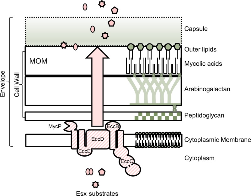
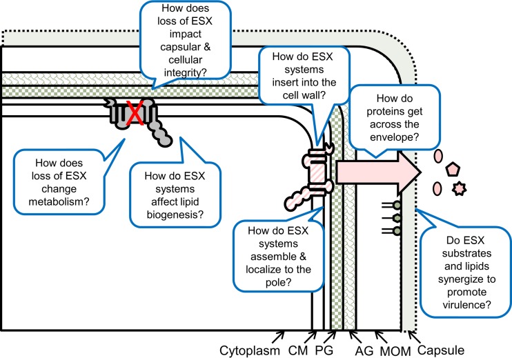
Mycobacterial 6-kDa early secreted antigenic target (ESAT-6) system (ESX) exporters transport proteins across the cytoplasmic membrane. Many proteins transported by ESX systems are then translocated across the mycobacterial cell envelope and secreted from the cell. Although the mechanism underlying protein transport across the mycolate outer membrane remains elusive, the ESX systems are closely connected with and localize to the cell envelope. Links between ESX-associated proteins, cell wall synthesis, and the maintenance of cell envelope integrity have been reported. Genes encoding the ESX systems and those required for biosynthesis of the mycobacterial envelope are coregulated. Here, we review the interplay between ESX systems and the mycobacterial cell envelope.

Keywords: ESX system; cell envelope; lipids; mycobacteria; protein secretion; transport.


Figures

FIG 1
FIG 1
ESX exporters in the mycobacterial cell envelope. The model shows a cartoon representation of the complex mycobacterial cell envelope, spanning from the cytoplasmic membrane through the mycolate outer membrane (MOM) and capsule. Proteins are excluded from the cell envelope, other than ESX proteins, for simplicity. Components of the envelope are not drawn to scale. For a recent review on the mycobacterial cell envelope, please see references , , and . The ESX membrane complex and MycP are indicated in the cytoplasmic membrane (striped). The dotted line in the Ecc protein refers to the fact that specifically in the ESX-1 system, the EccC protein is split into two proteins (EccCa1 and EccCb1). Additional ESX proteins required for transport are excluded for simplicity (Esp and additional Ecc proteins, PE and PPE). For recent reviews on the molecular mechanisms of ESX export, please see references to . The cartoon was based on schematics in references and . Ecc, ESX-conserved component. Large arrow indicates that proteins traverse the cell wall via an unknown mechanism.
FIG 2
FIG 2
Unanswered questions regarding the interactions between ESX systems and the mycobacterial cell envelope. CM, cytoplasmic membrane; PG, peptidoglycan; AG, arabinogalactan; MOM, mycolate outer membrane.

References

    1. Chagnot C, Zorgani MA, Astruc T, Desvaux M. 2013. Proteinaceous determinants of surface colonization in bacteria: bacterial adhesion and biofilm formation from a protein secretion perspective. Front Microbiol 4:303. doi: 10.3389/fmicb.2013.00303. - DOI - PMC - PubMed
    1. Green ER, Mecsas J. 2016. Bacterial secretion systems: an overview. Microbiol Spectr 4:215–239. doi: 10.1128/microbiolspec.VMBF-0012-2015. - DOI - PMC - PubMed
    1. Hoffmann C, Leis A, Niederweis M, Plitzko JM, Engelhardt H. 2008. Disclosure of the mycobacterial outer membrane: cryo-electron tomography and vitreous sections reveal the lipid bilayer structure. Proc Natl Acad Sci U S A 105:3963–3967. doi: 10.1073/pnas.0709530105. - DOI - PMC - PubMed
    1. Sani M, Houben EN, Geurtsen J, Pierson J, de Punder K, van Zon M, Wever B, Piersma SR, Jimenez CR, Daffe M, Appelmelk BJ, Bitter W, van der Wel N, Peters PJ. 2010. Direct visualization by cryo-EM of the mycobacterial capsular layer: a labile structure containing ESX-1-secreted proteins. PLoS Pathog 6:e1000794. doi: 10.1371/journal.ppat.1000794. - DOI - PMC - PubMed
    1. Bansal-Mutalik R, Nikaido H. 2014. Mycobacterial outer membrane is a lipid bilayer and the inner membrane is unusually rich in diacyl phosphatidylinositol dimannosides. Proc Natl Acad Sci U S A 111:4958–4963. doi: 10.1073/pnas.1403078111. - DOI - PMC - PubMed

Publication types

MeSH terms

Substances

LinkOut - more resources