. 2017 May 16;114(20):E4002-E4009.
doi: 10.1073/pnas.1524845114. Epub 2017 May 1.

SP and IL-33 together markedly enhance TNF synthesis and secretion from human mast cells mediated by the interaction of their receptors

Affiliations

SP and IL-33 together markedly enhance TNF synthesis and secretion from human mast cells mediated by the interaction of their receptors

Alexandra Taracanova et al. Proc Natl Acad Sci U S A. .

Abstract

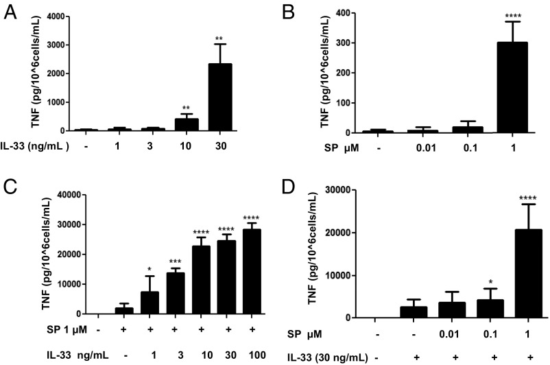
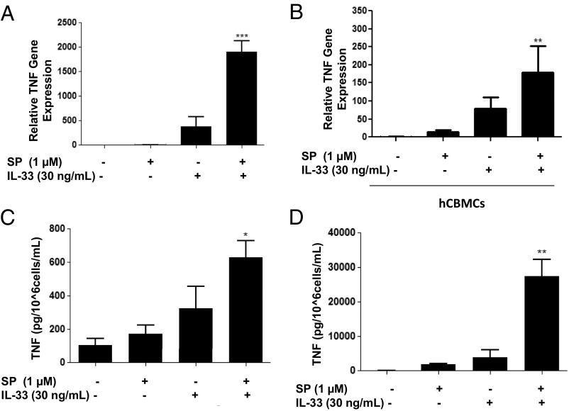
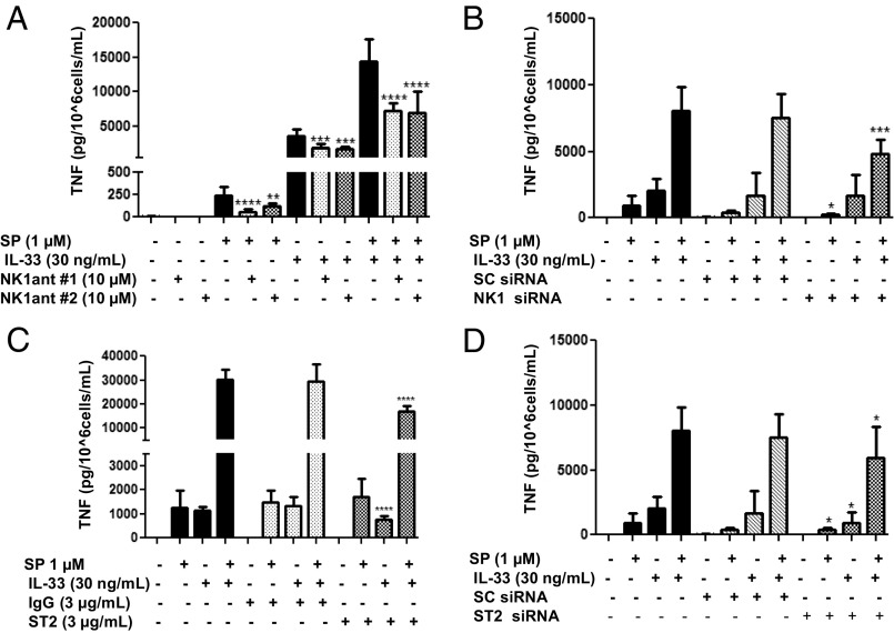
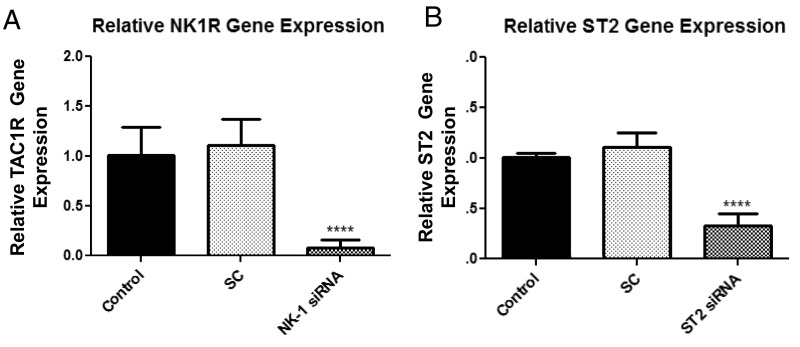
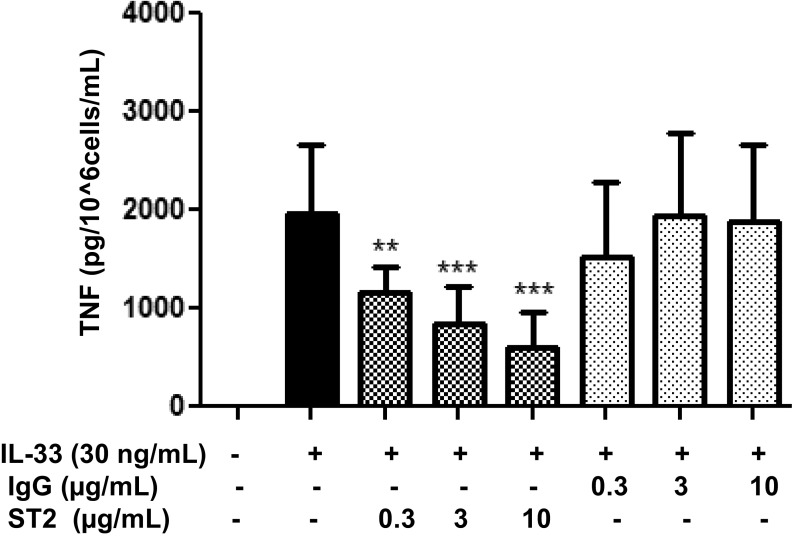
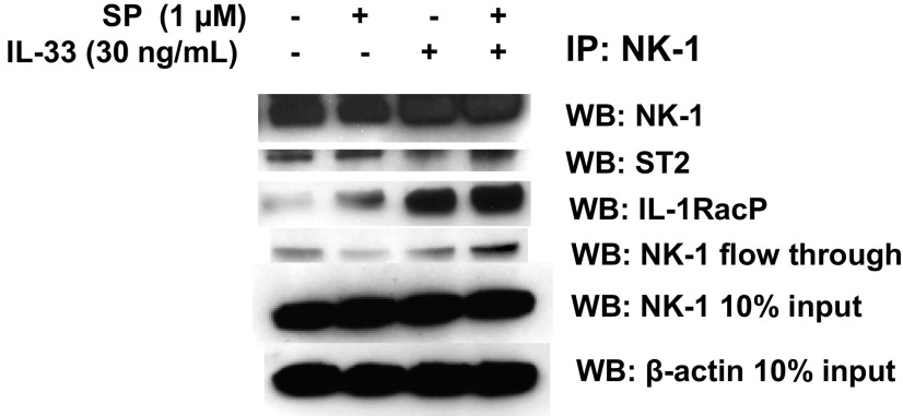
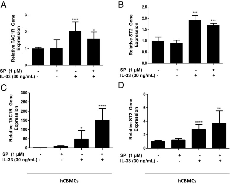
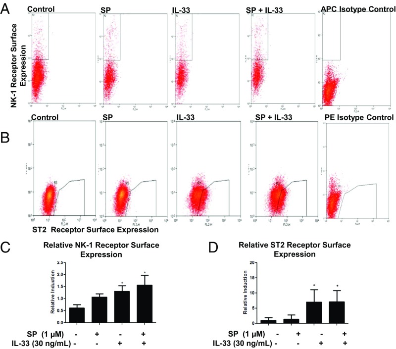
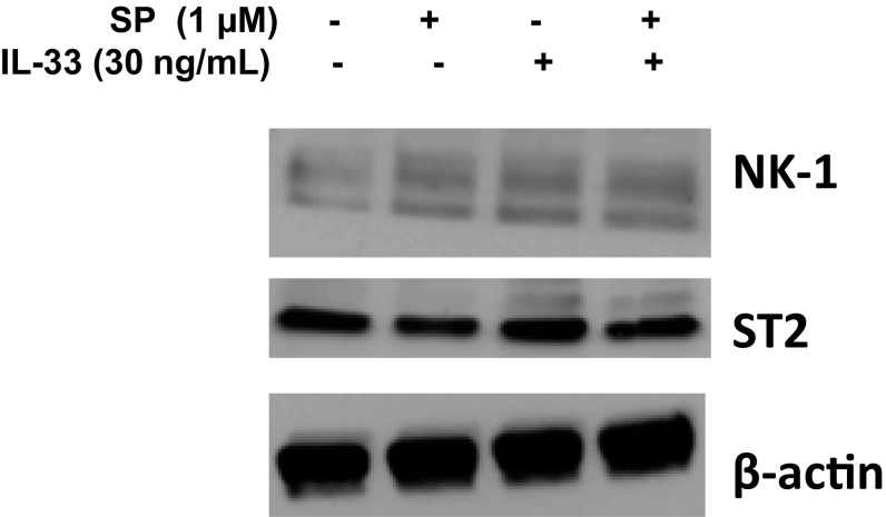
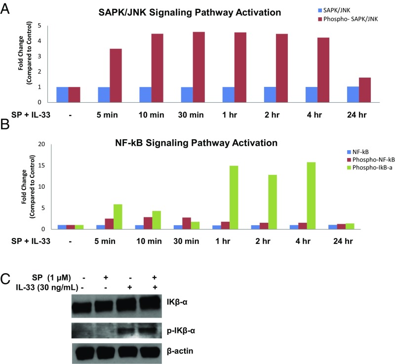
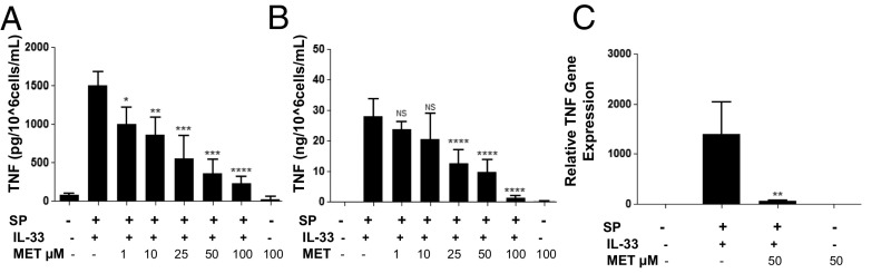
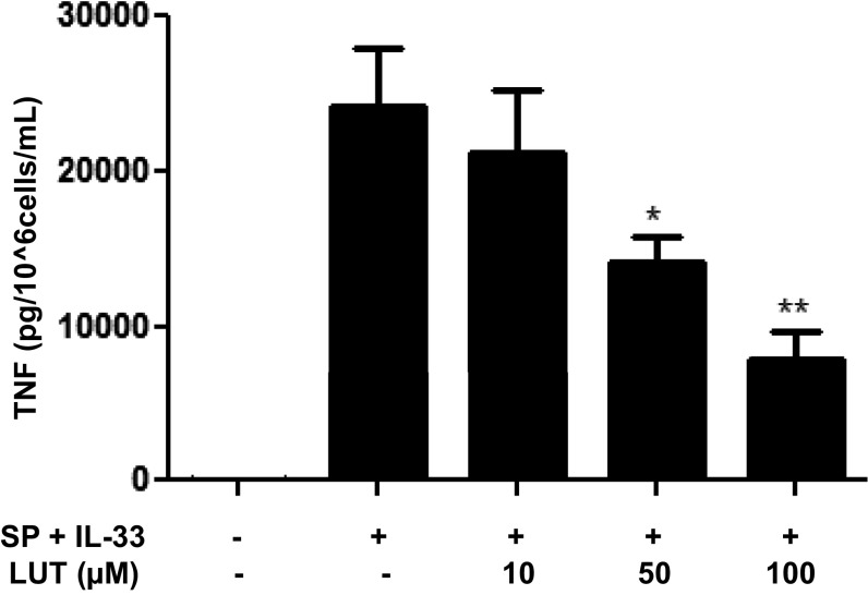
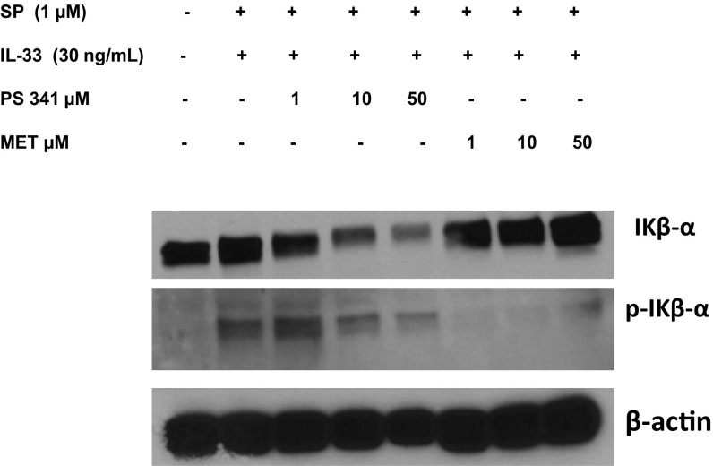
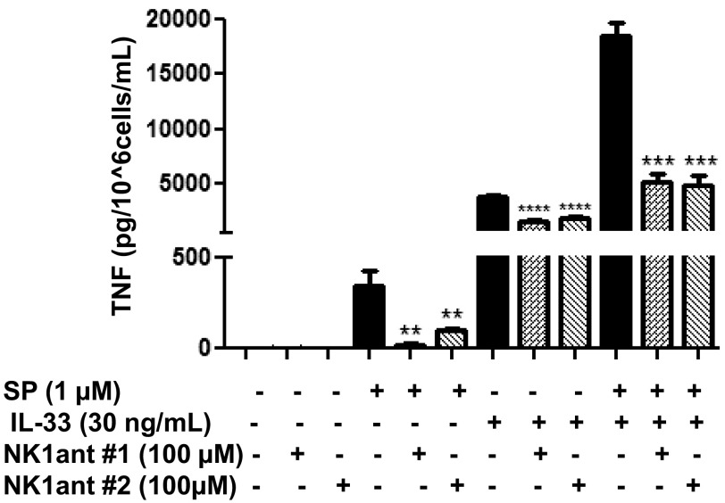
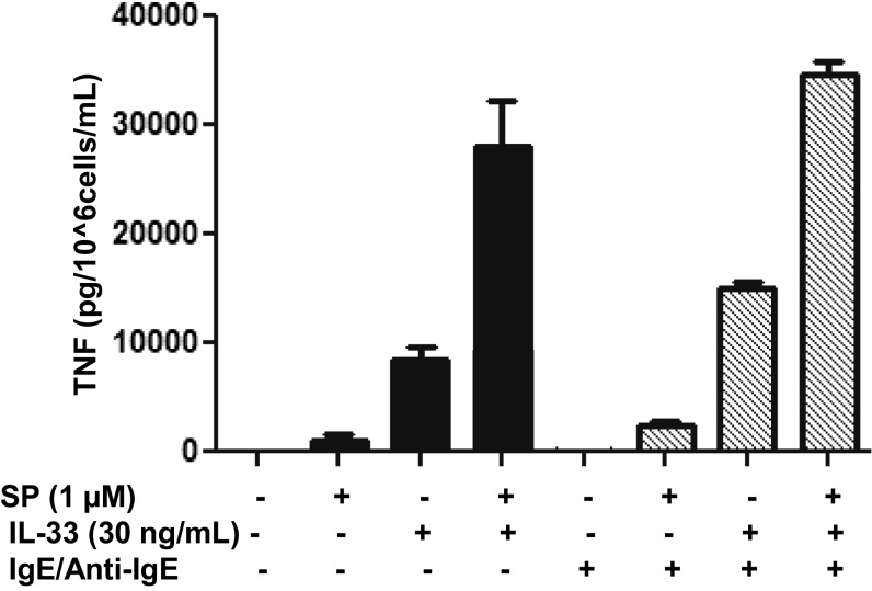
The peptide substance P (SP) and the cytokine tumor necrosis factor (TNF) have been implicated in inflammatory processes. Mast cells are recognized as important in inflammatory responses. Here, we report that IL-33 (30 ng/mL), a member of the IL-1 family of cytokines, administered in combination with SP (1 µM), markedly increase (by 1,000-fold) TNF gene expression in cultured human LAD2 and primary mast cells derived from umbilical cord blood. SP (0.01-1 μM) and IL-33 (1-100 ng/mL) in combination also greatly stimulate TNF secretion (by 4,500-fold). Pretreatment of LAD2 cells with two different neurokinin-1 (NK-1) receptor antagonists and siRNA inhibits TNF secretion by 50% (P < 0.001) when stimulated by SP and IL-33. Pretreatment of LAD2 cells with a neutralizing antibody for IL-33 receptor, ST2, inhibits TNF secretion by 50% (P < 0.001), and ST2 siRNA decreases TNF secretion by 30% (P < 0.05), when stimulated by SP and IL-33. Surprisingly, NK-1 antagonists also inhibit 50% of TNF secretion (P < 0.001) when stimulated only by IL-33, and ST2 receptor reduction also decreases SP-stimulated TNF secretion by 30% (P < 0.05), suggesting an interaction between NK-1 and ST2 receptors. Moreover, IL-33 increases NK-1 gene and surface protein expression, as well as IKβ-α phosphorylation. Pretreatment of LAD2 cells with 5,7,3',4'-tetramethoxyflavone (methoxyluteolin) (1-100 μM) inhibits (P < 0.001) TNF gene expression (98%) and secretion (64%) at 50 µM and phosphorylation of p-IKB-α at 1 μM when stimulated by SP and IL-33. These findings identify a unique amplification process of TNF synthesis and secretion via the interaction of NK-1 and ST2 receptors inhibitable by methoxyluteolin.

Keywords: inflammation; interleukin-33; mast cells; substance P; tumor necrosis factor.


Conflict of interest statement

Conflict of interest statement: T.C.T. and Cem Akin were coauthors on a 2015 publication. This publication was a review article and did not involve any research collaboration.

Figures

Fig. 1.
Fig. 1.
Selection of the optimal doses to study TNF secretion stimulated by SP and IL-33 when administered in combination. LAD2 cells (1 × 10⁵ cells per well) were seeded in 96-well culture plates and stimulated with IL-33 (1–30 ng/mL) alone (A), with SP (0.1–1 µM) alone (B), or with SP (0.01–1 μΜ), IL-33 (1-100 ng/mL), or their combination as shown (C and D) for 24 h. Supernatant fluids were collected at the end of the incubation period and assayed by TNF ELISA. n = 3.*P < 0.05; **P < 0.01; ***P < 0.001; ****P < 0.0001.
Fig. 2.
Fig. 2.
SP and IL-33 markedly enhance TNF gene expression and secretion in human mast cells. (A and B) LAD2 cells (1 × 10⁶ cells per well) (A) and hCBMCs (0.3 × 10⁶ cell per well) (B) were seeded in 12-well culture plates and stimulated with SP (1 μΜ), IL-33 (30 ng/mL), or their combination for 6 h. TNF mRNA expression levels were measured by qRT-PCR and normalized to human GAPDH endogenous control. (C and D) LAD2 cells (1 × 10⁵ cells per well) were seeded in 96-well culture plates and stimulated with SP (1 μΜ), IL-33 (30 ng/mL) or their combination as shown for 24 h. Control cells were treated with culture media only. Supernatant fluids (D) and cell lysates (C) were collected at the end of the incubation period and assayed by ELISA. n = 3. *P < 0.05; **P < 0.01.
Fig. 3.
Fig. 3.
NK-1 receptor antagonists and ST2 neutralizing antibody inhibit TNF secretion. (A) LAD2 cells were pretreated with NK-1R antagonists L-733,060 (10 μM) and CP-96345 (10 µM) for 30 min and then stimulated with SP (1 μΜ), IL-33 (30 ng/mL) or their combination for 24 h. (C) LAD2 cells were preincubated with ST2 neutralizing antibody (3 ng/mL) or IgG control (3 ng/mL) for 2 h, then stimulated with SP (1 μΜ), IL-33 (30 ng/mL), or their combination for 24 h. (B and D) LAD2 cells were pretreated with NK-1 receptor siRNA (50 µM) (B), ST2 receptor siRNA (D), or scrambled siRNA (SC) (50 µM) (B and D) for 72–96 h and then stimulated with SP (1 μΜ), IL-33 (30 ng/mL), or their combination for 24 h. Collected supernatant fluids were assayed by TNF ELISA. n = 3. *P < 0.05; **P < 0.01; ***P < 0.001; ****P < 0.0001.
Fig. S1.
Fig. S1.
NK-1 and ST2 transient gene knockdown. LAD2 cells were treated with NK-1, ST2, or scramble (SC) siRNA (50 µM) for 72–96 h. Expression levels of NK-1 (A) and ST2 mRNA (B) were measured by qRT-PCR and normalized to human GAPDH endogenous control. n = 3. ****P < 0.0001.
Fig. S2.
Fig. S2.
Dose-dependent blockade of ST2 receptor. LAD2 cells (1 × 10⁵ cells per well) were seeded in 96-well culture plates, preincubated with ST2 neutralizing antibody (0.3–10 µg/mL) or IgG control (0.3–10 µg/mL) for 2 h, and then stimulated with IL-33 (30 ng/mL) for 24 h. Supernatant fluids were collected at the end of the incubation period and assayed by TNF ELISA. n = 3. **P < 0.01; ***P < 0.001.
Fig. S3.
Fig. S3.
NK-1 receptor coimmunoprecipitates with ST2 and IL-1RacP. LAD2 cells (5 × 10⁶ cells) were stimulated with the combination of SP and IL-33. Cell lysates were collected after 30 min and immunoprecipitated for an antibody against NK-1 receptor. IP samples as well as 10% loading control samples were assayed for protein levels of NK-1, ST2, IL-1RacP, and β-actin by Western blot analysis. n = 3.
Fig. 4.
Fig. 4.
Effect of SP and IL-33 on NK-1 and ST2 gene expression. LAD2 cells (1 × 10⁶ cells per well) (A and B) and hCBMCs (0.3 × 10⁶ cell per well) (C and D) were seeded in 12-well culture plates and stimulated with SP (1 μΜ), IL-33 (30 ng/mL), or their combination for 24 h. NK-1 and ST2mRNA expression levels were measured by qRT-PCR and normalized to human GAPDH endogenous control. n = 3. *P < 0.05; **P < 0.01; ***P < 0.001; ****P < 0.0001.
Fig. 5.
Fig. 5.
IL-33 increases NK-1 and ST2 expression. (A and B) LAD2 cells (1 × 10⁶ cells per well) were seeded in 12-well culture plates and stimulated with SP (1 μΜ), IL-33 (30 ng/mL), or their combination for 24 h. Collected cells were probed for allophylocyanin (APC)-conjugated NK-1 receptor (A) and phycoerythrin (PE)-conjugated ST2 receptor (B). Data are from a representative experiment. (C and D) Relative NK-1 (C) and ST2 (D) expression on cell the surface compared with control. n = 3. *P < 0.05.
Fig. S4.
Fig. S4.
IL-33 increases NK-1 total protein. LAD2 cells (1 × 10⁶ cells) were stimulated with the combination of SP (1 µM) and IL-33 (30 ng/mL). Cell lysates were collected after 24 h, and protein levels of NK-1 and ST2 were measured by Western blot analysis. β-actin served as a loading control. n = 3.
Fig. 6.
Fig. 6.
NF-κB and SAPK/JNK signaling pathways are involved in TNF gene expression and secretion. (A and B) LAD2 cells (2 × 10⁶ cells) were stimulated with SP (1 μΜ) and IL-33 (30 ng/mL) for 5 min, 10 min, 30 min, 1 h, 2 h, 4 h, and 24 h. Phosphorylation of SAPK/JNK (Th183/Tyr185) (A), NF-κB p65 (Ser536), and IΚβ-α (Ser32) (B) was detected using the PathScan Inflammation Mutli-Target Sandwich ELISA Kit. Whole-cell lysates were assayed at a protein concentration of 1 mg/mL. (C) LAD2 MCs were stimulated with SP (1 μM), IL-33 (30 ng/mL), and their combination. Cell lysates were collected after 1 h, and protein levels of IΚβ-α and phospho-IΚβ-α were measured by Western blot analysis. β-actin served as a loading control. n = 3.
Fig. 7.
Fig. 7.
Methoxyluteolin inhibits TNF gene expression and secretion. (A and B) LAD2 cells (1 × 10⁵ cells per well) were seeded in 96-well culture plates and preincubated with methoxyluteolin (MET; 1–100 μM), then stimulated with the combination of SP (1 μΜ) and IL-33 (30 ng/mL) for 24 h. Control cells were treated with 0.1% DMSO, the highest concentration corresponding to that of 100 μΜ methoxyluteolin. Collected supernatant fluids (A) and cell lysates (B) were assayed by TNF ELISA. (C) LAD2 cells (1 × 10⁶ cells per well) were seeded in 12-well culture plates and preincubated with methoxyluteolin (50 μM), then stimulated with the combination of SP (1 μΜ) and IL-33 (30 ng/mL) for 6 h. TNF mRNA expression levels were measured by qRT-PCR and normalized to human GAPDH endogenous control. n = 3. *P < 0.05; **P < 0.01; ***P < 0.001; ****P < 0.0001.
Fig. S5.
Fig. S5.
Luteolin inhibits TNF secretion. LAD2 cells (1 × 10⁵ cells per well) were seeded in 96-well culture plates and preincubated with luteolin (Lut) (10–100 μM), then stimulated with the combination of SP (1 μΜ) and IL-33 (30 ng/mL) for 24 h. Control cells were treated with 0.1% DMSO, the highest concentration corresponding to that of 100 μΜ Lut. Supernatant fluids were assayed for TNF by ELISA. n = 3. *P < 0.05; **P < 0.01, Student’s t test.
Fig. 8.
Fig. 8.
Methoxyluteolin inhibits IKβ-α phosphorylation. LAD2 cells (1 × 10⁶ cells) were preincubated with proteasome inhibitor PS 341 (1, 10, and 50 μM) or methoxyluteolin (1, 10, and 50 μM) and then stimulated with the combination of SP and IL-33. Cell lysates were collected after 1 h, and protein levels of IKβ-α and phospho-IΚβ-α were measured by Western blot analysis. β-actin served as a loading control. n = 3.
Fig. S6.
Fig. S6.
NK-1 receptor antagonists at 100 µM do not completely inhibit TNF secretion. LAD2 cells were pretreated with NK-1R antagonists L-733,060 (100 μM) and CP-96345 (100 µM) for 30 min and then stimulated with SP (1 μΜ), IL-33 (30 ng/mL), or their combination for 24 h. Collected supernatant fluids were assayed by TNF ELISA. n = 3. **P < 0.01; ***P < 0.001; ****P < 0.0001.
Fig. S7.
Fig. S7.
hCBMCs contain tryptase and express surface c-kit receptor. Photomicrographs of hCBMCs stained by immunohistochemistry at 12 wk for tryptase content (A) and c-kit receptor surface expression (B). (Magnification: 1,000×.)
Fig. S8.
Fig. S8.
Lack of secretory granule content maturity in hCBMCs. Transmission electron micrographs of individual human mast cells: (A) hCBMCs, 9-wk culture; (B) hCBMCs, 15-wk culture; (C) normal human detrusor muscle mast cell. (Magnification: 10,000×.) Note the differences in quality, content, and texture of the secretory granules.
Fig. S9.
Fig. S9.
IgE/anti-IgE does not increase SP and IL-33 stimulated secretion of TNF. LAD2 cells (1 × 10⁵ cells per well) were seeded in 96-well culture plates and preincubated with human IgE (1 µg/mL) or medium overnight, and the next day stimulated with anti-IgE (10 ng/mL) for 2 h and/or SP (1 µM) and IL-33 (30 ng/mL) for 24 h. Supernatant fluids were collected at the end of the incubation period and assayed by TNF ELISA. n = 3.

Comment in

  • Reply to Fattori et al.: Action of SP and IL-33 on mast cells.
    Theoharides TC, Taracanova A, Tsilioni I, Conti P, Leeman SE. Theoharides TC, et al. Proc Natl Acad Sci U S A. 2017 Nov 21;114(47):E10036. doi: 10.1073/pnas.1716233114. Epub 2017 Nov 6. Proc Natl Acad Sci U S A. 2017. PMID: 29109296 Free PMC article. No abstract available.
  • IL-33/ST2 signaling boosts inflammation and pain.
    Fattori V, Borghi SM, Verri WA Jr. Fattori V, et al. Proc Natl Acad Sci U S A. 2017 Nov 21;114(47):E10034-E10035. doi: 10.1073/pnas.1716120114. Epub 2017 Nov 6. Proc Natl Acad Sci U S A. 2017. PMID: 29109297 Free PMC article. No abstract available.

References

    1. Chang MM, Leeman SE. Isolation of a sialogogic peptide from bovine hypothalamic tissue and its characterization as substance P. J Biol Chem. 1970;245:4784–4790. - PubMed
    1. Saraceno R, Kleyn CE, Terenghi G, Griffiths CE. The role of neuropeptides in psoriasis. Br J Dermatol. 2006;155:876–882. - PubMed
    1. Douglas SD, Leeman SE. Neurokinin-1 receptor: Functional significance in the immune system in reference to selected infections and inflammation. Ann N Y Acad Sci. 2011;1217:83–95. - PMC - PubMed
    1. O’Connor TM, et al. The role of substance P in inflammatory disease. J Cell Physiol. 2004;201:167–180. - PubMed
    1. Steinhoff MS, von Mentzer B, Geppetti P, Pothoulakis C, Bunnett NW. Tachykinins and their receptors: Contributions to physiological control and the mechanisms of disease. Physiol Rev. 2014;94:265–301. - PMC - PubMed

Publication types

MeSH terms

LinkOut - more resources