. 2017 Jun 1;198(11):4435-4447.
doi: 10.4049/jimmunol.1601717. Epub 2017 May 1.

Kinase Activities of RIPK1 and RIPK3 Can Direct IFN-β Synthesis Induced by Lipopolysaccharide

Affiliations

Kinase Activities of RIPK1 and RIPK3 Can Direct IFN-β Synthesis Induced by Lipopolysaccharide

Danish Saleh et al. J Immunol. .

Abstract

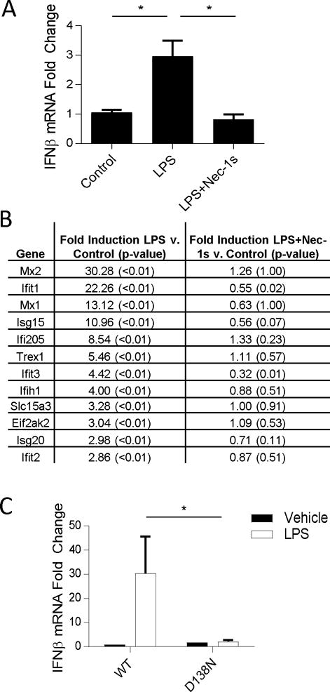
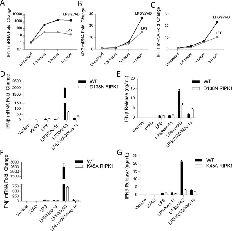
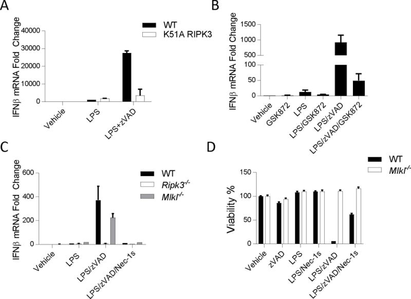
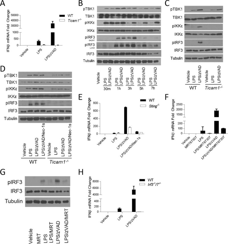
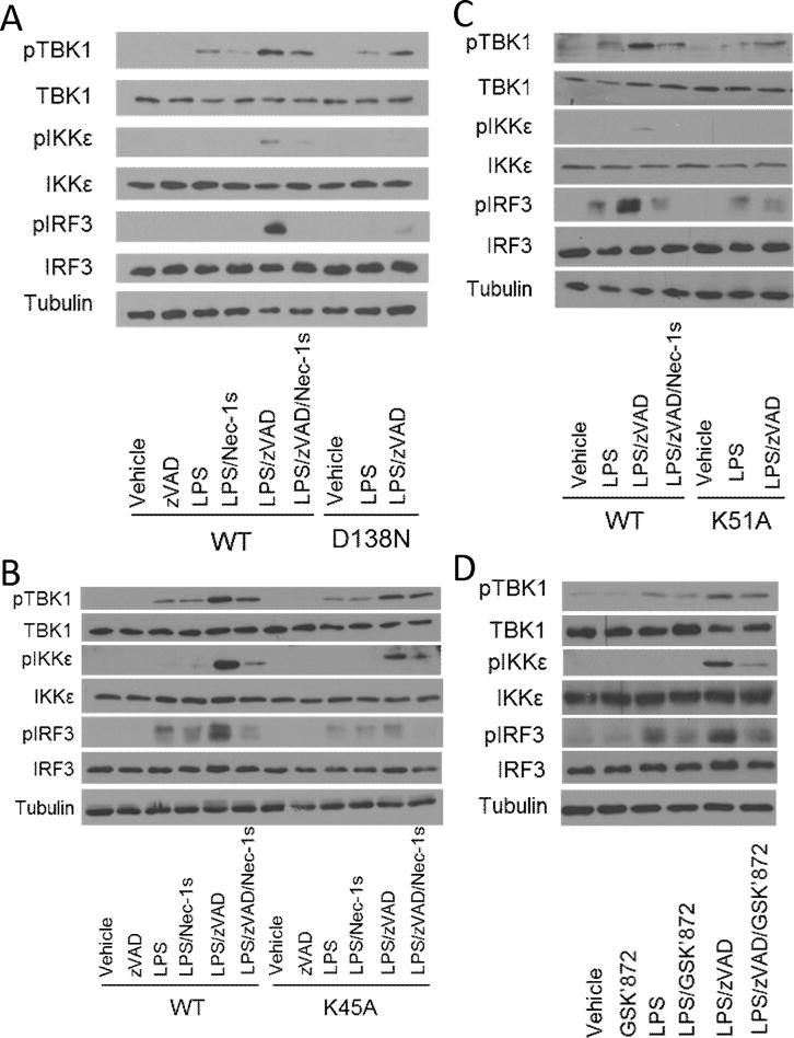
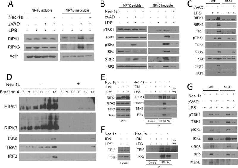
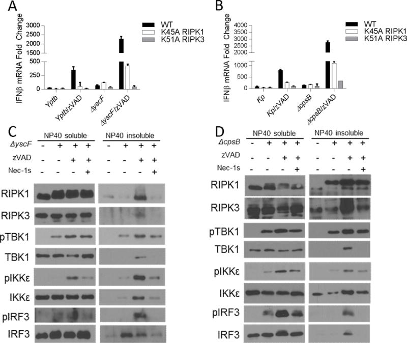
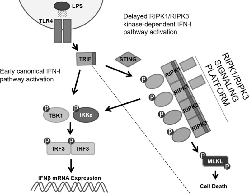
The innate immune response is a central element of the initial defense against bacterial and viral pathogens. Macrophages are key innate immune cells that upon encountering pathogen-associated molecular patterns respond by producing cytokines, including IFN-β. In this study, we identify a novel role for RIPK1 and RIPK3, a pair of homologous serine/threonine kinases previously implicated in the regulation of necroptosis and pathologic tissue injury, in directing IFN-β production in macrophages. Using genetic and pharmacologic tools, we show that catalytic activity of RIPK1 directs IFN-β synthesis induced by LPS in mice. Additionally, we report that RIPK1 kinase-dependent IFN-β production may be elicited in an analogous fashion using LPS in bone marrow-derived macrophages upon inhibition of caspases. Notably, this regulation requires kinase activities of both RIPK1 and RIPK3, but not the necroptosis effector protein, MLKL. Mechanistically, we provide evidence that necrosome-like RIPK1 and RIPK3 aggregates facilitate canonical TRIF-dependent IFN-β production downstream of the LPS receptor TLR4. Intriguingly, we also show that RIPK1 and RIPK3 kinase-dependent synthesis of IFN-β is markedly induced by avirulent strains of Gram-negative bacteria, Yersinia and Klebsiella, and less so by their wild-type counterparts. Overall, these observations identify unexpected roles for RIPK1 and RIPK3 kinases in the production of IFN-β during the host inflammatory responses to bacterial infection and suggest that the axis in which these kinases operate may represent a target for bacterial virulence factors.


Figures

Figure 1
Figure 1. LPS induced IFN-I production is dependent on kinase-activity of RIPK1
(A) qRT-PCR analysis of Ifnb mRNA expression in wild type (WT) mice injected with Nec-1s (iv) 15 min prior to LPS (ip). n= 6 animals per group and *p<0.05. Values reflect mean ± SE. (B) RNA seq analysis of a panel of IFN-I response genes in CD11b+ cells isolated from mice injected as in (A). Values reflect mean. P values marked in parentheses. GSE73836. (C) qRT-PCR analysis of Ifnb mRNA expression WT and D138N mice injected with LPS (ip). n= 3–7 animals per group and *p<0.05. Values reflect mean across biological variants and error bars reflect SE.
Figure 2
Figure 2. LPS with zVAD induces IFNβ synthesis in a RIPK1 kinase-dependent manner
(A–C) Time course of mRNA expression changes in Ifnb (A), Mx2 (B), and Ifit1 (C) evaluated by qRT-PCR in wild type BMDMs. Black squares – LPS/zVAD; Grey circles – LPS. (D-E) qRT-PCR and ELISA analysis of Ifnb mRNA expression and IFNβ protein release in WT and D138N BMDMs treated for 5–7 hrs. (F-G) qRT-PCR and ELISA analysis of Ifnb mRNA expression and IFNβ protein release in WT and K45A BMDMs treated for 5–7 hrs. Data are representative. Error bars reflect SD from the mean. BMDMs were treated with LPS=10 ng/mL, zVAD=50 μM, and/or Nec-1s=30μM where indicated.
Figure 3
Figure 3. Kinase-activity of RIPK3, but not MLKL, is required for RIPK1 kinase-dependent IFNβ synthesis
(A) qRT-PCR analysis of Ifnb mRNA expression in WT and K51A BMDMs treated for 5–7 hrs. (B) qRT-PCR of Ifnb mRNA expression in wild type BMDMs treated with RIPK3 kinase inhibitor (GSK872= 5μM) for 5–7 hrs. (C) qRT-PCR of Ifnb mRNA expression in wild type (WT), RIPK3 knockout (Ripk3−/−) and MLKL knockout (Mlkl−/−) BMDMs treated for 7 hrs. (D) Cell viability of WT and Mlkl−/− BMDMs treated for 24 hours and evaluated by CellTiterGlo ATP assay. Experiments were repeated 3 times. Data are representative. Error bars reflect SD from the mean. BMDMs were treated with LPS=10 ng/mL, zVAD=50 μM, GSK’872=5μM, and/or Nec-1s=30μM where indicated.
Figure 4
Figure 4. RIPK1 kinase-dependent IFNβ synthesis requires TRIF, STING, TBK1/IKKε, and IRF3/7
(A) qRT-PCR of Ifnb mRNA expression in WT and Ticam1−/− BMDMs treated for 5–7 hrs. (B) Time course of TBK1, IKKε, and IRF3 phosphorylation evaluated by Western analysis in wild type BMDMs. (C) Western analysis of TBK1, IKKε, and IRF3 phosphorylation in wild type (WT) and Ticam1−/− BMDMs treated for 3–4 hours. (D) Western analysis of TBK1, IKKε, and IRF3 phosphorylation in wild type (WT) and Ticam1−/− BMDMs treated for 1 hour. (E) qRT-PCR analysis of Ifnb mRNA expression in WT and Sting−/− BMDMs treated for 5–7 hrs. (F) qRT-PCR of Ifnb mRNA expression in wild type BMDMs treated with TBK1/IKKε inhibitor (MRT67307, 2μM) for 5–7 hrs. (G) Western analysis of IRF3 phosphorylation in wild type BMDMs treated with MRT67307 for 3–4 hrs. (H) qRT-PCR of Ifnb mRNA expression in WT and Irf3−/−7−/− BMDMs treated for 5–7 hours. Data are representative. Error bars reflect SD from the mean. BMDMs were treated with LPS=10 ng/mL, zVAD=50 μM, Nec-1=30μM and/or MRT67307=2μM where indicated.
Figure 5
Figure 5. IFN-I pathway activation by LPS with zVAD is RIPK1 and RIPK3 kinase-dependent
(A) Western analysis of TBK1, IKKε, and IRF3 phosphorylation in wild type (WT) and D138N RIPK1 (D138N) BMDMs treated for 3–4 hrs. (B) Western analysis of TBK1, IKKε, and IRF3 phosphorylation in wild type (WT) and K45A RIPK1 (K45A) BMDMs treated for 3–4 hrs. (C) Western analysis of TBK1, IKKε, and IRF3 phosphorylation in wild type (WT) and K51A RIPK3 (K51A) BMDMs treated for 3–4 hrs. (D) Western analysis of TBK1, IKKε, and IRF3 phosphorylation in wild type BMDMs treated with RIPK3 kinase inhibitor (GSK’872) for 3–4 hrs. BMDMs were treated with LPS=10 ng/mL, zVAD=50 μM, Nec-1=30μM and/or GSK’872=5μM where indicated.
Figure 6
Figure 6. Kinase activity of RIPK1 and RIPK3 are required for localization and activation of IFN-I pathway intermediaries in detergent-insoluble cellular fractions
(A) Western analysis of localization and modification of RIPK1 and RIPK3 in NP40-soluble and NP40-insoluble fractions from wild type BMDMs treated for 3–4 hrs. (B) Western analysis of localization and phosphorylation of TRIF, TBK1, IKKε, IRF3 in NP40-soluble and NP40-insoluble fractions from wild type BMDMs treated for 3–4 hrs. (C) Western analysis of RIPK1, RIPK3, TRIF, TBK1, IKKε, and IRF3 in NP40-insoluble fractions from wild type (WT) and K51A RIPK3 (K51A) BMDMs treated for 3–4 hrs.(D) Western analysis of RIPK1, RIPK3, TBK1, IKKε, and IRF3 in RAW264.7 macrophages treated with LPS and IDN6556 (10μM) +/− Nec-1s for 3–4 hrs. Lysates were fractionated on a linear sucrose-gradient by velocity sedimentation and protein was collected following chloroform-methanol precipitation. (E) Co-immunoprecipitation analysis of RIPK3, TBK1, and IKKε following immunoprecipitation of RIPK1 from RAW264.7 macrophages treated for 3–4 hrs. (F) Co-immunoprecipitation analysis of TRIF following immunoprecipitation of IKKε from RAW264.7 macrophages treated for 3–4 hrs. Control=Beads only. (G) Western analysis of RIPK1, RIPK3, TRIF, TBK1, IKKε, and IRF3 in NP40-insoluble fractions from wild type (WT) and Mlkl−/− BMDMs treated for 3–4 hrs. BMDMs were treated with LPS=10 ng/mL, zVAD=50 μM, IDN6556=10μM, and/or Nec-1s=30μM where indicated.
Figure 7
Figure 7. RIPK1 and RIPK3 kinase-dependent IFNβ synthesis is augmented by attenuated strains of Yersinia pseudotuberculosis and Klebsiella pneumonia
(A) qRT-PCR of Ifnb mRNA expression in wild type (WT), K45A RIPK1 (K45A), and K51A RIPK3 (K51A) BMDMs infected with wild type Yersinia pseudotuberculosis (Yptb) or a mutant strain unable to inject pathogenicity factors into macrophages (ΔyscF), at an MOI of 40–60. (B) qRT-PCR of Ifnb mRNA expression in WT, K45A, and K51A BMDMs infected with wild type Klebsiella pneumonia (Kp), or a mutant strain lacking its out polysaccharide capsule virulence factor (ΔcpsB) at an MOI of 40–60. (C) Western analysis of localization and phosphorylation of TRIF, TBK1, IKKε, IRF3 in NP40-soluble and NP40-insoluble fractions from wild type BMDMs treated as described in (A). (D) Western analysis of localization and phosphorylation of TRIF, TBK1, IKKε, IRF3 in NP40-soluble and NP40-insoluble fractions from wild type BMDMs treated as described in (B). Data are representative. Error bars reflect SD from the mean. BMDMs were treated with zVAD=50 μM and/or Nec-1s=30μM where indicated. Gentamicin (100μg/mL) was added 2 hours post-infection in each experiment.
Figure 8
Figure 8
RIPK1 and RIPK3 kinases engage the IFN-I pathway downstream of the adapter protein TRIF.

References

    1. Degterev A, Hitomi J, Germscheid M, Ch’en IL, Korkina O, Teng X, Abbott D, Cuny GD, Yuan C, Wagner G, Hedrick SM, Gerber SA, Lugovskoy A, Yuan J. Identification of RIP1 kinase as a specific cellular target of necrostatins. Nat Chem Biol. 2008;4:313–21. - PMC - PubMed
    1. Christofferson DE, Li Y, Yuan J. Control of Life-or-Death Decisions by RIP1 Kinase. Annu Rev Physiol. 2014;76:129–50. - PubMed
    1. Kaczmarek A, Vandenabeele P, Krysko DV. Necroptosis: the release of damage-associated molecular patterns and its physiological relevance. Immunity. 2013;38:209–23. - PubMed
    1. Vanden Berghe T, Linkermann A, Jouan-Lanhouet S, Walczak H, Vandenabeele P. Regulated necrosis: the expanding network of non-apoptotic cell death pathways. Nat Rev Mol Cell Biol. 2014;15:135–47. - PubMed
    1. Linkermann A, Green DR. Necroptosis. N Engl J Med. 2014;370:455–65. - PMC - PubMed

Publication types

MeSH terms