Review
. 2017 May 1;9(5):a025817.
doi: 10.1101/cshperspect.a025817.

Microtubule-Based Transport and the Distribution, Tethering, and Organization of Organelles

Affiliations
Review

Microtubule-Based Transport and the Distribution, Tethering, and Organization of Organelles

Kari Barlan et al. Cold Spring Harb Perspect Biol. .

Abstract

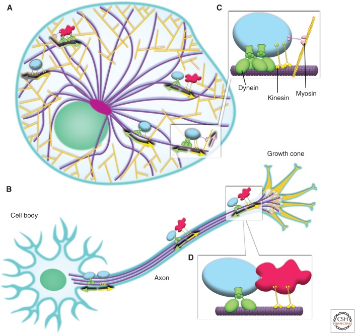
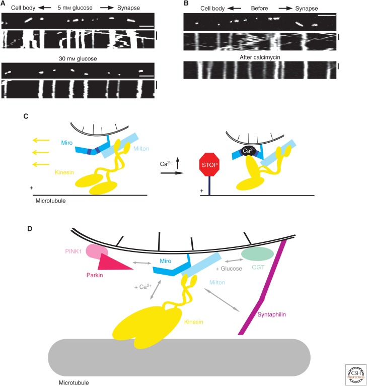
SUMMARYMicrotubules provide long tracks along which a broad range of organelles and vesicles are transported by kinesin and dynein motors. Motor protein complexes also tether cargoes to cytoskeletal filaments, helping facilitate their interaction and communication. The generation of biochemically distinct microtubule subpopulations allows subsets of motors to recognize a given microtubule identity, allowing further organization within the cytoplasm. Both transport and tethering are spatiotemporally regulated through multiple modes, including acute modification of both motor-cargo and motor-track associations by various physiological signals. Strict regulation of intracellular transport is particularly important in specialized cell types such as neurons. Here, we review general mechanisms by which cargo transport is controlled and also highlight examples of transport regulated by multiple mechanisms.


Figures

Figure 1.
Figure 1.
Membrane organelles require multiple motors and cytoskeletal filaments for their distribution. (A) In a steady-state eukaryotic cell, the molecular motors kinesin and dynein transport cargo over long distances along radially arranged microtubules (purple). Actin filaments (dark yellow) are denser near the cell periphery and primarily support short-range transport events by myosin motors. (B) The architecture of the cytoskeletal transport machinery in a neuron is somewhat analogous, with bundles of microtubules (purple tracks) extending from the cell body into the axon and dendrites (not highlighted in this figure), and with actin concentrated in the growth cone at the axon terminal. (C) Organelles are often moved by multiple motors, including motors of opposite polarity and on different cytoskeletal tracks. (D) Microtubules and actin also provide scaffolding where organelle interactions can take place, as attachment to a filament restricts three-dimensional diffusion of organelles to movement in one dimension. The activity of these motors, and the way in which individual cargoes are transported, is likely regulated by molecular factors specific to individual organelles to allow for rapid changes in distribution and motility. (Modified from Barlan et al. 2013b.) Note that the various motors, cargoes, and cellular constituents are not illustrated here to scale.
Figure 2.
Figure 2.
Multiple physiological inputs regulate the transport of mitochondria in neurons. (A,B) Kymographs showing mitochondria moving in axons of mouse hippocampal neurons. The y-axis of each kymograph represents time, and the x-axis depicts the position of the organelles such that stationary organelles appear as vertical lines, whereas moving organelles are diagonals. The first frame of a time-lapse movie is shown above each kymograph. Scale bars in kymographs are 10 µm and 100 sec. Mitochondria motility is arrested in response to (A) increased glucose, as a result of the activity of O-GlcNAc transferase (OGT) on the kinesin-binding protein Milton, and (B) treatment with the ion-carrier antibiotic calcimycin, which increases intracellular Ca2+ levels. (C) Cartoon showing how Ca2+ inhibits the transport of mitochondria. When Ca2+ is bound to the mitochondrial protein Miro, the interaction of Miro with the kinesin motor domain prevents kinesin from binding to the microtubule. (D) Schematic showing some of the numerous factors regulating mitochondrial motility in neurons, including the E3 ubiquitin ligase Parkin, the serine/threonine-protein kinase PINK1, OGT, the receptor Miro and its partner Milton, and the mitochondria-associated protein syntaphilin. (Images courtesy of Gulcin Pekkurnaz and Tom Schwarz; a portion of the figure was modified from Wang and Schwarz 2009, with permission from Elsevier.)
Figure 3.
Figure 3.
Obtaining a uniform distribution of melanosomes in cultured melanocytes requires a tripartite actin-tethering complex. The two rows are matched bright-field (AD) images showing pigmented melanosomes, and phase-contrast (EH) images showing the shapes of the cells. Melanocytes are from (A,E) wild-type mice, (B,F) ashen mice with a mutation in the small GTPase Rab27a, (C,G) leaden mice with a mutation of the adaptor protein melanophilin, and (D,H) dilute mice with a mutation of the motor myosin-Va. Melanosomes are uniformly distributed in melanocytes from wild-type mice, but are not anchored to actin filaments in the cell periphery of melanocytes from the mutant mice. This change in pigment distribution compromises melanosome transfer from melanocytes to keratinocytes and results in a lightening of the coat colors of mutant mice. Bar, 12 µm. (Images courtesy of Xufeng Wu and John Hammer. This work originally appeared in Wu et al. 2002; G, reprinted, with permission from Macmillan Publishers Ltd., from Wu et al. 2002.)

Similar articles

Cited by

References

    1. Akhmanova A, Hammer JA III. 2010. Linking molecular motors to membrane cargo. Curr Opin Cell Biol 22: 479–487. - PMC - PubMed
    1. Baas PW, Qiang L. 2005. Neuronal microtubules: When the MAP is the roadblock. Trends Cell Biol 15: 183–187. - PubMed
    1. Barlan K, Gelfand VI. 2010. Intracellular transport: ER and mitochondria meet and greet along designated tracks. Curr Biol 20: R845–R847. - PubMed
    1. Barlan K, Lu W, Gelfand VI. 2013a. The microtubule-binding protein ensconsin is an essential cofactor of kinesin-1. Curr Biol 23: 317–322. - PMC - PubMed
    1. Barlan K, Rossow MJ, Gelfand VI. 2013b. The journey of the organelle: Teamwork and regulation in intracellular transport. Curr Opin Cell Biol 25: 483–488. - PMC - PubMed

Substances

LinkOut - more resources