Review
. 2017 Mar 13;1(12):420-430.
doi: 10.1302/2058-5241.1.160005. eCollection 2016 Dec.

Classification of full-thickness rotator cuff lesions: a review

Affiliations
Review

Classification of full-thickness rotator cuff lesions: a review

Alexandre Lädermann et al. EFORT Open Rev. .

Abstract

Rotator cuff lesions (RCL) have considerable variability in location, tear pattern, functional impairment, and repairability.Historical classifications for differentiating these lesions have been based upon factors such as the size and shape of the tear, and the degree of atrophy and fatty infiltration. Additional recent descriptions include bipolar rotator cuff insufficiency, 'Fosbury flop tears', and musculotendinous lesions.Recommended treatment is based on the location of the lesion, patient factors and associated pathology, and often includes personal experience and data from case series. Development of a more comprehensive classification which integrates historical and newer descriptions of RCLs may help to guide treatment further. Cite this article: Lädermann A, Burkhart SS, Hoffmeyer P, et al. Classification of full thickness rotator cuff lesions: a review. EFORT Open Rev 2016;1:420-430. DOI: 10.1302/2058-5241.1.160005.

Keywords: classification; massive rotator cuff tear; repair; repairable and non-repairable; rotator cuff lesion; shoulder imaging; tear pattern.


Conflict of interest statement

Conflict of Interest: One or more of the authors has declared the following potential conflict of interest or source of funding: S.S.B. is a consultant for and receives royalties from Arthrex, Inc. (Naples, Florida). P.J.D. is a consultant for and receives research support from Arthrex, Inc.

Figures

Fig. 1
Fig. 1
Classification of the rotator cuff, representing a) the bone, b) the tendon, c) the musculotendinous junction and d) the muscle, respectively.
Fig. 2
Fig. 2
a) Schema of right greater tuberosity malunion resulting in dysfunction of the rotator cuff because the length-tension relationship is altered. b) Post-operative anteroposterior radiograph of a right shoulder showing proximal humeral screw osteosynthesis of an unreduced greater tuberosity (A2 RCL (rotator cuff lesion)) and an inferior subluxation due to operative nerve block. c) Post-operative anteroposterior radiograph of the same patient after revision surgery that included arthroscopic hardware removal, arthrolysis, rotator cuff detachment, tuberoplasty with a burr, and repair with restoration of the normal length-tension relationship of the rotator cuff. Lateral acromioplasty should have been added.
Fig. 3
Fig. 3
a) Schema of an A3B1 rotator cuff lesion (RCL) (combined bony and tendinous insufficiency). b) Example of a 46-year-old man who sustained a left fracture of the greater tuberosity treated by plating. Plate removal revealed massive humeral head bone loss. A tentative repair had been unsuccessful with persistent pain and pseudoparalysis. c) A fresh frozen allograft of calcaneum and Achilles tendon was used to compensate for this deficiency.
Fig. 4
Fig. 4
a) In the Collin et al classification, the rotator cuff is divided into five components: supraspinatus; superior subscapularis; inferior subscapularis; infraspinatus; and teres minor. b) Rotator cuff tears classified by the involved components: type A, supraspinatus and superior subscapularis tears; type B, supraspinatus and entire subscapularis tears; type C, supraspinatus, superior subscapularis, and infraspinatus tears; type D, supraspinatus and infraspinatus tears; and type E, supraspinatus, infraspinatus, and teres minor tears (reprinted with permission from Elsevier from Collin P, Matsumura N, Lädermann A, Denard PJ, Walch G. Relationship between massive chronic rotator cuff tear pattern and loss of active shoulder range of motion. J Shoulder Elbow Surg 2014;23:1195–1202).
Fig. 5
Fig. 5
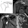
a) Schema of a B2 rotator cuff lesion (RCL) (full-thickness defects medial to an intact footprint of the rotator cuff). b) Coronal T2-weighted FATSAT MRI image after a right arthroscopic double-row rotator cuff repair showing an intact rotator cuff footprint but full-thickness defect in the rotator cuff medial to an intact rotator cuff footprint.
Fig. 6
Fig. 6
Sagittal and axial T2-weighted FATSAT MRI images demonstrating oedema of the infraspinatus. During surgery, a tendon stump confirming a B2 lesion was found and allowed a side-to-side repair.
Fig. 7
Fig. 7
a) Illustration of a right B3 rotator cuff lesion (RCL) (‘Fosbury flop tear’). b) Coronal T2-weighted FATSAT MRI image of the right shoulder. Adherences between the bursal tendon side and the wall of the subacromial bursa, fluid in the subacromial bursa, and abnormal orientation of the fibres in the tendon stump (write arrow) are noted.
Fig. 8
Fig. 8
a) A coronal view of a right shoulder CT arthrogram shows a probable B2 rotator cuff lesion (RCL) with a Patte 3 retraction. b) The arthroscopic view through lateral portal revealed after partial debridment that the tendon was not retracted but actually had adhered under the acromion (B4 RCL).
Fig. 9
Fig. 9
Axial and sagittal T2-weighted FATSAT MRI images demonstrating a type C rotator cuff lesion (RCL) with an intact tendon, a stage 3 rupture of the musculotendinous junction, and huge oedema of the muscle.
Fig. 10
Fig. 10
a) Schema of a D rotator cuff insufficiency. b) Coronal T2-weighted SPAIR MRI image of a right shoulder showing a B2D3 lesion with an intramuscular arthrosynovial cyst. c) Coronal T2-weighted PD and d) sagittal T1-weighted image (demonstrating D3 rotator cuff lesions with a calcified haematoma in the supraspinatus and an intramuscular lipoma of the subscapularis, respectively.

Similar articles

Cited by

References

    1. Court-Brown CM, Garg A, McQueen MM. The epidemiology of proximal humeral fractures. Acta Orthop Scand 2001;72:365-371. - PubMed
    1. Neer CS., II Displaced proximal humeral fractures. I. Classification and evaluation. J Bone Joint Surg [Am] 1970;52-A:1077-1089. - PubMed
    1. Bono CM, Renard R, Levine RG, Levy AS. Effect of displacement of fractures of the greater tuberosity on the mechanics of the shoulder. J Bone Joint Surg [Br] 2001;83-B:1056-1062. - PubMed
    1. Lädermann A, Denard PJ, Burkhart SS. Arthroscopic management of proximal humerus malunion with tuberoplasty and rotator cuff retensioning. Arthroscopy 2012;28:1220-1229. - PubMed
    1. Greiner S, Scheibel M. Bony avulsions of the rotator cuff: arthroscopic concepts. Der Orthopade 2011;40:21-4,26-30. [Article in German]. - PubMed

LinkOut - more resources