. 2016:1:0003.
doi: 10.1038/s41551-016-0003. Epub 2016 Dec 5.

Long-term self-renewing human epicardial cells generated from pluripotent stem cells under defined xeno-free conditions

Affiliations

Long-term self-renewing human epicardial cells generated from pluripotent stem cells under defined xeno-free conditions

Xiaoping Bao et al. Nat Biomed Eng. 2016.

Abstract

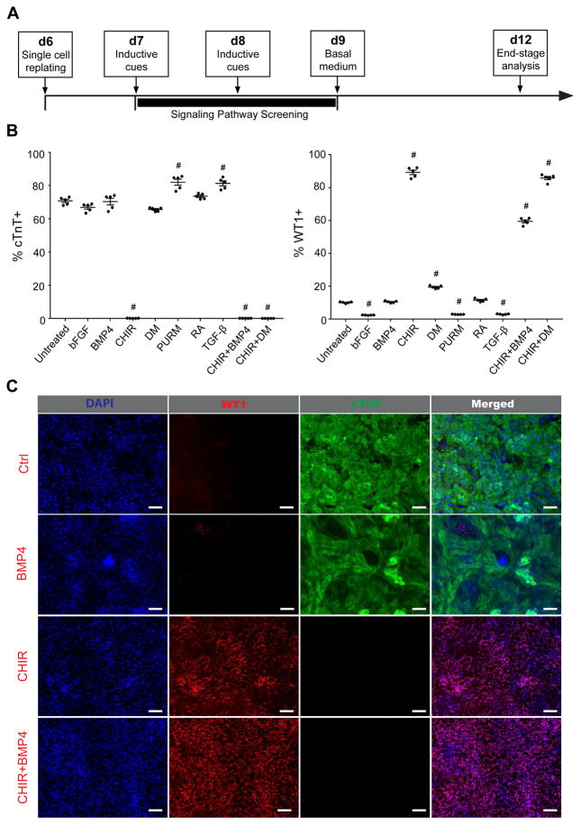
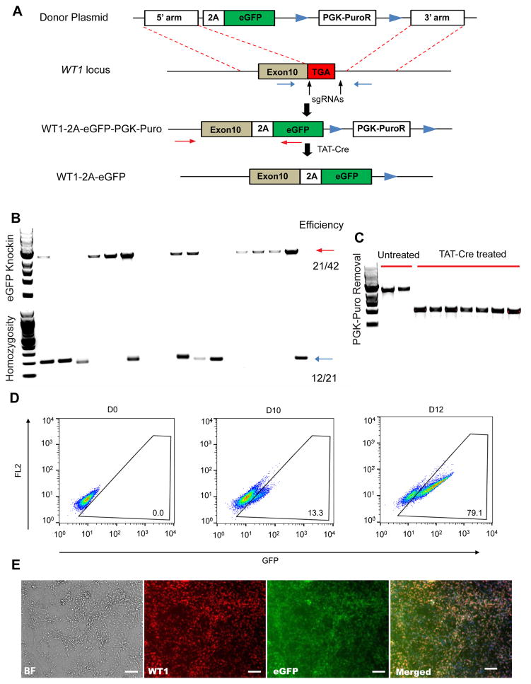
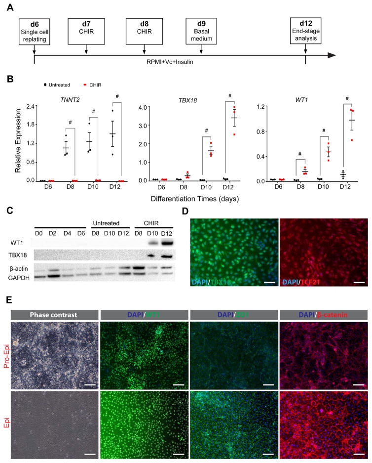
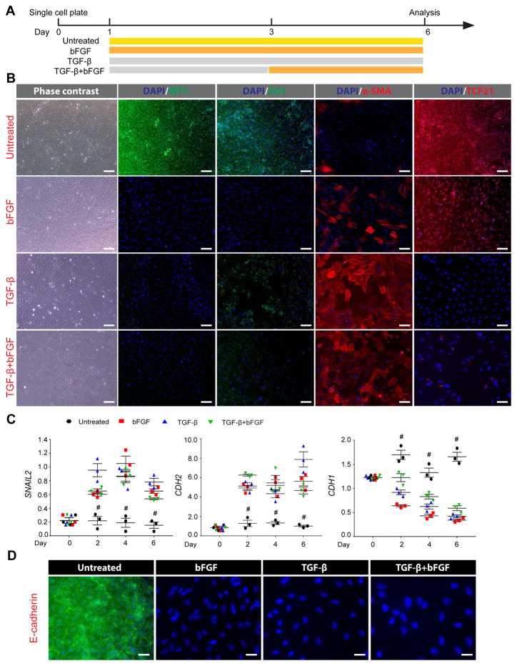
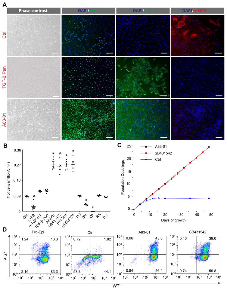
The epicardium contributes both multi-lineage descendants and paracrine factors to the heart during cardiogenesis and cardiac repair, underscoring its potential for cardiac regenerative medicine. Yet little is known about the cellular and molecular mechanisms that regulate human epicardial development and regeneration. Here, we show that the temporal modulation of canonical Wnt signaling is sufficient for epicardial induction from 6 different human pluripotent stem cell (hPSC) lines, including a WT1-2A-eGFP knock-in reporter line, under chemically-defined, xeno-free conditions. We also show that treatment with transforming growth factor beta (TGF-β)-signalling inhibitors permitted long-term expansion of the hPSC-derived epicardial cells, resulting in a more than 25 population doublings of WT1+ cells in homogenous monolayers. The hPSC-derived epicardial cells were similar to primary epicardial cells both in vitro and in vivo, as determined by morphological and functional assays, including RNA-seq. Our findings have implications for the understanding of self-renewal mechanisms of the epicardium and for epicardial regeneration using cellular or small-molecule therapies.


Conflict of interest statement

COMPETING FINANCIAL INTERESTS The authors declare no competing financial interests.

Figures

Figure 1
Figure 1
Wnt/β-catenin signaling directs the specification of WT1+ epicardial lineages from hPSC-derived cardiac progenitors. (A) Schematic of the protocol to differentiate NKX2.5+ISL1+ hPSC-derived cardiac progenitors toward the epicardial lineage. H13 hESC-derived cultures differentiated as shown in (A) in the presence of the indicated molecular signaling regulators were subjected to flow cytometry analysis (B) and immunostaining analysis (C) for WT1 and cTnT at day 12. Scale bars, 100 μm. Data are represented as mean ± SEM of five independent replicates. # p<0.05, indicated treatment versus untreated condition. CHIR: CHIR99021; DM: Dorsomorphin; PURM: Purmorphamine; RA: Retinoic acid.
Figure 2
Figure 2
Construction of WT1-2A-eGFP knockin ES03 hESC line using Cas9 nuclease. (A) Schematic diagram of the knockin strategy at the stop codon of the WT1 locus. Vertical arrows indicate sgRNA1 and sgRNA2 targeting sites. Red and blue horizontal arrows are PCR primers for assaying WT1 locus targeting and homozygosity, respectively. (B) Representative PCR genotyping of hESC clones after puromycin selection is shown, and the expected PCR product for correctly targeted WT1 locus is ~3 kbp (red arrows) with an efficiency of 21/44. A homozygosity assay was performed on the knockin clones, and those without ~200 bp PCR products were homozygous (blue arrows). (C) PCR genotyping of hESC clones after TAT-Cre mediated excision of the PGK-Puro cassette. Clones with the PCR products of ~1 kbp are PGK-Puro free, and those with ~3 kbp contain PGK-Puro. (D) Live cell flow analysis of GFP+ cells at day 0, day 10 and day 12 during CHIR treatment of WT1-2A-eGFP knockin ES03. (E) Phase contrast images and corresponding eGFP fluorescent images of WT1-2A-eGFP hPSC-derived epicardial cells after excision of the PGK-Puro cassette. Scale bars, 100 μm.
Figure 3
Figure 3
Molecular analysis of hPSC-derived epicardial cells under chemically-defined, albumin-free conditions. (A) Schematic of the optimized protocol for differentiation of hPSCs to epicardial cells in RPMI basal medium. (B–D) H13 hESC-derived cardiac progenitors were differentiated as illustrated in (A). Gene expression was assessed by quantitative RT-PCR (B). Data are represented as mean ± SEM of three independent replicates. # p<0.05, CHIR-treated condition versus untreated condition at indicated time; t test. (C) At different time points, WT1 and TBX18 expression was assessed by western blot. (D) At day 12, immunostaining for TBX18 and TCF21 was performed. Scale bars, 50 μm. (E) Representative phase contrast microscopy and fluorescence immunostaining for WT1, ZO1 and β-catenin of day 12 pro-epicardium (Pro-Epi) and day 18 epicardium (Epi). Scale bars, 100 μm.
Figure 4
Figure 4
hPSC-derived epicardial cells undergo EMT in response to bFGF and TGF-β1 treatment, yielding epicardium-derived cells that display characteristics of fibroblasts and vascular smooth muscle cells. (A) Schematic of the protocols used for the EMT induction of H13 hESC-derived epicardial cells with 10 ng/mL bFGF and 5 ng/mL TGF-β1. (B) At day 18, phase contrast images displaying cell morphology and fluorescence images showing the presence of WT1, ZO1, α-SMA and TCF21. Scale bars, 100 μm. (C) qPCR analysis of EMT related genes SNAIL2, CDH2 and CDH1 and (D) immunostaining analysis of E-cadherin expression after the indicated bFGF and TGF-β1 treatments. Data are represented as mean ± SEM of three independent replicates. # p<0.05, untreated condition versus bFGF-, TGF-β1- and bFGF+TGF-β1- treated conditions at indicated time. Scale bars, 50 μm.
Figure 5
Figure 5
Long-term expansion of hPSC-derived epicardial cells. (A–B) H13 hESC-derived day 18 epicardial cells were seeded at a density of 0.05 million cells/cm2 and treated with the indicated small molecules for 3 days (concentrations provided in Table S1). At day 4, representative phase contrast microscopy and fluorescence immunostaining for WT1, ZO1 and α-SMA (A) and the total cell numbers were assessed (B). Data are represented as mean ± SEM of five independent replicates. # p<0.05, indicated treatment versus untreated condition; one-way ANOVA with Tukey’s honest significant difference (HSD) as post-hoc analysis. α-TGF-β I: TGF-β receptor 1 antibody; TGF-β Pan: TGF-β pan specific antibody. Scale bars, 100 μm. (C–D) H13 hESC-derived epicardial cells were passaged and counted every four days in the absence or presence of the indicated TGF-β inhibitors: 0.5 μM A83-01 or 2 μM SB431542. The population doublings were calculated and shown in (C), and day 48 cultures were subjected to flow analysis of WT1 and Ki67 expression (D).
Figure 6
Figure 6
hPSC-derived epicardial cells were similar to primary epicardial cells. (A) Hierarchical clustering analysis of RNA-seq expression data of hPSCs, hPSC-derived endoderm (Endo), ectoderm (Ecto), mesoderm (Mes), CMs, day 12 epicardial cells (19-9-7-Epi), day 48 epicardial cells (H9-Epi, ES03-Epi, 19-9-11-Epi), and primary epicardial cells (donor 9605, 9633, 9634, and 9635). (B) 3D scores plot of first 3 principal components (PCs) from the PCA. The ellipses show the 95% confidence limit and each data point correspond to different biological samples. Black arrows show the development transition from hPSCs to mesoderm, from which CMs and epicardial cells arise. (C) Before transplantation to mouse heart, ES03-eGFP cells were differentiated as illustrated in Figure 3A, cultured for 5 passages in A83-01 containing medium, and subjected to flow cytometry analysis for WT1 and GFP expression. (D–E) After 12 days, hearts were harvested, and representative hematoxilin and eosin stain (H&E) staining and dual immunostaining plots of smooth muscle actin (SMA) and human-specific mitochondria (Mito) (D) and SMA and GFP (E) of cross-sections of the hearts are shown. Arrows denote the corresponding sites in the H&E images. Scale bar, 50 μm. (F) Model highlighting the specification of hPSCs to epicardial lineages by stage-specific modulation of canonical WNT signaling and the long-term maintenance of hPSC-derived epicardial cells using TGF-β signaling inhibitors.

References

    1. Brade T, Pane LS, Moretti A, Chien KR, Laugwitz KL. Embryonic heart progenitors and cardiogenesis. Cold Spring Harb Perspect Med. 2013;3:a013847. - PMC - PubMed
    1. Männer J, Ruiz-Lozano P. Development and Function of the Epicardium. Adv Dev Biol. 2007;18:333–357.
    1. Pérez-Pomares JM, et al. Origin of coronary endothelial cells from epicardial mesothelium in avian embryos. Int J Dev Biol. 2002;46:1005–13. - PubMed
    1. Smart N, et al. Thymosin beta4 induces adult epicardial progenitor mobilization and neovascularization. Nature. 2007;445:177–82. - PubMed
    1. Zhou B, et al. Adult mouse epicardium modulates myocardial injury by secreting paracrine factors. J Clin Invest. 2011;121:1894–1904. - PMC - PubMed