Review
. 2017 Feb 20;3(3):331-338.
doi: 10.1016/j.jcmgh.2017.01.013. eCollection 2017 May.

The Origins of Gastric Cancer From Gastric Stem Cells: Lessons From Mouse Models

Affiliations
Review

The Origins of Gastric Cancer From Gastric Stem Cells: Lessons From Mouse Models

Yoku Hayakawa et al. Cell Mol Gastroenterol Hepatol. .

Abstract

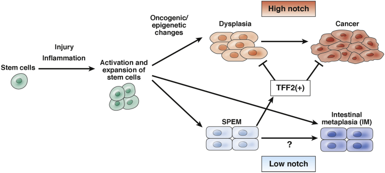
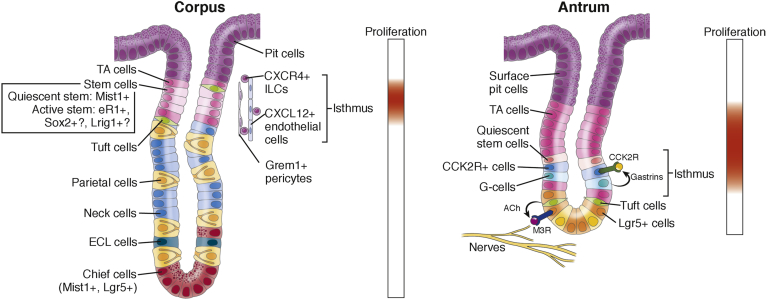
The cellular origin of digestive cancers has been a long-standing question in the cancer field. Mouse models have identified long-lived stem cells in most organ systems, including the luminal gastrointestinal tract, and numerous studies have pointed to tissue resident stem cells as the main cellular origin of cancer. During gastric carcinogenesis, chronic inflammation induces genetic and epigenetic alterations in long-lived stem cells, along with expansion of stem cell niches, eventually leading to invasive cancer. The gastric corpus and antrum have distinct stem cells and stem cell niches, suggesting differential regulation of cancer initiation at the 2 sites. In this short review, we discuss recent experimental models and human studies, which provide important insights into the pathogenesis of gastric cancer.

Keywords: CCK2R; CCK2R, cholecystokinin receptor 2; Gastric Cancer; IM, intestinal metaplasia; Lgr5; Mist1; SPEM, spasmolytic polypeptide-expressing metaplasia; Stem Cell; Stem Cell Niche.


Figures

Figure 1
Figure 1
Model of stem cell–derived gastric carcinogenesis. During gastric carcinogenesis, long-lived stem cells and their niche are activated and expanded in response to tissue injury and inflammation. Activated stem cells give rise to metaplasia and dysplasia after accumulation of genetic and epigenetic changes. During Barrett’s esophagus development, dysplasia can progress to cancer with high Notch expression, while metaplasia appears to be postmitotic and a distinct lineage with low Notch expression. Given that Barrett’s esophagus may originate from gastric cardia glands, this may be the case in gastric metaplasia/dysplasia development. Indeed, notch signaling has been shown to increase proliferation and decrease differentiation, and thus clearly regulate mucous cell phenotypes in the stomach., , , Aberrant notch activation leads to hyperplasia and dysplasia both in the corpus and antrum., , SPEM cells express TFF2, which inhibits cancer progression.,
Figure 2
Figure 2
Gastric corpus and antrum stem cells and their niche. Stem cells are thought to reside within proliferative isthmus (upper part of the corpus glands and lower part of the antral glands). In the corpus glands, the isthmus is located between abundant transit-amplifying (TA) cells. There is a slow-cycling stem cell that expresses Mist1 and a rapidly cycling stem cell that may express eR1, Sox2, or Lrig1, and they can differentiate into various cell types such as pit, parietal, neck, enterochromaffin-like (ECL), and tuft cells. C-X-C motif chemokine ligand 12 (CXCL12)+ endothelial cells, C-X-C motif chemokine receptor type 4 (CXCR4)+ innate lymphoid cells (ILCs), and Grem1+ pericytes appear to compose the corpus stem cell niche. Chief cells at the base express Mist1 and Lgr5, but they normally are postmitotic and do not divide. In the antrum, several rapidly cycling stem cells are reported just below the major TA cell zone, including Lgr5+ cells at the base and CCK2R+ cells at +4. Lgr5+ cells can be activated by acetylcholine (ACh)-producing nerves and tuft cells through muscarinic acetylcholine receptor subtype 3 (M3R). Gastrins secreted from antral G cells can regulate CCK2R+ stem cell function in a paracrine manner. Slow-cycling stem cells in the antrum have not been identified yet.

References

    1. Correa P. Human gastric carcinogenesis: a multistep and multifactorial process–First American Cancer Society Award Lecture on Cancer Epidemiology and Prevention. Cancer Res. 1992;52:6735–6740. - PubMed
    1. Rossi D.J., Jamieson C.H., Weissman I.L. Stems cells and the pathways to aging and cancer. Cell. 2008;132:681–696. - PubMed
    1. Zeuner A., Todaro M., Stassi G. Colorectal cancer stem cells: from the crypt to the clinic. Cell Stem Cell. 2014;15:692–705. - PubMed
    1. Greaves M., Maley C.C. Clonal evolution in cancer. Nature. 2012;481:306–313. - PMC - PubMed
    1. Graham T.A., McDonald S.A., Wright N.A. Field cancerization in the GI tract. Future Oncol. 2011;7:981–993. - PubMed

LinkOut - more resources