

Oxidative Stress Contributes to Status Epilepticus Associated Mortality
- PMID: 28462450
- PMCID: PMC5640323
- DOI: 10.1007/s11064-017-2273-1
Oxidative Stress Contributes to Status Epilepticus Associated Mortality
Abstract
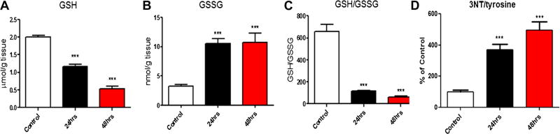
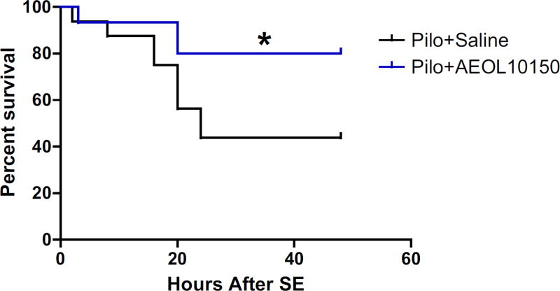
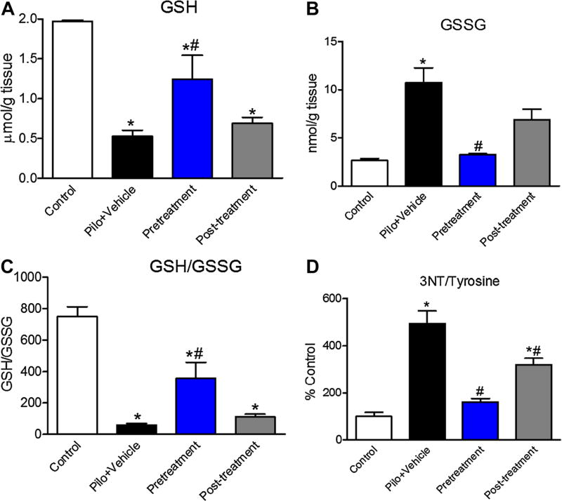
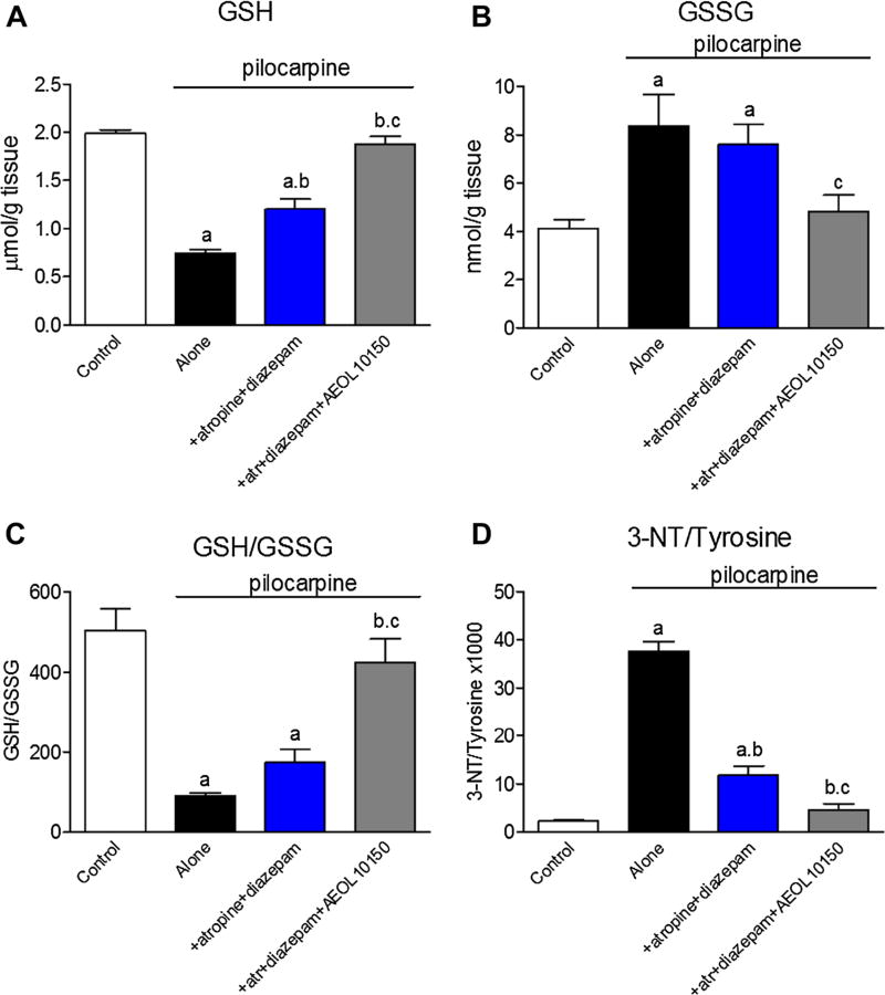
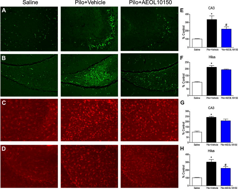
Status epilepticus is a common manifestation of nerve agent toxicity and represents a serious medical emergency with high rates of mortality and neurologic injury in those that survive. The aim of the current study was to determine if targeting oxidative stress with the catalytic antioxidant, AEOL10150, would reduce pilocarpine-induced mortality and attenuate neuronal death and neuroinflammation. We found that treatment with AEOL10150 in conjunction with scopolamine and diazepam following pilocarpine-induced SE was able to significantly reduce mortality compared to treatment with just scopolamine and diazepam. Mortality was further reduced when AEOL10150 was used in conjunction with atropine and diazepam which is considered the standard of care for nerve agent exposures. Both treatment paradigms offered significant protection against SE-induced oxidative stress. Additionally, treatment with scopolamine, AEOL10150 and diazepam attenuated SE-induced neuronal loss and neuroinflammation. Taken together, the data suggest that pharmacological targeting of oxidative stress can improve survival and attenuate secondary neurological damage following SE induced by the nerve agent surrogate pilocarpine.
Keywords: Epilepsy; Glutathione; Nerve agent toxicity; Pilocarpine.
Figures
References
-
- Trinka E, Höfler J, Zerbs A. Causes of status epilepticus. Epilepsia. 2012;53:127–138. - PubMed
-
- de Araujo Furtado M, Rossetti F, Chanda S, Yourick D. Exposure to nerve agents: from status epilepticus to neuroinflammation, brain damage, neurogenesis and epilepsy. Neurotoxicology. 2012;33(6):1476–1490. - PubMed
-
- Hauser WA. Status epilepticus: epidemiologic considerations. Neurology. 1990;40(5 Suppl 2):9–13. - PubMed
-
- DeLorenzo RJ, Pellock JM, Towne AR, Boggs JG. Epidemiology of status epilepticus. J Clin Neurophysiol. 1995;12(4):316–325. - PubMed
-
- Logroscino G, Hesdorffer DC, Cascino G, Annegers JF, Hauser WA. Short-term mortality after a first episode of status epilepticus. Epilepsia. 1997;38(12):1344–1349. - PubMed
MeSH terms
Substances
Grants and funding
LinkOut - more resources
Full Text Sources
Other Literature Sources
Medical
