Review
. 2017 Jul;28(7):506-518.
doi: 10.1016/j.tem.2017.03.005. Epub 2017 Apr 24.

Sphingolipids and Lipoproteins in Health and Metabolic Disorders

Affiliations
Review

Sphingolipids and Lipoproteins in Health and Metabolic Disorders

Jahangir Iqbal et al. Trends Endocrinol Metab. 2017 Jul.

Abstract

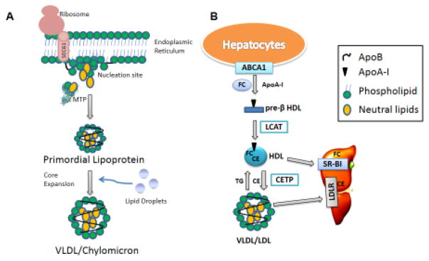
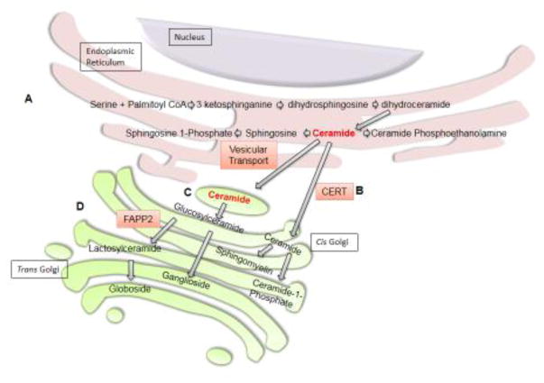
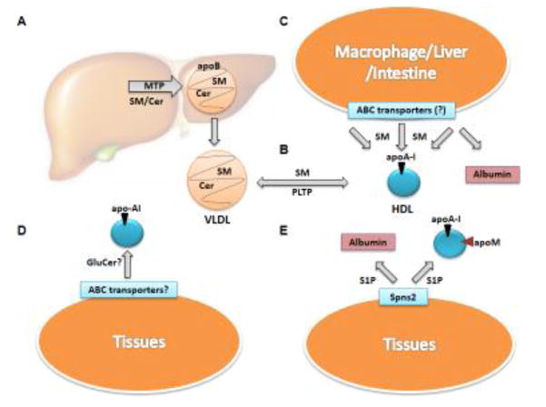
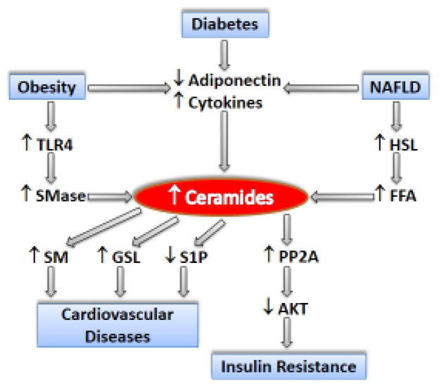
Sphingolipids are structurally and functionally diverse molecules with significant physiologic functions and are found associated with cellular membranes and plasma lipoproteins. The cellular and plasma concentrations of sphingolipids are altered in several metabolic disorders and may serve as prognostic and diagnostic markers. Here we discuss various sphingolipid transport mechanisms and highlight how changes in cellular and plasma sphingolipid levels contribute to cardiovascular disease, obesity, diabetes, insulin resistance, and nonalcoholic fatty liver disease (NAFLD). Understanding of the mechanisms involved in intracellular transport, secretion, and extracellular transport may provide novel information that might be amenable to therapeutic targeting for the treatment of various metabolic disorders.

Keywords: MTP; ceramide; lipoproteins; metabolic disorders; sphingolipids; sphingomyelin.


Conflict of interest statement

Conflicts of interest

The authors declare that they have no conflicts of interest with the contents of this article.

Figures

Figure 1
Figure 1. Lipoprotein Assembly
(A) VLDL and chylomicron follow similar biosynthesis pathways in the liver and intestine, respectively. While apoB is being translated, MTP physically binds apoB to possibly help it translocate into the endoplasmic reticulum lumen and to provide structural stability for further lipidation. As hydrophobic sequences are translated, apoB associates with the lumenal side of the ER membrane to form nucleation sites. MTP may transfer cholesteryl esters, triglycerides, free cholesterol, ceramide, and sphingomyelin to these nucleation sites and the translating apoB to form primordial lipoproteins. In the second step of lipoprotein assembly, the primordial lipoproteins fuse with lumenal lipid droplets to form VLDL or chylomicron that are secreted into the blood and lymph, respectively. (B) HDL Assembly. Apolipoprotein A-I (apoA-I) interacts with plasma membrane-embedded ABCA1 and accepts free cholesterol (FC) to form pre-β HDL extracellularly. Lecithin – cholesterol acyltransferase (LCAT) converts FC in pre-β HDL to cholesteryl esters (CE), which move to the core of HDL. HDL’s CE may be selectively delivered to the liver or steroidogenic tissues by scavenger receptor BI (SR-BI). Cholesteryl ester transfer protein (CETP) exchanges CE for triglycerides from HDL to apoB-containing lipoproteins. CE transferred to apoB-containing lipoproteins are delivered to the liver via receptor mediated endocytosis involving receptors such as LDL receptors.
Figure 2
Figure 2. Sphingolipid Biosynthesis
(A) De novo ceramide synthesis begins in the endoplasmic reticulum with the condensation of serine and palmitoyl CoA by serine palmitoyltransferase to form 3-ketosphinganine. Next, 3-ketosphinganine reductase catalyzes the reduction of 3-ketosphinganine to dihydrosphingosine which is N-acylated by ceramide synthases to form dihydroceramide. Finally, ceramide is formed by the dehydrogenation of dihydroceramide by dihydroceramide desaturase 1. Ceramides are precursors for the biosynthesis of more complex sphingolipids. In the ER, ceramides can be converted to ceramide phosphoethanolamine or deacylated by ceramidase to form sphingosine. Sphingosine can be phosphorylated to form sphingosine 1-phosphate. Ceramides can also be transferred from the endoplasmic reticulum to the Golgi by either vesicular transport or ceramide transfer protein (CERT). (B) Ceramide transfer by CERT predestines ceramide for sphingomyelin or ceramide 1-phosphate synthesis. In the Golgi, sphingomyelin synthase can convert ceramide to sphingomyelin. Alternatively, ceramide kinase can phosphorylate ceramide to form ceramide 1-phosphate. (C) Ceramide may also be transferred by vesicular transport to the cytoplasmic side of the Golgi, where it can be glycosylated to form glucosylceramide. Glucosylceramide can be transported to the lumenal side of the cis Golgi and converted to gangliosides. (D) FAPP2 can also transfer glucosylceramide from the cis Golgi to the trans Golgi, where it is converted to lactosylceramide and then to globosides.
Key Figure 3
Key Figure 3. Sphingolipid Transport to Plasma
(A) Ceramide (Cer) and sphingomyelin (SM) are incorporated into VLDL during lipoprotein assembly by microsomal triglyceride transfer protein (MTP). (B) Phospholipid transfer protein (PLTP) may transfer SM between VLDL and HDL. It is unknown whether PLTP can transfer Cer, glucosylceramide (GluCer), or other complex sphingolipids. (C) SM may also be incorporated into HDL via efflux from different cells. ATP-binding cassette transporters on the plasma membrane of macrophages, liver, intestine, or other peripheral tissues may efflux SM to free apolipoprotein (apoA-I), HDL, albumin or other acceptors. (D) The mechanism for GluCer incorporation into lipoproteins is not defined. It is likely that GluCer is incorporated into HDL during synthesis or effluxed from the plasma membrane. (E) In the plasma, sphingosine 1-phosphate (S1P) is present on HDL and albumin. It is effluxed from the plasma membrane of peripheral tissues, mainly erythrocytes and platelets, by Spinster2 (Spns2) to apoM-containing HDL and albumin.
Figure 4
Figure 4. Schematic Diagram Depicting Molecular Events in Sphingolipid Associated Pathologies
In diabetes, obesity and non-alcoholic fatty liver disease (NAFLD) decreased synthesis of anti-inflammatory adiponectin and increased production of pro-inflammatory cytokines lead to increased production of ceramides. Obesity also causes activation of TLR4 signaling pathway which leads to activation of sphingomyelinase (SMase). SMase hydrolyzes sphingomyelin (SM) to ceramide. In NAFLD, insulin is not able to inhibit hormone-sensitive lipase (HSL) activity in adipose tissue leading to increased production of free fatty acids (FFAs), specially palmitic acid, that are shunted for ceramides synthesis. Ceramides, in turn, enhance the activity of protein phosphatase 2A (PP2A) which causes dephosphorylation and inactivation of AKT leading to insulin resistance. Increased ceramides may be a substrate for increased SM and glycosphingolipids (GSL) synthesis that have been shown to increase the risk of cardiovascular diseases. Synthesis of sphinogsine 1-phosphate (S1P), which is cardioprotective, is decreased in cardiovascular diseases.

Similar articles

Cited by

References

    1. Borodzicz S, et al. Sphingolipids in cardiovascular diseases and metabolic disorders. Lipids Health Dis. 2015;14:55. - PMC - PubMed
    1. Feingold KR, Grunfeld C. Introduction to Lipids and Lipoproteins. In: De Groot LJ, Chrousos G, Dungan K, et al., editors. Endotext [Internet] South Dartmouth (MA): MDText.com, Inc; 2000. Updated 2015 Jun 10. Available from: https://www.ncbi.nlm.nih.gov/books/NBK305896/
    1. Walsh MT, Hussain MM. Targeting microsomal triglyceride transfer protein and lipoprotein assembly to treat homozygous familial hypercholesterolemia. Crit Rev Clin Lab Sci. 2016:1–23. - PubMed
    1. Hussain MM, et al. Multiple functions of microsomal triglyceride transfer protein. Nutr Metab (Lond) 2012;9:14. - PMC - PubMed
    1. Tiwari S, Siddiqi SA. Intracellular trafficking and secretion of VLDL. Arterioscler Thromb Vasc Biol. 2012;32:1079–1086. - PMC - PubMed

Publication types