. 2017 May 2:8:15157.
doi: 10.1038/ncomms15157.

Variations in brain defects result from cellular mosaicism in the activation of heat shock signalling

Affiliations

Variations in brain defects result from cellular mosaicism in the activation of heat shock signalling

Seiji Ishii et al. Nat Commun. .

Abstract

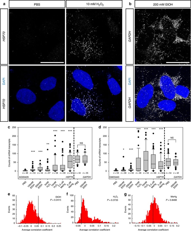
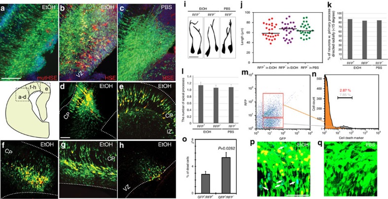
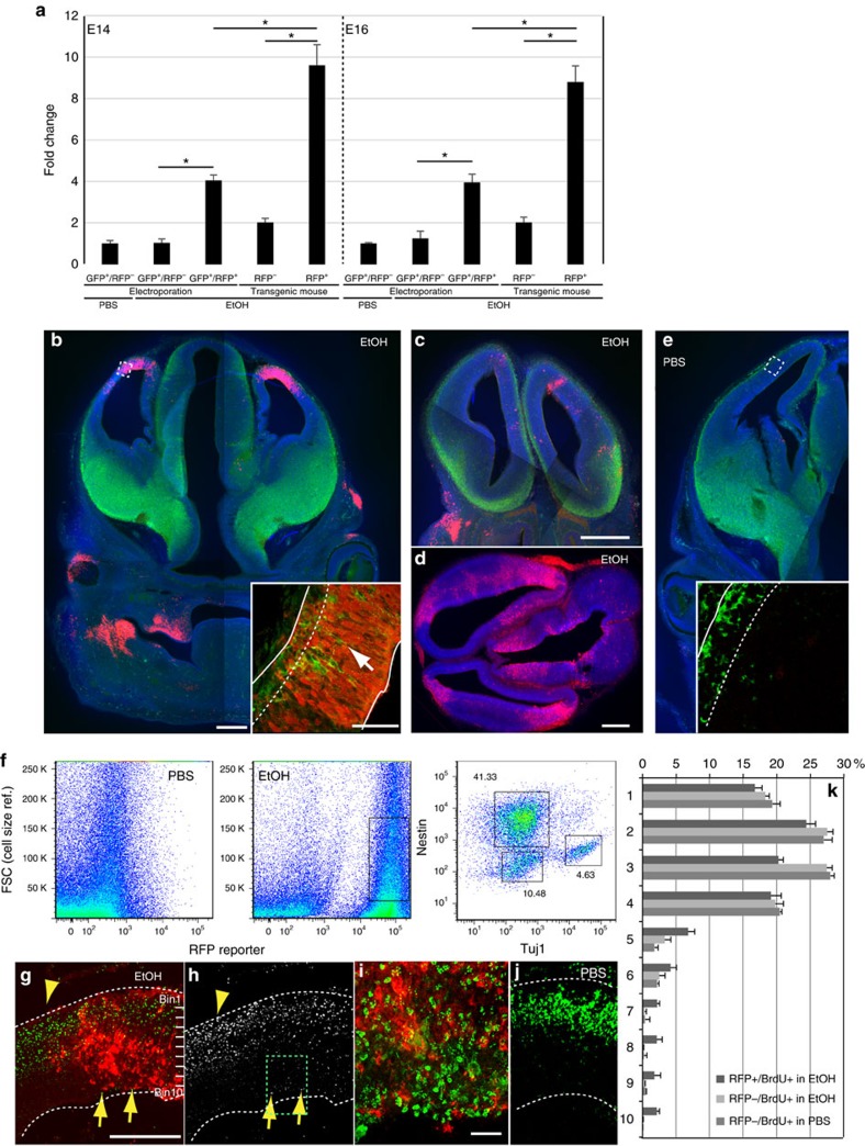
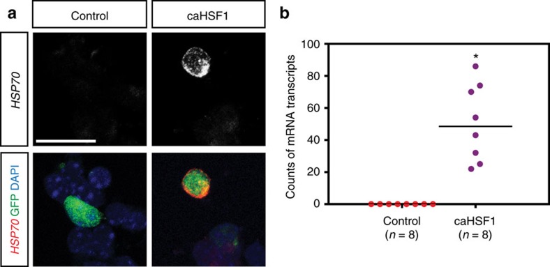
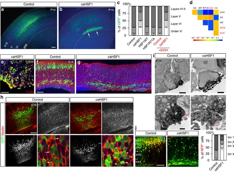
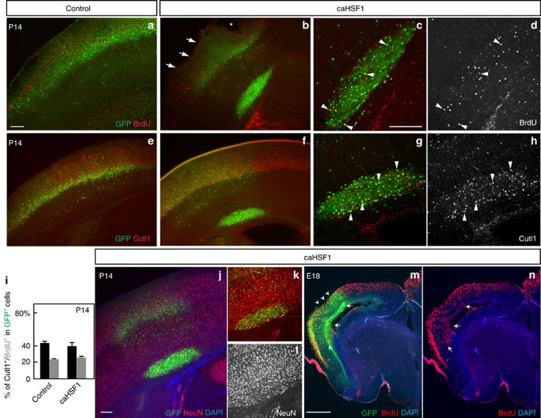
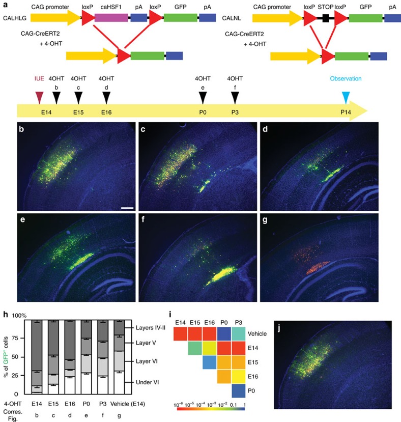
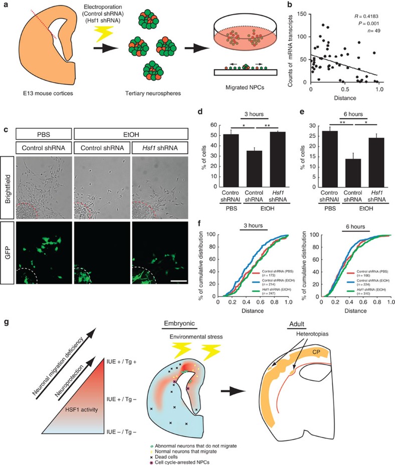
Repetitive prenatal exposure to identical or similar doses of harmful agents results in highly variable and unpredictable negative effects on fetal brain development ranging in severity from high to little or none. However, the molecular and cellular basis of this variability is not well understood. This study reports that exposure of mouse and human embryonic brain tissues to equal doses of harmful chemicals, such as ethanol, activates the primary stress response transcription factor heat shock factor 1 (Hsf1) in a highly variable and stochastic manner. While Hsf1 is essential for protecting the embryonic brain from environmental stress, excessive activation impairs critical developmental events such as neuronal migration. Our results suggest that mosaic activation of Hsf1 within the embryonic brain in response to prenatal environmental stress exposure may contribute to the resulting generation of phenotypic variations observed in complex congenital brain disorders.


Conflict of interest statement

The authors declare no competing financial interests.

Figures

Figure 1
Figure 1. Variable levels of activation of Hsf1–Hsp signalling by environmental stress.
(a,b) Representative smFISH images of HSP70 mRNA (a) and GAPDH mRNA (b) in an hNPC line (AG09429). (c,d) Quantification of HSP70 and GAPDH mRNA puncta in hNPCs (AG09429 and GM03440) exposed to the indicated reagents. In the box plot, the line within the box indicates the median; the upper and lower edges of the box represent the 25th and 75th percentiles, respectively. Error bars indicate the 10th and 90th percentiles and each dot indicates cells within the top/bottom 10th percentiles. The variability of HSP70 mRNA particles was significantly increased in response to all these reagents compared to that with PBS exposure (*P<0.05, **P<0.01, ***P<0.001 by Levene's test. More than 35 cells in different positions within the dishes were measured in each group). (eg) The correlation coefficients of cellular positions in the dishes and Hsp70 mRNA levels are compared between the experimental data (broken lines) and simulated data sets representing probabilistic events (red histograms). The presented P values of one-sample Z-test indicate that the levels of the Hsf1–Hsp 70 signalling activation in each cell are randomly determined regardless of the positions in the culture dish. Scale bars, 0.02 mm.
Figure 2
Figure 2. Normal development of HSE–RFP reporter+ neurons and increased cell death of reporter cells in IUE-based reporter analysis.
(ac) pHSE- (b,c) or pmutHSE- (a) RFP was electroporated with pCAG–GFP into the mouse (C57NL/6J) lateral cortex at E14, followed by daily EtOH (a,b) or PBS (control, c) administration to the dam until E16. The reporter expression (red) was observed in NPCs and neurons by electroporation of p HSE–RFP followed by EtOH administration (b). Electroporation of pmut HSE–RFP plasmid that contains point mutations in HSF1 binding sites in the promoter (Supplementary Fig. 3) resulted in reporter expression in less than 0.1% of total electroporated (GFP+) cells in the cortex exposed to EtOH (n=22). Similarly, electroporation of p HSE–RFP in Hsf1 KO mice resulted in no reporter induction by EtOH exposure (n=3/3). (dh) The HSE–RFP reporter expression was observed in NPCs and neurons throughout the cortex, including lateral (d), medial (e) and dorsal (fh) regions. CP: cortical plate, IZ; intermediate zone, VZ: ventricular zone. Scale bar, 250 μm. (i) Normal morphology of HSE–RFP + neurons in IUE approach. Representative morphologies of RFP+/GFP+ and RFP/GFP+ neurons in the CP exposed to EtOH or PBS (control) as indicated. (j) The length of the primary process was plotted for neurons in the indicated groups, showing no significant differences between the groups. (k) The percentage of neurons with a radially-directed (<15 degrees) primary process. (l) The number of apical processes was quantified for neurons in the indicated groups. No significant differences were found between the groups. (mo) Flow cytometric analysis of the electroporated cells (m), revealed that HSE–RFP + cells (orange in n) contain less dead cells than HSE–RFP cells (gray in n) in the cortex exposed to EtOH. The mice were electroporated with p HSE–RFP and pCAG–GFP at E14 followed by daily EtOH administration until E16. Cells were then dissociated and stained with a cell death marker (7-AAD). (o) Percentages of the cells positive for cell death marker in HSE–RFP -positive and –negative cells. P<0.01 by t-test (n=7 embryos). (p,q) Consistent with the data quantified with 7-AAD (mo), activated Caspase-3 (blue) is observed frequently in HSE–RFP/GFP+ electroporated cells (p, shown in white arrows), but not in HSE–RFP+/GFP+ in EtOH (p) or HSE–RFP/GFP+ in PBS-exposed (q) cortices. Scale bar, 50 μm.
Figure 3
Figure 3. Heterogeneity in the reporter expression of Hsf1–Hsp signalling and the migration deficiency in reporter+ cells in the cortex.
(a) Different sensitivity for detection of Hsf1–Hsp signalling activation between IUE- and transgenic-based reporter assays. qRT–PCR of Hsp70 was performed with the total RNA extracted from reporter-positive and -negative cells sorted by a flow cytometer 8 h after administration at E14 (left) or E16 (right). Data are represented as mean±s.e.m. *P<0.0001 by t-test from three independent experiments using multiple embryos from several dams. (b,c,d) Heterogeneous reporter expressions of Hsf1–Hsp signalling driven by EtOH exposure at E15 (b) or 16 (c,d) in three HSE–RFP reporter transgenic mice 1 day after last exposure. Each sample shows a distinct reporter expression pattern. Reporter-expressing regions contain Tuj1+ immature neurons (green) incorrectly positioned in the ventricular zone (arrows in the inset in b). The square in b indicates the region from which the inset was taken. The dotted line in the inset in b shows the boundary of the cortical plate and ventricular zone. Approximately 9% of the total analysed embryos showed robust reporter expression (n=10/115 embryos) (green: Tuj1, red: HSE–RFP, blue: DAPI). (e) No reporter expression after PBS (control) exposure. (0/137 embryos). (f) Flow cytometric analysis of reporter expressing cells dissociated from E16 cortices 1 day after the last exposure. The percentages of Nestin+/Tuj1, Nestin/Tuj1+ and Nestin/Tuj1 cells were determined after gating of the RFP+ cells as indicated with a square in the EtOH-exposed sample. The objects showing larger FSC profiles are cellular debris. (gj) Birthdate labelling with BrdU (green in (g,i,j), white in (h)) at E15 shows delayed migration of BrdU+ cells at E18 in the region containing HSE–RFP + cells (arrows) compared with the surrounding region (arrowheads), after daily administration of EtOH at E14-16. i is a higher magnification view of square in h. (k) Quantification was performed by identifying the domains that contain RFP+ cells throughout radial axis in somatosensory cortical regions. The RFP cells were counted in the adjacent regions in EtOH exposed samples and the corresponding regions in PBS-exposed samples (RFP/BrdU+ in PBS). *P<0.001, repeated measures ANOVA, n=3 each. Scale bars, 0.5 mm (bh,j), and 50 μm (in the inset in b,e,i).
Figure 4
Figure 4. IUE-mediated expression of caHSF1 recapitulates the physiologically relevant levels of HSF1 activation elicited by environmental challenges.
(a) Representative images of Hsp70 mRNA expression in pCAGIG (control) or pCAGIG-caHSF1 electroporated cells. The plasmids were introduced into mouse frontal cortices by IUE at E14. The electroporated cells were dissociated 24 h post-electroporation, and plated on culture dishes. 3 h later, Hsp70 mRNAs were detected by smFISH. (b) The graph shows the number of Hsp70 mRNA particles in the control- and caHSF1-electroporated cells. caHSF1 overexpression results in significantly higher numbers of Hsp70 mRNAs compared with the control (*P<0.001 by Mann-Whitney U-Test). Importantly, these numbers fall in the range of the top 5% and 20% of those induced by EtOH and MeHg, respectively (Fig. 1 and Supplementary Fig. 1). Scale bar, 0.02 mm.
Figure 5
Figure 5. Excess activation of Hsf1 causes heterotopia formation.
(a,b) The P14 cortex electroporated with the control (a) or caHSF1 expression (b) plasmid at E14. caHSF1 expression causes the formation of periventricular heterotopia. Blue: nuclei counter staining. (c) Distribution of GFP+ electroporated cells transfected with indicated constructs. Data are represented as mean±s.e.m. F(3,48)=1197, P<0.0001 by repeated measures ANOVA among groups without EtOH exposure. P<0.001 by post hoc Tukey test between caHSF1 and other HSF1 forms or control. F(3,24)=80.59, P<0.0001 between caHSF1 and control comparison under EtOH exposure. (d) Pairwise comparisons of cumulative radial distributions using Kolmogorov–Smirnov (K–S) test. Each P-value is shown in a color according to the scale at the bottom. (e) Confirmation of caHSF1 (with a FLAG-tag) expression in the GFP+ electroporated cells by FLAG immunohistochemistry. Blue: nuclei counter staining. (fh) Tuj1 (f,g) and Nestin (h) immunohistochemistry at indicated ages following IUE at E14. Arrows and arrowheads in h indicate oval-shaped, radially oriented NPCs in the control, and abnormal round-shaped, non-radially oriented caHSF1-expressing NPCs, respectively in the VZ. (i) Immuno-electron microscopy of control- and caHSF1-electroporated NPCs at the ventricular surface. Electroporated cells were visualized by GFP immunohistochemistry using DAB (black). Complete loss of adherens junctions is accompanied by the detachment of caHSF1-expressing NPCs from surrounding non-electroporated cells. The remaining disorganized adherens junctions are revealed by decreased electron density (arrowheads) compared with normal adherens junctions in the control (arrows). (j) Using the CALSL/Tα1-Cre system, exogenous genes (RFP only (control) or RFP plus caHSF1) were expressed exclusively in immature neurons. The GFP plasmid (pCAGIG) also was introduced to label all electroporated cells (n=6 embryos). caHSF1 expression interferes radial migration of neurons cell-autonomously. Radial distribution of RFP+ neurons (graph) shows significant difference between control and caHSF1 electroporation (F(3,24)=244.3, P<0.0001 by repeated measures ANOVA. P<0.0001 by K-S test). Data are represented as mean±s.e.m. Scale bars, 0.3 (a,b), 0.04 (e) and 0.2 (fh,j) mm, and 5 (i, upper panels) and 0.2 (i, lower panels) μm.
Figure 6
Figure 6. caHSF1 disturbs neuronal positioning without affecting differentiation.
(ah) Mice were electroporated with pCAGIG (control) or pCAGIG-caHSF1 at E14 and stained for BrdU (injected at E15) or Cutl1 (a marker for upper layer neurons) (red or white) at P14. caHSF1-electroporated (GFP+) cells in the heterotopia include Cutl1+ and BrdU+ cells. Most non-electroporated BrdU+ neurons were localized in upper layers (layers II-IV). Arrows and an asterisk indicate disruptions in the marginal zone and pial surface above the heterotopia (b). Arrowheads indicate representative double-labelled cells (c,d,g,h). (i) The percentages of Cutl1+ (black) and BrdU+ (gray) cells within caHSF1-electroporated cells are comparable to those within control-electroporated cells, indicating that caHSF1 does not affect differentiation. Data are represented as mean±s.e.m. P=n.s. by t-test (n=5 each). (jl) Immunostaining for NeuN (a marker for mature neurons, red or white) on the P14 cortex shows normal neuronal differentiation of caHSF1-electroporated (at E14) cells (labelled with GFP) even within the heterotopia. (m,n) BrdU+ caHSF1-electroporated (at E14) cells (GFP+) were found in deep layers at E18 (arrows). BrdU was incorporated at E15. Disruption in the cortical surface (arrowheads) was already found at E18 in some caHSF1-electroporated samples. Scale bars=0.2 (al) and 0.5 (m,n) mm.
Figure 7
Figure 7. Heterotopias formed by temporal prenatal expression of caHSF1 persist after birth.
(a) Scheme describing the experiments. CAG-promoter-loxP-FLAGcaHSF1-loxp-GFP (CALHLG) with CAG-RFP and CAG-CreERT2 was electroporated at E14. 4-OHT was administered at the indicated time points, and all samples were fixed at P14. 4-OHT administration at any time point consistently resulted in excision of the FLAGcaHSF1 unit (indicated by GFP expression) in over 95% of RFP+ electroporated cells. FLAG immunohistochemistry confirmed complete sequestration of FLAG-tagged caHSF1 by 2 days after 4-OHT injection. (bi) Distribution of the electroporated cells was observed at P14 after administration of 4-OHT or vehicle only (control) at different time points (bg), and quantitatively analysed (h,i). Scale bar, 0.5 mm. Panel i shows pairwise comparisons of cumulative radial distributions using Kolmogorov–Smirnov (K–S) test. Each P value is shown in a color according to the scale at the bottom. Without 4-OHT (vehicle only), CALHLG expression causes heterotopia formation (g,h), similar to pCAG-caHSF1 introduction. Delay of caHSF1 until after birth (4-OHT injection at P0 or P3) causes the formation of large heterotopias (e,f,h) (i: P=NS, K–S test). Expression of caHSF1 at low levels for a very short period (4-OHT injection 6 h after IUE at E14), did not form heterotopias (b,h) (P=NS compared with pCALNL-GFP, n=6). Expression of caHSF1 until E15 or E16 was sufficient for heterotopia formation (c,d,h) (P<0.05 compared with the vehicle control or pCALNL-GFP (see j), n=13, 5, respectively). Data are represented as mean±s.e.m. (j) No migration defects by electroporation of pCALNL-GFP with CAG-CreERT2 and pCAG-RFP followed by 4-OHT injection at E16 (see a).
Figure 8
Figure 8. Reducing excessive Hsf1 activation mitigates EtOH-induced migration deficits.
(a) Schematic drawing of the migration assay. (b) The levels of Hsp70 expression (smFISH) are negatively correlated with the migration distance of cells from the center of the neurosphere after 3-hour culture. The position of the cell that had migrated the longest distance from the center (0.0) was set as 1.0, and was used for determining the relative positions of the other cells. (c) Representative images of the migration assays after a 6-h culture. Scale bar, 0.1 mm. (d,e) The percentages of GFP+ cells within the outer half of the maximum migration distance from the center of the neurosphere in total GFP+ cells. Four independent experiments were performed per group. Data are represented as mean±s.e.m. One-way ANOVA, F(2,9)=9.15, P=0.007 (d), F(2,9)=9.59 P=0.006 (e). *P<0.05, **P<0.01 by post hoc Tukey test. (f) Cumulative distribution of the cells shows the alleviation of EtOH-induced migration deficits by Hsf1 shRNA (n>150 cells from 4 biological replicates per group, P<0.05 by K-S test for all pairs except the pair of Control shRNA (PBS) (red) versus Hsf1 shRNA (EtOH)(green). (g) A model for the production of various cortical dysplasia due to probabilistic Hsf1–Hsp signalling activation elicited by prenatal environmental stressors. Excessive activation of HSF1 in a subpopulation of cortical cells, as detected in the reporter transgenic mouse (left), disrupts their normal developmental processes such as migration (green cells in middle panel). Moderate activation of the signalling that is detectable by IUE-based method, but not in the transgenic mouse (Fig. 3), serves to protect the cells from the environmental stressors (Fig. 2, left), and allows the cells develop normally (yellow cells in middle). Meanwhile, another subpopulation of cells may not have enough HSF1 activation to protect them from damage elicited by environmental challenges, thereby resulting in focal accumulation of dead cells or NPCs with impaired proliferation (Fig. 2, the cells indicated by X in middle panel). These heterogeneous events of abnormal development occur probabilistically (Fig. 1), accounting for individually distinct pattern of focal cortical malformations in the cortex exposed to similar levels of environmental challenges (right panel).

Similar articles

Cited by

References

    1. Rakic P. Specification of cerebral cortical areas. Science 241, 170–176 (1988). - PubMed
    1. Gao P., Sultan K. T., Zhang X. J. & Shi S. H. Lineage-dependent circuit assembly in the neocortex. Development 140, 2645–2655 (2013). - PMC - PubMed
    1. Thompson B. L., Levitt P. & Stanwood G. D. Prenatal exposure to drugs: effects on brain development and implications for policy and education. Nat. Rev. Neurosci. 10, 303–312 (2009). - PMC - PubMed
    1. Ben-Ari Y. Neuro-archaeology: pre-symptomatic architecture and signature of neurological disorders. Trends Neurosci. 31, 626–636 (2008). - PubMed
    1. Ishii S. & Hashimoto-Torii K. Impact of prenatal environmental stress on cortical development. Front. Cell Neurosci. 9, 207 (2015). - PMC - PubMed

Publication types

MeSH terms

LinkOut - more resources