. 2017 Jun 21;28(6):1722-1733.
doi: 10.1021/acs.bioconjchem.7b00182. Epub 2017 May 10.

Developing Targeted Hybrid Imaging Probes by Chelator Scaffolding

Affiliations

Developing Targeted Hybrid Imaging Probes by Chelator Scaffolding

Dominik Summer et al. Bioconjug Chem. .

Abstract

Positron emission tomography (PET) as well as optical imaging (OI) with peptide receptor targeting probes have proven their value for oncological applications but also show restrictions depending on the clinical field of interest. Therefore, the combination of both methods, particularly in a single molecule, could improve versatility in clinical routine. This proof of principle study aims to show that a chelator, Fusarinine C (FSC), can be utilized as scaffold for novel dimeric dual-modality imaging agents. Two targeting vectors (a minigastrin analogue (MG11) targeting cholecystokinin-2 receptor overexpression (CCK2R) or integrin αVβ3 targeting cyclic pentapeptides (RGD)) and a near-infrared fluorophore (Sulfo-Cyanine7) were conjugated to FSC. The probes were efficiently labeled with gallium-68 and in vitro experiments including determination of logD, stability, protein binding, cell binding, internalization, and biodistribution studies as well as in vivo micro-PET/CT and optical imaging in U-87MG αVβ3- and A431-CCK2R expressing tumor xenografted mice were carried out. Novel bioconjugates showed high receptor affinity and highly specific targeting properties at both receptors. Ex vivo biodistribution and micro-PET/CT imaging studies revealed specific tumor uptake accompanied by slow blood clearance and retention in nontargeted tissues (spleen, liver, and kidneys) leading to visualization of tumors at early (30 to 120 min p.i.). Excellent contrast in corresponding optical imaging studies was achieved especially at delayed time points (24 to 72 h p.i.). Our findings show the proof of principle of chelator scaffolding for hybrid imaging agents and demonstrate FSC being a suitable bifunctional chelator for this approach. Improvements to fine-tune pharmacokinetics are needed to translate this into a clinical setting.


Conflict of interest statement

The authors declare no competing financial interest.

Figures

Scheme 1
Scheme 1. Synthesis of Hybrid Imaging Agents (Stereochemistry Omitted)
Figure 1
Figure 1
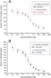
Binding affinity of metal-free and metal-bound Sulfo-Cy7-FSC-MG on A431-CCK2 cells (A) and of Sulfo-Cy7-FSC-RGD on human melanoma M21 (αvβ3-positive) cells (B); IC50 values are expressed as mean of three individual experiments in triplicates ± SD.
Figure 2
Figure 2
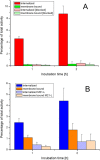
Radio cell internalization studies after 1 and 2 h incubation of [68Ga]Sulfo-Cy7-FSC-MG using A431-CCK2R cells (A) and of [68Ga]Sulfo-Cy7-FSC-RGD using human melanoma M21 (αvβ3-positive) and M21-L (αvβ3-negative) cells (B).
Figure 3
Figure 3
Fluorescence cell uptake studies after 2 h incubation of Sulfo-Cy7-FSC-MG using A431-CCK2R cells and of Sulfo-Cy7-FSC-RGD using human melanoma M21 (αvβ3-positive) and M21-L (αvβ3-negative) cells.
Figure 4
Figure 4
Ex vivo biodistribution studies in A431-CCK2R/A431-mock tumor xenografted BALB/c nude mice 1 and 2 h p.i. for [68Ga]Sulfo-Cy7-FSC-MG (A) and for [68Ga]Sulfo-Cy7-FSC-RGD in U-87MG xenograft-bearing mice 30 and 90 min p.i. (B). Corresponding tumor-to-organ ratios are shown for [68Ga]Sulfo-Cy7-FSC-MG (C) and [68Ga]Sulfo-Cy7-FSC-RGD (D) (error bars omitted).
Figure 5
Figure 5
Dual-modality animal imaging studies: Static microPET/CT images (coronal slices) and 3D volume rendered projections of fused microPET/CT (prone position) of [68Ga]Sulfo-Cy7-FSC-MG in A431-CCK2R/A431-mock tumor xenograft-bearing BALB/c nude mice 60 and 120 min p.i. (A) and [68Ga]Sulfo-Cy7-FSC-RGD 30 and 90 min p.i. in mice bearing U-87MG tumor xenografts (C). Corresponding static near-infrared fluorescence images (supine position) at various time intervals are shown in (B) for Sulfo-Cy7-FSC-MG and (D) for Sulfo-Cy7-FSC-RGD.

Similar articles

Cited by

References

    1. Mankoff D. A.; Link J. M.; Linden H. M.; Sundararajan L.; Krohn K. A. (2008) Tumor Receptor Imaging. J. Nucl. Med. 49, 149S–163S. 10.2967/jnumed.107.045963. - DOI - PubMed
    1. Laverman P.; Sosabowski J. K.; Boerman O. C.; Oyen W. J. G. (2012) Radiolabelled peptides for oncological diagnosis. Eur. J. Nucl. Med. Mol. Imaging 39, S78–S92. 10.1007/s00259-011-2014-7. - DOI - PMC - PubMed
    1. Ambrosini V.; Fani M.; Fanti S.; Forrer F.; Maecke H. R. (2011) Radiopeptide Imaging and Therapy in Europe. J. Nucl. Med. 52, 42S–55S. 10.2967/jnumed.110.085753. - DOI - PubMed
    1. Velikyan I. (2014) Prospective of 68Ga-Radiopharmaceutical development. Theranostics 4, 47–80. 10.7150/thno.7447. - DOI - PMC - PubMed
    1. van Leeuwen F. W. B.; de Jong M.; Evangelista L.; Barbet J.; del Vecchio S.; Schibli R. (2014) Molecular imaging: the emerging role of optical imaging in nuclear medicine. Eur. J. Nucl. Med. Mol. Imaging 41, 2150–2153. 10.1007/s00259-014-2845-0. - DOI - PubMed

Publication types

MeSH terms

LinkOut - more resources