. 2018 Jan;24(1-2):81-93.
doi: 10.1089/ten.TEA.2016.0205. Epub 2017 Jun 13.

Meniscal Tissue Engineering Using Aligned Collagen Fibrous Scaffolds: Comparison of Different Human Cell Sources

Affiliations

Meniscal Tissue Engineering Using Aligned Collagen Fibrous Scaffolds: Comparison of Different Human Cell Sources

Jihye Baek et al. Tissue Eng Part A. 2018 Jan.

Abstract

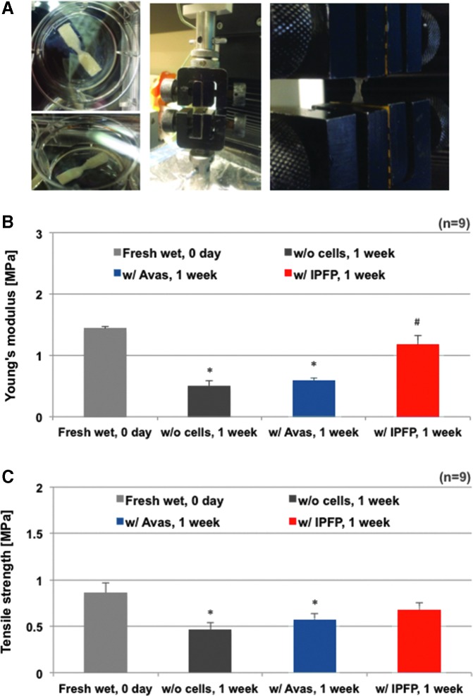
Hydrogel and electrospun scaffold materials support cell attachment and neotissue development and can be tuned to structurally and mechanically resemble native extracellular matrix by altering either electrospun fiber or hydrogel properties. In this study, we examined meniscus tissue generation from different human cell sources including meniscus cells derived from vascular and avascular regions, human bone marrow-derived mesenchymal stem cells, synovial cells, and cells from the infrapatellar fat pad (IPFP). All cells were seeded onto aligned electrospun collagen type I scaffolds and were optionally encapsulated in a tricomponent hydrogel. Single or multilayered constructs were generated and cultivated in defined medium with selected growth factors for 2 weeks. Cell viability, cell morphology, and gene-expression profiles were monitored using confocal microscopy, scanning electron microscopy, and quantitative polymerase chain reaction (qPCR), respectively. Multilayered constructs were examined with histology, immunohistochemistry, qPCR, and for tensile mechanical properties. For all cell types, TGFβ1 and TGFβ3 treatment increased COL1A1, COMP, Tenascin C (TNC), and Scleraxis (SCX) gene expression and deposition of collagen type I protein. IPFP cells generated meniscus-like tissues with higher meniscogenic gene expression, mechanical properties, and better cell distribution compared to other cell types studied. We show proof of concept that electrospun collagen scaffolds support neotissue formation and IPFP cells have potential for use in cell-based meniscus regeneration strategies.

Keywords: adult stem cells; biomimetic materials; meniscus.


Conflict of interest statement

No competing financial interests exist.

Figures

<b>FIG. 1.</b>
FIG. 1.
Schematic and cell density of three-dimensional cultures on electrospun collagen scaffolds with or without hydrogel, and pictures of the constructs as representative. (A) Schematic representation of the multilayered constructs of collagen-aligned fibrous scaffolds with cells encapsulated within the tricomponent hydrogel with meniscus cells. (B) Schematic representation of the multilayered constructs of collagen-aligned fibrous scaffolds with cells excluding tricomponent hydrogel. (C) Macro top-view showing half a disc (scale in millimeters). (D) Macro side-view of half a disc of hMSCs was encapsulated in tricomponent hydrogel between electrospun collagen scaffold sheets. (E) Cell density in the multilayered collagen constructs with or without hydrogel. *p < 0.05, **p < 0.005, ***p < 0.0005; ++p < 0.05 compared to vas, avas, MSC, ##p < 0.05 compared to vas, avas, MSC, and synovial. §p < 0.05 compared to avas, #p < 0.05 compared to vas and avas, and +p < 0.05 compared to vas. hMSC, human bone marrow-derived mesenchymal stem cell.
<b>FIG. 2.</b>
FIG. 2.
Cellular response to single aligned collagen fibrous scaffolds. Scanning electron microscopy of (A) vascular, (B) avascular human meniscus cells, (C) MSCs, (D) synovial, and (E) IPFP cells cultivated on aligned electrospun collagen fibers (Mag. 625 × ; scale bar: 5 μm). (F) vascular, (G) avascular human meniscus cells, (H) MSCs, (I) synovial, and (J) IPFP cells on aligned scaffolds demonstrating viability (live/dead) and aligned cells cultivated on collagen scaffolds (Mag. 10 × ; scale bar: 200 μm in confocal images). White arrows indicate fiber direction on each image. IPFP, infrapatellar fat pad.
<b>FIG. 3.</b>
FIG. 3.
Histological analysis of three-dimensional cultures of human meniscus vascular and avascular cells, hMSCs, synovial, and IPFP cells on electrospun collagen scaffolds without hydrogel (A) H&E, (B) Safranin O fast green, (C) DAPI, and (D) IHC collagen type I stain of all cell types for multilayered constructs without hydrogel. (Mag. = 40 × , scale bar: 100 μm). H&E, hematoxylin and eosin; DAPI, 4′, 6-diamidino-2-phenylindole; IHC, immunohistochemistry.
<b>FIG. 4.</b>
FIG. 4.
Histological analysis of three-dimensional cultures of human meniscus vascular and avascular cells, hMSCs, synovial, and IPFP cells on electrospun collagen scaffolds embedded in the tricomponent hydrogel. (A) H&E, (B) Safranin O fast green, (C) DAPI, and (D) IHC collagen type I stain of all cell types encapsulated within hydrogel for multilayered constructs. (Mag. = 40 × , scale bar: 100 μm).
<b>FIG. 5.</b>
FIG. 5.
Relative fold change in gene expression of human vascular and avascular meniscus cells, MSCs, synovial, and IPFP cells of single collagen scaffolds and multilayered collagen constructs without hydrogel or embedded in the tricomponent hydrogel. (A) Gene expression of different cell types on single collagen scaffolds, (B) of different cell types encapsulated without or (C) within the tricomponent hydrogel for multilayered constructs (n = 3 donors, with duplicates). Expression levels are relative to monolayer controls (dotted line). Each (i) COL1A1, (ii) COMP, (iii) TNC, and (iv) SCX gene expression of all different type of cells encapsulated within or without hydrogel for multilayered constructs (n = 3 donors, with duplicates). Line = p < 0.05 compared to different condition (CNT = without growth factors, TGFβ1, and TGFβ3); ++p < 0.05 compared to vas, avas, MSC, and synovial cells, ##p < 0.05 compared vas, avas, and MSC cells, lllp < 0.05 compared to vas, MSC, and synovial cells, *+p < 0.05 compared to vas, avas, and synovial cells, §+p < 0.05 compared to vas, MSC, and synovial cells, #+p < 0.05 compared to vas, and synovial cells, #$p < 0.05 compared to avas, and synovial cells, §§p < 0.05 compared to vas, and MSC cells, llp < 0.05 compared to avas, and MSC cells,**p < 0.05 compared to vas, and avascular cells, +p < 0.05 compared to synovial cells, §p < 0.05 compared to MSC cells #p < 0.05 compared to avascular cells, *p < 0.05 compared to vascular cells.
<b>FIG. 6.</b>
FIG. 6.
Mechanical strength of three-dimensional cultures of human meniscus and IPFP cells on electrospun collagen scaffolds embedded in the tricomponent hydrogel. The mechanical properties of multilayered constructs were quantified via tensile testing (n = 1 donor, nine replicates group). (A) Images of the multilayered collagen construct specimen and the tensile strength testing process and (B) Young's modulus (p = 0.0007 among experimental groups), Kruskal–Wallis test; *p < 0.005 versus freshly wet acellular multilayered construct's Young's modulus, Mann–Whitney test; #p < 0.005 versus Young's modulus of IPFP-cell encapsulated within hydrogel on multilayered constructs cultured for 1 week, Mann–Whitney test and (C) ultimate tensile strength of three-dimensional constructs (p < 0.05 among experimental groups, Kruskal–Wallis test; *p < 0.05 vs. freshly wet multilayered constructs' tensile strength, Mann–Whitney test).

References

    1. Sweigart M.A., and Athanasiou K.A. Toward tissue engineering of the knee meniscus. Tissue Eng 7, 111, 2001 - PubMed
    1. Baker P., Coggon D., Reading I., Barrett D., McLaren M., and Cooper C. Sports injury, occupational physical activity, joint laxity, and meniscal damage. J Rheumatol 3, 557, 2002 - PubMed
    1. Boyd K.T., and Myers P.T. Meniscus preservation; rationale, repair techniques and results. Knee 10, 1, 2003 - PubMed
    1. Poulsen M.R., and Johnson D.L. Meniscal injuries in the young, athletically active patient. Phys Sportsmed 39, 123, 2011 - PubMed
    1. Setton L.A., Guilak F., Hsu E.W., and Vail T.P. Biomechanical factors in tissue engineered meniscal repair. Clin Orthop Relat Res 367(Suppl), S254, 1999 - PubMed

Publication types