

Isolation of CD248-expressing stromal vascular fraction for targeted improvement of wound healing
- PMID: 28464475
- PMCID: PMC5568953
- DOI: 10.1111/wrr.12542
Isolation of CD248-expressing stromal vascular fraction for targeted improvement of wound healing
Abstract
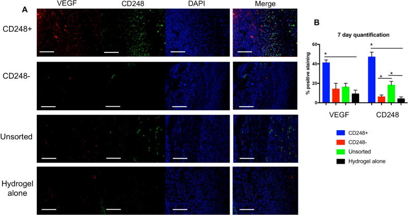
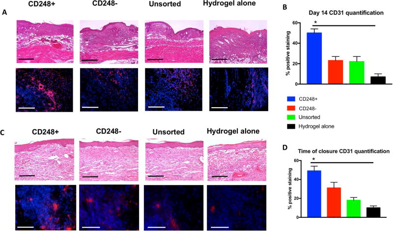
Wound healing remains a global issue of disability, cost, and health. Addition of cells from the stromal vascular fraction (SVF) of adipose tissue has been shown to increase the rate of full thickness wound closure. This study aimed to investigate the angiogenic mechanisms of CD248+ SVF cells in the context of full thickness excisional wounds. Single cell transcriptional analysis was used to identify and cluster angiogenic gene-expressing cells, which was then correlated with surface marker expression. SVF cells isolated from human lipoaspirate were FACS sorted based on the presence of CD248. Cells were analyzed for angiogenic gene expression and ability to promote microvascular tubule formation in vitro. Following this, 6mm full thickness dermal wounds were created on the dorsa of immunocompromised mice and then treated with CD248+, CD248-, or unsorted SVF cells delivered in a pullalan-collagen hydrogel or the hydrogel alone. Wounds were measured every other day photometrically until closure. Wounds were also evaluated histologically at 7 and 14 days post-wounding and when fully healed to assess for reepithelialization and development of neovasculature. Wounds treated with CD248+ cells healed significantly faster than other treatment groups, and at 7 days, had quantitatively more reepithelialization. Concurrently, immunohistochemistry of CD31 revealed a much higher presence of vascularity in the CD248+ SVF cells treated group at the time of healing and at 14 days post-op, consistent with a pro-angiogenic effect of CD248+ cells in vivo. Therefore, using CD248+ pro-angiogenic cells obtained from SVF presents a viable strategy in wound healing by promoting increased vessel growth in the wound.
© 2017 by the Wound Healing Society.
Figures


Similar articles
-
Adipose Extracellular Matrix/Stromal Vascular Fraction Gel Secretes Angiogenic Factors and Enhances Skin Wound Healing in a Murine Model.Biomed Res Int. 2017;2017:3105780. doi: 10.1155/2017/3105780. Epub 2017 Aug 1. Biomed Res Int. 2017. PMID: 28835892 Free PMC article.
-
Extracellular matrix/stromal vascular fraction gel conditioned medium accelerates wound healing in a murine model.Wound Repair Regen. 2017 Nov;25(6):923-932. doi: 10.1111/wrr.12602. Epub 2018 Feb 6. Wound Repair Regen. 2017. PMID: 29240284
-
Stromal vascular fraction shows robust wound healing through high chemotactic and epithelialization property.Cytotherapy. 2017 Apr;19(4):543-554. doi: 10.1016/j.jcyt.2017.01.006. Epub 2017 Feb 13. Cytotherapy. 2017. PMID: 28209525
-
A systematic review of autologous adipose-derived stromal vascular fraction (SVF) for the treatment of acute cutaneous wounds.Arch Dermatol Res. 2022 Jul;314(5):417-425. doi: 10.1007/s00403-021-02242-x. Epub 2021 May 28. Arch Dermatol Res. 2022. PMID: 34047823
-
Biotechnological Management of Angiopathic Wounds: Challenges and Perspectives.Int J Low Extrem Wounds. 2018 Dec;17(4):214-217. doi: 10.1177/1534734618813232. Epub 2018 Nov 25. Int J Low Extrem Wounds. 2018. PMID: 30474446 Review.
Cited by
-
Deconstructing Fat to Reverse Radiation Induced Soft Tissue Fibrosis.Bioengineering (Basel). 2023 Jun 20;10(6):742. doi: 10.3390/bioengineering10060742. Bioengineering (Basel). 2023. PMID: 37370673 Free PMC article. Review.
-
Adipose-Derived Stromal Cell-Based Therapies for Radiation-Induced Fibrosis.Adv Wound Care (New Rochelle). 2024 May;13(5):235-252. doi: 10.1089/wound.2022.0103. Epub 2022 Dec 9. Adv Wound Care (New Rochelle). 2024. PMID: 36345216 Free PMC article. Review.
-
Microvascular fragment spheroids: Three-dimensional vascularization units for tissue engineering and regeneration.J Tissue Eng. 2021 Aug 27;12:20417314211035593. doi: 10.1177/20417314211035593. eCollection 2021 Jan-Dec. J Tissue Eng. 2021. PMID: 34471514 Free PMC article.
-
The Essential Role of Angiogenesis in Adenosine 2A Receptor Deficiency-mediated Impairment of Wound Healing Involving c-Ski via the ERK/CREB Pathways.Int J Biol Sci. 2024 Aug 19;20(11):4532-4550. doi: 10.7150/ijbs.98856. eCollection 2024. Int J Biol Sci. 2024. PMID: 39247808 Free PMC article.
-
High throughput screening of mesenchymal stem cell lines using deep learning.Sci Rep. 2022 Oct 20;12(1):17507. doi: 10.1038/s41598-022-21653-y. Sci Rep. 2022. PMID: 36266301 Free PMC article.
References
-
- Kuo YR, Wang CT, Cheng JT, Kao GS, Chiang YC, Wang CJ. Adipose-Derived Stem Cells Accelerate Diabetic Wound Healing Through the Induction of Autocrine and Paracrine Effects. Cell transplantation. 2016;25(1):71–81. - PubMed
MeSH terms
Substances
Grants and funding
LinkOut - more resources
Full Text Sources
Other Literature Sources
Medical
Miscellaneous
