Immunohistochemical evaluation of epithelial ovarian carcinomas identifies three different expression patterns of the MX35 antigen, NaPi2b
- PMID: 28464843
- PMCID: PMC5414119
- DOI: 10.1186/s12885-017-3289-2
Immunohistochemical evaluation of epithelial ovarian carcinomas identifies three different expression patterns of the MX35 antigen, NaPi2b
Abstract
Background: To characterize the expression of the membrane transporter NaPi2b and antigen targeted by the MX35 antibody in ovarian tumor samples. The current interest to develop monoclonal antibody based therapy of ovarian cancer by targeting NaPi2b emphasizes the need for detailed knowledge and characterization of the expression pattern of this protein. For the majority of patients with ovarian carcinoma the risk of being diagnosed in late stages with extensive loco-regional spread disease is substantial, which stresses the need to develop improved therapeutic agents.
Methods: The gene and protein expression of SLC34A2/NaPi2b were analyzed in ovarian carcinoma tissues by QPCR (n = 73) and immunohistochemistry (n = 136). The expression levels and antigen localization were established and compared to the tumor characteristics and clinical data.
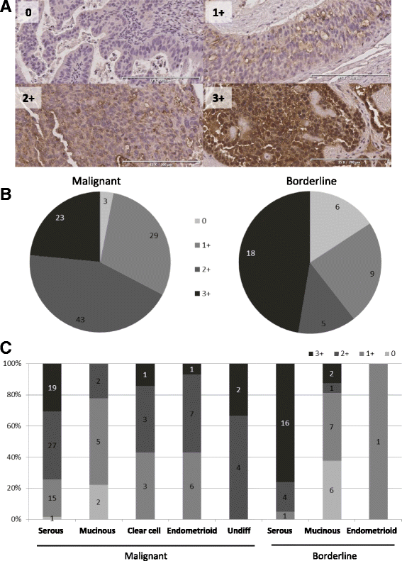
Results: Positive staining for the target protein, NaPi2b was detected for 93% of the malignant samples, and we identified three separate distribution patterns of the antigen within the tumors, based on the localization of NaPi2b. There were differences in the staining intensity as well as the distribution pattern when comparing the tumor grade and histology, the mucinous tumors presented a significantly lower expression of both the targeted protein and its related gene.
Conclusion: Our study identified differences regarding the level of the antigen expression between tumor grade and histology. We have identified differences in the antigen localization between borderline tumors, type 1 and type 2 tumors, and suggest that a pathological evaluation of NaPi2b in the tumors would be helpful in order to know which patients that would benefit from this targeted therapy.
Keywords: Monoclonal antibody; NaPi2b expression; Ovarian cancer; Radiotherapy.
Figures


Similar articles
-
Immunohistochemical analysis of NaPi2b protein (MX35 antigen) expression and subcellular localization in human normal and cancer tissues.Exp Oncol. 2011 Sep;33(3):157-61. Exp Oncol. 2011. PMID: 21956469
-
Monoclonal antibody MX35 detects the membrane transporter NaPi2b (SLC34A2) in human carcinomas.Cancer Immun. 2008 Feb 6;8:3. Cancer Immun. 2008. PMID: 18251464 Free PMC article.
-
In silico analysis and immunohistochemical characterization of NaPi2b protein expression in ovarian carcinoma with monoclonal antibody Mx35.Appl Immunohistochem Mol Morphol. 2012 Mar;20(2):165-72. doi: 10.1097/pai.0b013e318228e232. Appl Immunohistochem Mol Morphol. 2012. PMID: 22553815
-
Morphologic, Immunophenotypic, and Molecular Features of Epithelial Ovarian Cancer.Oncology (Williston Park). 2016 Feb;30(2):166-76. Oncology (Williston Park). 2016. PMID: 26892153 Review.
-
Targeting NaPi2b in ovarian cancer.Cancer Treat Rev. 2023 Jan;112:102489. doi: 10.1016/j.ctrv.2022.102489. Epub 2022 Nov 14. Cancer Treat Rev. 2023. PMID: 36446254 Review.
Cited by
-
Antibody-drug conjugates come of age in oncology.Nat Rev Drug Discov. 2023 Aug;22(8):641-661. doi: 10.1038/s41573-023-00709-2. Epub 2023 Jun 12. Nat Rev Drug Discov. 2023. PMID: 37308581 Review.
-
Targeted radiopharmaceuticals: an underexplored strategy for ovarian cancer.Theranostics. 2024 Sep 30;14(16):6281-6300. doi: 10.7150/thno.99782. eCollection 2024. Theranostics. 2024. PMID: 39431018 Free PMC article. Review.
-
Value of Antibody Drug Conjugates for Gynecological Cancers: A Modern Appraisal Following Recent FDA Approvals.Int J Womens Health. 2023 Aug 28;15:1353-1365. doi: 10.2147/IJWH.S400537. eCollection 2023. Int J Womens Health. 2023. PMID: 37663226 Free PMC article. Review.
-
Analysis of blood group antigens on MUC5AC in mucinous ovarian cancer tissues using in situ proximity ligation assay.Glycobiology. 2021 Dec 18;31(11):1464-1471. doi: 10.1093/glycob/cwab090. Glycobiology. 2021. PMID: 34459484 Free PMC article.
-
Establishment and Mechanism Study of a Primary Ovarian Insufficiency Mouse Model Using Lipopolysaccharide.Anal Cell Pathol (Amst). 2021 Nov 16;2021:1781532. doi: 10.1155/2021/1781532. eCollection 2021. Anal Cell Pathol (Amst). 2021. PMID: 34824967 Free PMC article.
References
-
- Gryshkova V, Goncharuk I, Gurtovyy V, Khozhayenko Y, Nespryadko S, Vorobjova L, Usenko V, Gout I, Filonenko V, Kiyamova R. The study of phosphate transporter NAPI2B expression in different histological types of epithelial ovarian cancer. Exp Oncol. 2009;31(1):37–42. - PubMed
MeSH terms
Substances
LinkOut - more resources
Full Text Sources
Other Literature Sources
Medical

