. 2017 May 2;14(1):89.
doi: 10.1186/s12985-017-0758-9.

Pathogenesis of virulent and attenuated foot-and-mouth disease virus in cattle

Affiliations

Pathogenesis of virulent and attenuated foot-and-mouth disease virus in cattle

Jonathan Arzt et al. Virol J. .

Abstract

Background: Understanding the mechanisms of attenuation and virulence of foot-and-mouth disease virus (FMDV) in the natural host species is critical for development of next-generation countermeasures such as live-attenuated vaccines. Functional genomics analyses of FMDV have identified few virulence factors of which the leader proteinase (Lpro) is the most thoroughly investigated. Previous work from our laboratory has characterized host factors in cattle inoculated with virulent FMDV and attenuated mutant strains with transposon insertions within Lpro.

Methods: In the current study, the characteristics defining virulence of FMDV in cattle were further investigated by comparing the pathogenesis of a mutant, attenuated strain (FMDV-Mut) to the parental, virulent virus from which the mutant was derived (FMDV-WT). The only difference between the two viruses was an insertion mutation in the inter-AUG region of the leader proteinase of FMDV-Mut. All cattle were infected by simulated-natural, aerosol inoculation.

Results: Both viruses were demonstrated to establish primary infection in the nasopharyngeal mucosa with subsequent dissemination to the lungs. Immunomicroscopic localization of FMDV antigens indicated that both viruses infected superficial epithelial cells of the nasopharynx and lungs. The critical differences between the two viruses were a more rapid establishment of infection by FMDV-WT and quantitatively greater virus loads in secretions and infected tissues compared to FMDV-Mut. The slower replicating FMDV-Mut established a subclinical infection that was limited to respiratory epithelial sites, whereas the faster replication of FMDV-WT facilitated establishment of viremia, systemic dissemination of infection, and clinical disease.

Conclusion: The mutant FMDV was capable of achieving all the same early pathogenesis landmarks as FMDV-WT, but was unable to establish systemic infection. The precise mechanism of attenuation remains undetermined; but current data suggests that the impaired replication of the mutant is more responsible for attenuation than differences in host immunological factors. These results complement previous studies by providing data of high-granularity describing tissue-specific tropism of FMDV and by demonstrating microscopic localization of virulent and attenuated clones of the same field-strain FMDV.

Keywords: Bovine; Cattle; FMD; FMDV; Foot-and-mouth; Pathogenesis; Virulence; Virus.


Figures
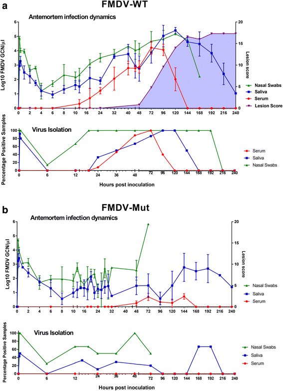
Fig. 1
Fig. 1
Antemortem infection dynamics in steers following aerosol-inoculation with distinct strains of FMDV-A24-Cruzeiro: a FMDV-WT (n = 20) or b FMDV-Mut (n = 9). a & b FMDV RNA detection in serum, oral and nasal swabs was performed through qRT-PCR, and is presented as log10 genome copy numbers (GCN)/ul. Data presented are average values (mean +/− SEM) based on samples collected from all cattle included at each time point. Virus isolation (VI) was performed on BHK-21 cells and is presented as the percentage of VI positive samples per time point. High GCN/ul of RNA and VI percentage positivity immediately after inoculation indicates detection of residual inoculated virus. * X-axes scales are non-linear. For FMDV-Mut, nasal swab data is derived entirely from animals 699, 9106, 9107, 1003, 1024, and 1025
Fig. 2
Fig. 2
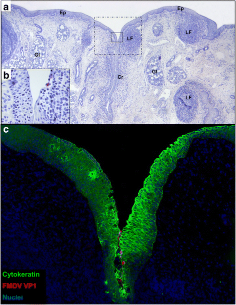
Detection of FMDV-WT by immunomicroscopy in bovine nasopharynx at 6 h post aerosol-inoculation. a Low magnification view demonstrates tissue architecture of the caudal aspect of the dorsal soft palate including surface epithelium (Ep), epithelial crypts (Cr), lymphoid follicles (LF) of mucosa-associated lymphoid tissue, and secretory glands (Gl). Two regions of interest (ROI) are centered on an epithelial crypt with a prominent subepithelial lymphoid follicle: solid-lined box indicates ROI highlighted in 1b; dashed box indicates ROI highlighted in 1c. Immunohistochemistry (IHC) with anti-FMDV capsid monoclonal antibody, micropolymer alkaline phosphatase detection system with hematoxylin counterstain. b Earliest microscopic detection of FMDV consisting of few cells containing FMDV VP1 within a shallow depression within the wall of an epithelial crypt. IHC with anti-FMDV capsid monoclonal antibody, micropolymer alkaline phosphatase detection system. c Serial section of tissue shown in a-b demonstrating few cells containing FMDV VP1 (red) within epithelium (green) or adhered to epithelial surface of crypt. Multichannel immunofluorescence microcopy, (animal ID 1002, tissue ID Dorsal soft palate -Caudal) (magnification: a 2×, b 40×, c 10×)
Fig. 3
Fig. 3
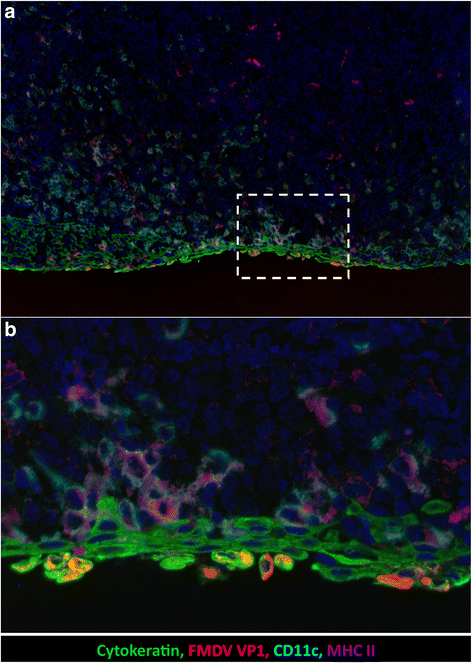
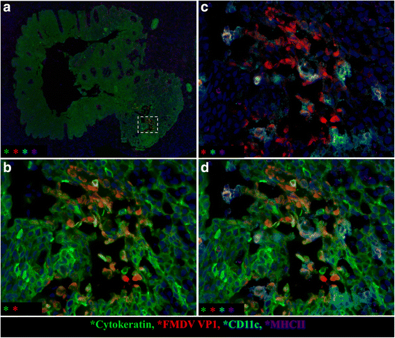
Detection of FMDV-WT by immunomicroscopy in bovine nasopharynx at 12 h post aerosol-inoculation. a Cells containing FMDV VP1 antigen (red) are within the cytokeratin-positive epithelium (green). Epithelium overlies an expansive field of mucosa-associated lymphoid tissue with numerous cells containing CD11c (turquoise) and MHCII (purple). Dashed-lined box indicates region of interest (ROI) shown at higher magnification in 3b. b FMDV-containing cells express cytokeratin and are in the superficial-most layer of epithelium. These cells have morphology consistent with acantholytic degeneration including swelling and dissociation from adjacent cells. Multichannel immunofluorescence microcopy, (animal ID 1027, tissue ID Dorsal nasopharynx -Rostral) (magnification: a 10×, b 40×)
Fig. 4
Fig. 4
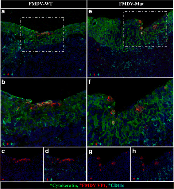
At 24 hpa, distribution and morphology of primary infection is similar for FMDV-WT and FMDV-Mut. Microscopic distribution of FMDV structural protein VP1 (red) cytokeratin (green), and CD11c (turquoise) in nasopharyngeal mucosa of cattle infected with FMDV-WT (a-d) or FMDV-Mut (e-h). a & e Both viruses similarly infected superficial cytokeratin-positive nasopharyngeal mucosal epithelial cells. Dashed-lined boxes indicate regions of interest (ROI) shown at higher magnification in b-d, f-h. b-d and f-h Higher magnification and selective channel combinations demonstrate similar co-localization patterns for both viruses. FMDV VP1 (red) predominantly localizes within cytokeratin-positive epithelial cells (green), but not with CD11c (turquoise). Few infected cells contain neither cytokeratin nor CD11c (phenotype undetermined). Multichannel immunofluorescence microcopy, (FMDV-WT animal ID 930, tissue ID Dorsal nasopharynx –Rostral; FMDV-Mut animal ID 699, tissue ID Dorsal soft palate -Caudal) (magnification: a & e 20×, b-d & f-h 40×)
Fig. 5
Fig. 5
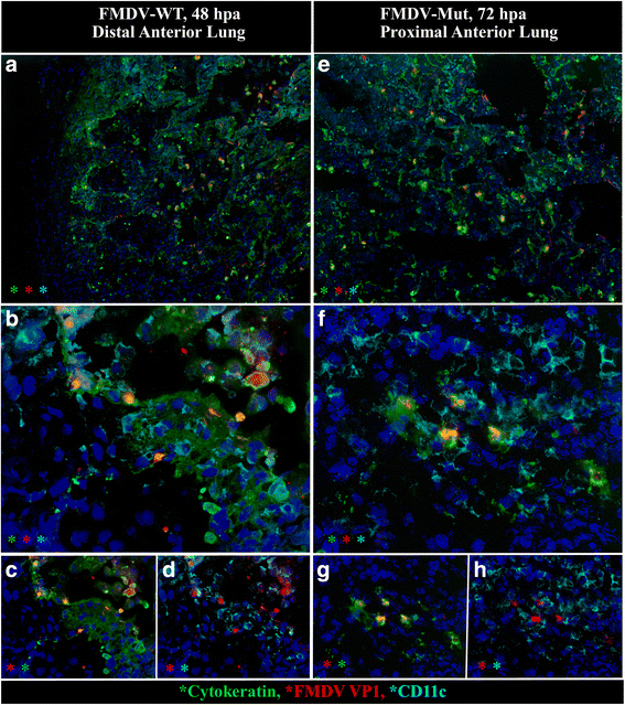
Distribution and morphology of pulmonary infection was similar for FMDV-WT at 48hpa and FMDV-Mut at 72hpa. Microscopic distribution of FMDV structural protein VP1 (red), cytokeratin (green), and CD11c (turquoise) in lungs of cattle infected with FMDV-WT (a-d) or FMDV-Mut (e-h). a & e Between 48 and 72hpa both viruses generated poorly demarcated foci of infected cells, and similarly formed vesicle-like cavitations comprised of acantholytic cells and debris. Foci of infection were more abundant in animals infected with FMDV-WT. b-d and f-h Higher magnification and selective channel combinations demonstrate similar co-localization patterns for both viruses. FMDV VP1 (red) predominantly localizes within cytokeratin-positive pulmonary epithelial cells (green), but not with CD11c monocytoid cells (turquoise). Acantholytic cells are cytokeratin-positive. Few infected cells contain neither cytokeratin nor CD11c (phenotype undetermined). Multichannel immunofluorescence microcopy. (FMDV-WT animal ID 960, tissue ID Lung; FMDV-Mut animal ID 1027, tissue ID Lung) (magnification: a & e 10×, b-d & f-h 40×)
Fig. 6
Fig. 6
FMDV generates microvesicles within the palatine tonsil of cattle during the clinical phase of disease. a Low magnification image demonstrates the architecture of a large tonsillar crypt delineated by cytokeratin-positive (green) squamous epithelium. Dashed box indicates region of interest (ROI) within crypt wall containing a focus of FMDV (red)-infected cells forming a microvesicle. ROI is featured at higher magnification in 6b-d including different detection channels in each panel. b Inclusion of FMDV VP1 (red) and cytokeratin (green) channels demonstrates disruption of epithelial architecture with cavitation/vesiculation. Many of the FMDV-containing cells are also cytokeratin-positive (epithelial cells). c Inclusion of FMDV VP1 (red), CD11c (turquoise), and MHC II (purple) demonstrates that some of the FMDV-containing cells are also individually- or double-positive for these markers of monocytoid, phagocytic, and antigen presenting cells. d Simultaneous viewing of all 4 channels demonstrates that within the vesicular cavity, FMDV-containing cells of distinct phenotypes are interspersed and in close proximity. Multichannel immunofluorescence microcopy. (animal ID 938, tissue ID palatine tonsil) (magnification: a 4×, b-d 40×)

Similar articles

Cited by

References

    1. Brown CC, Piccone ME, Mason PW, McKenna TS, Grubman MJ. Pathogenesis of wild-type and leaderless foot-and-mouth disease virus in cattle. J Virol. 1996;70(8):5638–41. - PMC - PubMed
    1. Arzt J, Pacheco JM, Smoliga GR, Tucker MT, Bishop E, Pauszek SJ, Hartwig EJ, de los Santos T, Rodriguez LL. Foot-and-mouth disease virus virulence in cattle is co-determined by viral replication dynamics and route of infection. Virology. 2014;452–453:12–22. doi: 10.1016/j.virol.2014.01.001. - DOI - PubMed
    1. Piccone ME, Pacheco JM, Pauszek SJ, Kramer E, Rieder E, Borca MV, Rodriguez LL. The region between the two polyprotein initiation codons of foot-and-mouth disease virus is critical for virulence in cattle. Virology. 2010;396(1):152–9. doi: 10.1016/j.virol.2009.10.020. - DOI - PubMed
    1. Belsham GJ. Influence of the Leader protein coding region of foot-and-mouth disease virus on virus replication. J Gen Virol. 2013;94(Pt 7):1486–95. doi: 10.1099/vir.0.052126-0. - DOI - PubMed
    1. Fowler V, Bashiruddin JB, Belsham GJ, Stenfeldt C, Botner A, Knowles NJ, Bankowski B, Parida S, Barnett P. Characteristics of a foot-and-mouth disease virus with a partial VP1 G-H loop deletion in experimentally infected cattle. Vet Microbiol. 2014;169(1–2):58–66. doi: 10.1016/j.vetmic.2013.12.008. - DOI - PubMed

Publication types

MeSH terms

LinkOut - more resources