

Review
doi: 10.1186/s12915-017-0375-5.
The biological function of the cellular prion protein: an update
Affiliations
- PMID: 28464931
- PMCID: PMC5412054
- DOI: 10.1186/s12915-017-0375-5
Item in Clipboard
Review
The biological function of the cellular prion protein: an update
BMC Biol.
.
Abstract
The misfolding of the cellular prion protein (PrPC) causes fatal neurodegenerative diseases. Yet PrPC is highly conserved in mammals, suggesting that it exerts beneficial functions preventing its evolutionary elimination. Ablation of PrPC in mice results in well-defined structural and functional alterations in the peripheral nervous system. Many additional phenotypes were ascribed to the lack of PrPC, but some of these were found to arise from genetic artifacts of the underlying mouse models. Here, we revisit the proposed physiological roles of PrPC in the central and peripheral nervous systems and highlight the need for their critical reassessment using new, rigorously controlled animal models.
Figures
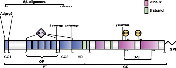
Structural organization of PrPC. Schematic representation of mature mouse PrPC, showing protein domains, sites of post-translational modification, and binding sites for divalent cations and protein interactors of functional relevance. CC1 charge cluster 1, OR octapeptide repeats, CC2 charge cluster 2, HD hydrophobic domain, FT flexible tail, GD globular domain. Structurally defined domains are depicted by pink (α-helix) and green (β-strand) boxes. GPI glycosylphosphatidylinositol anchor, CHO glycosylation site, S-S disulfide bridge. α, β, and γ cleavage sites are indicated. Copper binding sites (Cu
2+) within and outside the octapeptide region are reported as well as the sites involved in the interaction with Aβ oligomers and with the G-protein-coupled receptor Adgrg6
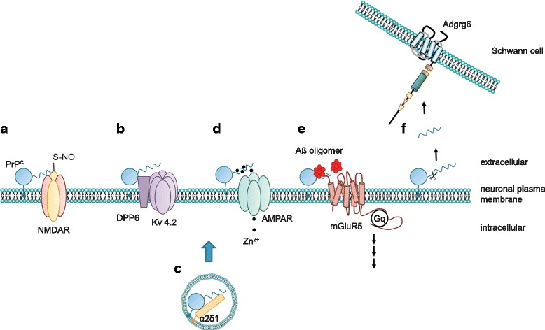
PrPC exerts its functions via distinct mechanisms. The cellular prion protein may utilize several mechanisms to modulate cellular functions. As schematically depicted in a, PrPC may directly alter the function of its target protein by mediating posttranslational modifications, for example, by promoting the S-nitrosylation of the NMDA receptor. Alternatively, PrPC modulates auxiliary proteins of ion channels, thereby regulating the biophysical properties of the channel (b) or its trafficking (c). Another function of PrPC arises from its ability to bind divalent cations such as zinc (Zn2+) or copper (Cu2+). It was claimed that PrPC may buffer these cations within the synaptic cleft and may facilitate their uptake (d) via AMPA receptors. Some better-defined actions of PrPC include its binding to misfolded oligomeric protein species and signaling in complex with other membrane receptors (e). Additionally, PrPC can signal in trans by its N-terminal cleavage products, which may bind to other receptors, prominently including the G-protein-coupled receptor Adgrg6 (f)
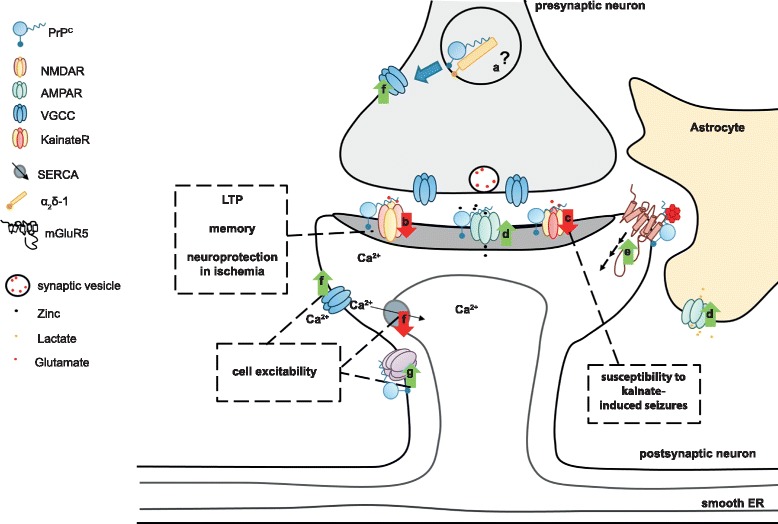
Schematic overview of possible physiological functions of PrPC and their effect in the central nervous system. PrPC regulates ion channels and neurotransmitter receptors at the pre- and postsynaptic levels. a PrPC might modulate VGCC trafficking at the presynapse via interaction with the α2δ-1 VGCC subunit. b Postsynaptically, PrPC dampens NMDA receptor-mediated currents by modulating various receptor subunits of this channel. It was speculated that control of NMDA receptor function might be related to certain reported phenotypes of PrPC-ablated mice. c PrPC may also control the glutamatergic system by modulating the subunit composition of kainate receptors. This possibly relates to increased susceptibility of PrPC-ablated mice to kainate-induced seizures. d PrPC associates with, and promotes cell surface localization of, AMPA receptor subunits. This facilitates zinc uptake at the synaptic cleft via AMPA receptors. On astrocytes, a PrPC–AMPA complex may be involved in the uptake of lactate. e PrPC binds to toxic oligomeric protein species. PrPC binds to Aβ oligomers and, in complex with metabotropic glutamate receptor 5 (mGluR5), was proposed to trigger intracellular signaling related to Alzheimer's disease pathology. f PrPC controls calcium influx via interaction with different ion channels. Additionally, PrPC was claimed to regulate calcium storage via the sarcoplasmic/endoplasmic reticulum calcium ATPase (SERCA). g PrPC positively modulates potassium currents as exemplified by association with DPP6, an auxiliary subunit of the Kv 4.2 potassium channel. Control of calcium and potassium channels might be related to the alleged function of PrPC in neuronal excitability. ER endoplasmic reticulum
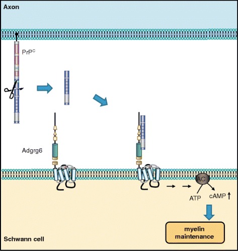
Axonal PrPC promotes myelin maintenance in trans via Adgrg6 on Schwann cells. Mice devoid of PrPC develop a chronic demyelinating neuropathy, which suggested a pro-myelinating function of PrPC. In the peripheral nervous system, the N1 fragment of axonal PrPC interacts with Adgrg6 expressed on Schwann cells. This binding elicits activation of Adgrg6, which signals via adenylyl cyclase, thereby leading to increased cellular levels of cAMP. This triggers a well-defined downstream signaling cascade promoting myelin maintenance
References
Publication types
MeSH terms
Substances
LinkOut - more resources
Full Text Sources
Other Literature Sources
Research Materials
