. 2017 May 2;8(1):104.
doi: 10.1186/s13287-017-0556-8.

Quiescence of human muscle stem cells is favored by culture on natural biopolymeric films

Affiliations

Quiescence of human muscle stem cells is favored by culture on natural biopolymeric films

Claire Monge et al. Stem Cell Res Ther. .

Abstract

Background: Satellite cells are quiescent resident muscle stem cells that present an important potential to regenerate damaged tissue. However, this potential is diminished once they are removed from their niche environment in vivo, prohibiting the long-term study and genetic investigation of these cells. This study therefore aimed to provide a novel biomaterial platform for the in-vitro culture of human satellite cells that maintains their stem-like quiescent state, an important step for cell therapeutic studies.

Methods: Human muscle satellite cells were isolated from two donors and cultured on soft biopolymeric films of controlled stiffness. Cell adhesive phenotype, maintenance of satellite cell quiescence and capacity for gene manipulation were investigated using FACS, western blotting, fluorescence microscopy and electron microscopy.

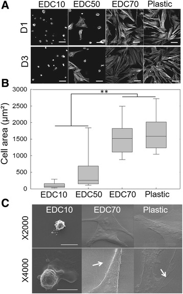
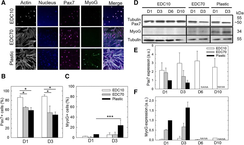
Results: About 85% of satellite cells cultured in vitro on soft biopolymer films for 3 days maintained expression of the quiescence marker Pax7, as compared with 60% on stiffer films and 50% on tissue culture plastic. The soft biopolymeric films allowed satellite cell culture for up to 6 days without renewing the media. These cells retained their stem-like properties, as evidenced by the expression of stem cell markers and reduced expression of differentiated markers. In addition, 95% of cells grown on these soft biopolymeric films were in the G0/G1 stage of the cell cycle, as opposed to those grown on plastic that became activated and began to proliferate and differentiate.

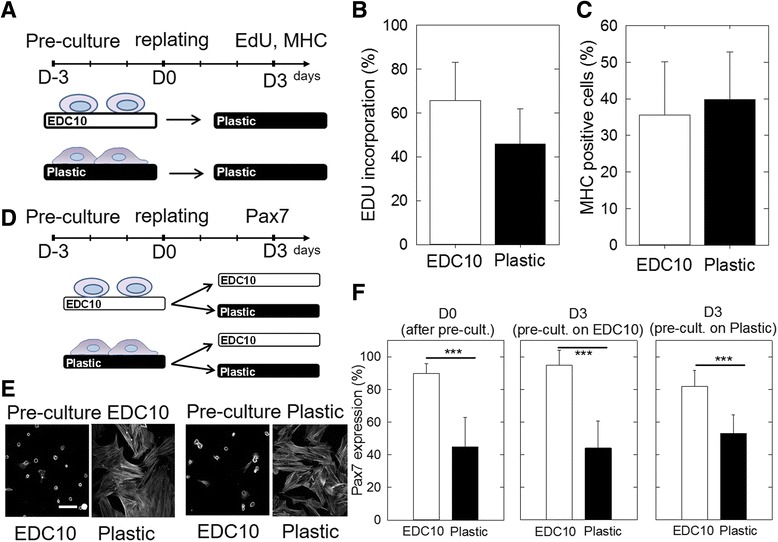
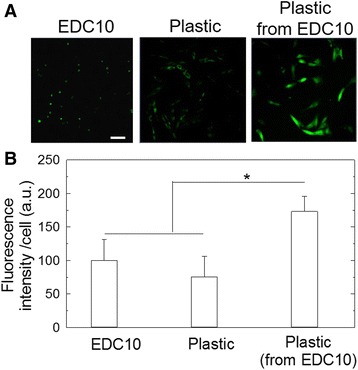
Conclusions: Our study identifies a new biomaterial made of a biopolymer thin film for the maintenance of the quiescence state of muscle satellite cells. These cells could be activated at any point simply by replating them onto a plastic culture dish. Furthermore, these cells could be genetically manipulated by viral transduction, showing that this biomaterial may be further used for therapeutic strategies.

Keywords: Biomimetism; Culture platform; Layer by layer; Polymeric biomaterial; Quiescence; Satellite cells.


Figures

Fig. 1
Fig. 1
SC morphology on biopolymeric films in comparison with plastic. Biopolymeric films were crosslinked at different levels: EDC10, EDC50 and EDC70. a Representative images of actin staining after 1 day (D1) or 3 days of culture (D3) on EDC10, EDC50 and EDC70 films and plastic. Scale bars: 50 μm. b Corresponding quantification of the cell spreading area. Data plotted from three independent experiments (n = 200 cells minimum per experimental condition). **p < 0.01. c Scanning electron microscopy imaging of SCs cultured on EDC10 films, EDC70 films and plastic. The white arrows indicate filopodia. Scale bars: 30 μm
Fig. 2
Fig. 2
SC morphology and ECM protein secretion on biopolymeric films. EDC10 and EDC70 films were compared with plastic. a Representative pictures of actin, fibronectin and collagen I staining after culture for 3, 10 and 21 days (respectively D3, D10 and D21) on EDC10 films, and for 3 days on EDC70 films and plastic. Scale bar: 50 μm. b Quantification of fibronectin and collagen I secretion by SCs cultured on EDC10 films (D3, D10, D21), EDC70 films and plastic (D3). Data are mean ± SD from two independent experiments (n = 100 cells minimum per condition). ***p <0.001. c Scanning electron microscopy micrographs of SCs seeded on EDC10 films for 10 days. Scale bars: 30 μm
Fig. 3
Fig. 3
Pax7 and myogenin expression in SCs cultured on biopolymeric films. a Fluorescent labeling of actin, Pax7, myogenin (MyoG) and nucleus (Hoechst) at D3 on EDC10 and EDC70 films in comparison with plastic. Scale bar: 50 μm. b Pax7 and c MyoG expression were quantified by immunofluorescence at D1 and D3 in GM. Data from three independent experiments (n = 100 cells minimum per experimental condition). *p < 0.05, ***p < 0.001. d Western blot analysis of Pax7 and MyoG expression and semi-quantitative determination of e Pax7 and f MyoG expression as compared with plastic control (the condition “plastic D1” was set to 1). Results of D6 and D10 are only given for the culture on EDC10 films because cell confluence was reached on EDC70 films and plastic as soon as day 3; for these conditions, D6 and D10 are thus referred to as “not applicable” (NA). Data are mean ± SD from three independent experiments. D1, D3, D6, D10 days 1, 3, 6, 10
Fig. 4
Fig. 4
Absence of SC proliferation on EDC10 films up to 10 days. a Quantification of SC proliferation by EdU assay: for D1, D3, D6 and D10 on EDC10 films and only for D1 and D3 on plastic, because confluence was reached (referred as “not applicable” (NA)). Data are box plots from three independent experiments (n = 200 cells in total per experimental condition). ***p < 0.001. b FACS analysis of the cell cycle at D3, D6 and D10 on EDC10 films and at D1 on plastic. Data plotted from two independent experiments (n = 5000 cells per condition). c Western blot of cyclin D1 and semi-quantitative analysis. The condition “plastic D1” was taken as the reference (value set to 1). Data are mean ± SD from three independent experiments. D1, D3, D6, D10 days 1, 3, 6, 10, EdU 5-ethynyl-2′-deoxyuridine
Fig. 5
Fig. 5
Reactivation of cells cultured on EDC10 films. a Schematic of experiment where SCs were precultured on EDC10 films or plastic for 3 days and then replated onto plastic. b Proliferation was assessed by EdU incorporation and c differentiation by the number of MHC+ cells after 3 days in DM. Data are mean ± SD from two different experiments (n = 50 cells per condition in each experiment). d Schematic of experiment where SCs were precultured either on EDC10 films or plastic for 3 days. SCs were then replated on EDC10 films and plastic for another 3 days. The cells were fixed and stained for actin and Pax7. e Representative images of actin staining. Scale bar: 50 μm. f Number of Pax7-positive cells: after the preculture (at D0), on EDC10 films and plastic, after replating for 3 days and depending on the preculture conditions (EDC10 films or plastic). Data are mean ± SD from three independent experiments (n = 100 cells). ***p < 0.001. D–3, D0, D3, days –3, 0, 3, EdU 5-ethynyl-2′-deoxyuridine, MHC skeletal myosin heavy chain
Fig. 6
Fig. 6
Transduction of SCs by lentivirus. a Confocal images of SC-transduced lentivirus GFP (MOI = 120) after 1 day of culture on EDC10 films or plastic. Scale bar: 100 μm. b Quantification of the fluorescence intensity on EDC10 films or plastic, measured 24 h after transduction and 24 h after replating on plastic (following a preculture on EDC10 films). *p < 0.05

References

    1. Schultz E, McCormick KM. Skeletal muscle satellite cells. Rev Physiol Biochem Pharmacol. 1994;123:213–57. doi: 10.1007/BFb0030904. - DOI - PubMed
    1. Charge SBP, Rudnicki MA. Cellular and molecular regulation of muscle regeneration. Physiol Rev. 2004;84:209–38. doi: 10.1152/physrev.00019.2003. - DOI - PubMed
    1. Hawke TJ, Garry DJ. Myogenic satellite cells: physiology to molecular biology. J Appl Physiol. 2001;91:534–51. - PubMed
    1. Peault B, Rudnicki M, Torrente Y, Cossu G, Tremblay JP, Partridge T, Gussoni E, Kunkel LM, Huard J. Stem and progenitor cells in skeletal muscle development, maintenance, and therapy. Mol Ther. 2007;15:867–77. doi: 10.1038/mt.sj.6300145. - DOI - PubMed
    1. Seale P, Sabourin LA, Girgis-Gabardo A, Mansouri A, Gruss P, Rudnicki MA. Pax7 is required for the specification of myogenic satellite cells. Cell. 2000;102:777–86. doi: 10.1016/S0092-8674(00)00066-0. - DOI - PubMed

Publication types