Review
. 2017 May 1;144(9):1588-1599.
doi: 10.1242/dev.139055.

Development of the hypothalamus: conservation, modification and innovation

Affiliations
Review

Development of the hypothalamus: conservation, modification and innovation

Yuanyuan Xie et al. Development. .

Abstract

The hypothalamus, which regulates fundamental aspects of physiological homeostasis and behavior, is a brain region that exhibits highly conserved anatomy across vertebrate species. Its development involves conserved basic mechanisms of induction and patterning, combined with a more plastic process of neuronal fate specification, to produce brain circuits that mediate physiology and behavior according to the needs of each species. Here, we review the factors involved in the induction, patterning and neuronal differentiation of the hypothalamus, highlighting recent evidence that illustrates how changes in Wnt/β-catenin signaling during development may lead to species-specific form and function of this important brain structure.

Keywords: Adult neurogenesis; Evolution; Hypothalamus; Wnt.


Conflict of interest statement

Competing interestsThe authors declare no competing or financial interests.

Figures

Fig. 1.
Fig. 1.
Location and structure of the hypothalamus. The anatomical position and basic anatomy of the hypothalamus is conserved throughout vertebrates. (A) Lateral view of the position of the hypothalamus primordium in a prototypical vertebrate during early brain development. The presumptive hypothalamus arises from the ventral forebrain at the rostroventral limit of the neural plate. (B) Lateral view of the forebrain showing conserved hypothalamic divisions in a prototypical vertebrate following initial patterning. Four rostrocaudal regions are delineated by solid gray lines, and selected hypothalamic nuclei are outlined by dashed red lines. ARC, arcuate nucleus; DMH, dorsomedial hypothalamus; LHA, lateral hypothalamic area; ME, median eminence; PVN, paraventricular nucleus; SCN, superchiasmatic nucleus; VMH, ventromedial hypothalamus.
Fig. 2.
Fig. 2.
Induction and patterning of the hypothalamus. Several morphogen signals induce and pattern the hypothalamus. (A) At neural plate stages, the hypothalamus primordium (gray oval) is induced by Wnt antagonists in the anterior neurectoderm that inhibit Wnt signals from the posterior mesoderm, and by Nodal and Shh from the prechordal axial mesoderm. Dorsal view is shown. (B) During hypothalamic patterning, a gradient of Wnt signaling signals creates discrete anterior/posterior regions of the hypothalamus (depicted by solid black lines). Hedgehog signaling provides dorsal/ventral polarity and establishes alar and basal zones within each region (delineated by dashed gray line), as well as floor plate (not shown). Orientation of boundaries is depicted according to current columnar models of hypothalamic anatomy. Lateral view is shown.
Fig. 3.
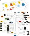
Fig. 3.
Factors involved in hypothalamic differentiation. (A) Schematic of hypothalamic nuclei in coronal rodent brain sections. A, anterior; D, dorsal; P, posterior; V, ventral. (B) Known factors involved in hypothalamic specification and differentiation. Red arrows indicate direct transcriptional activation. See Table 1 for references. aPV, anterior periventricular nucleus; ARC, arcuate nucleus; DMH, dorsomedial hypothalamus; LHA, lateral hypothalamic area; MMN, medial mammillary nucleus; PMN, premammillary nucleus; PVN, paraventricular nucleus; SCN, suprachiasmatic nucleus; SON, supraoptic nucleus; VMH, ventromedial hypothalamus; 3V, third ventricle.
Fig. 4.
Fig. 4.
Wnt signaling in the zebrafish posterior recess. (A) Ventral view of a whole larval zebrafish brain at 10 days post-fertilization stained for Wnt activity with the transgenic reporter TCFSiam:GFP (Wang et al., 2012) (green), Gfap+ radial glia (red) and cell nuclei (blue). Wnt signaling is present in the posterior ventricular recess of the hypothalamus (arrow) and is also observed in blood vessels throughout the brain. Anterior is to the left. (B) Schematic of Wnt function in hypothalamic progenitor differentiation. Wnt activity promotes neurogenesis by activating quiescent radial glia to produce committed neural progenitors.
Fig. 5.
Fig. 5.
Wnt activity in the adult hypothalamus. β-galactosidase staining in a coronal section (dorsal at the top) through the tuberal hypothalamus of a 6-month-old BAT-lacZ mouse (Wang et al., 2012). Wnt activity is present in ventricular and parenchymal regions.

References

    1. Acampora D., Postiglione M. P., Avantaggiato V., Di Bonito M., Vaccarino F. M., Michaud J. and Simeone A. (1999). Progressive impairment of developing neuroendocrine cell lineages in the hypothalamus of mice lacking the Orthopedia gene. Genes Dev. 13, 2787-2800. 10.1101/gad.13.21.2787 - DOI - PMC - PubMed
    1. Anthwal N., Pelling M., Claxton S., Mellitzer G., Collin C., Kessaris N., Richardson W. D., Gradwohl G. and Ang S.-L. (2013). Conditional deletion of neurogenin-3 using Nkx2.1iCre results in a mouse model for the central control of feeding, activity and obesity. Dis. Model. Mech. 6, 1133-1145. 10.1242/dmm.011916 - DOI - PMC - PubMed
    1. Aponte Y., Atasoy D. and Sternson S. M. (2011). AGRP neurons are sufficient to orchestrate feeding behavior rapidly and without training. Nat. Neurosci. 14, 351-355. 10.1038/nn.2739 - DOI - PMC - PubMed
    1. Atkin S. D., Owen B. M., Bookout A. L., Cravo R. M., Lee C., Elias C. F., Elmquist J. K., Kliewer S. A. and Mangelsdorf D. J. (2013). Nuclear receptor LRH-1 induces the reproductive neuropeptide kisspeptin in the hypothalamus. Mol. Endocrinol. 27, 598-605. 10.1210/me.2012-1371 - DOI - PMC - PubMed
    1. Aujla P. K., Naratadam G. T., Xu L. and Raetzman L. T. (2013). Notch/Rbpjkappa signaling regulates progenitor maintenance and differentiation of hypothalamic arcuate neurons. Development 140, 3511-3521. 10.1242/dev.098681 - DOI - PMC - PubMed

Publication types