. 2017 Jun 30;292(26):11021-11033.
doi: 10.1074/jbc.M116.770941. Epub 2017 May 2.

Myostatin inhibits osteoblastic differentiation by suppressing osteocyte-derived exosomal microRNA-218: A novel mechanism in muscle-bone communication

Affiliations

Myostatin inhibits osteoblastic differentiation by suppressing osteocyte-derived exosomal microRNA-218: A novel mechanism in muscle-bone communication

Yiwen Qin et al. J Biol Chem. .

Abstract

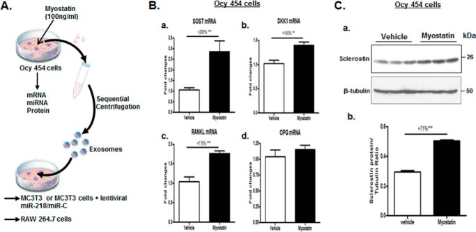
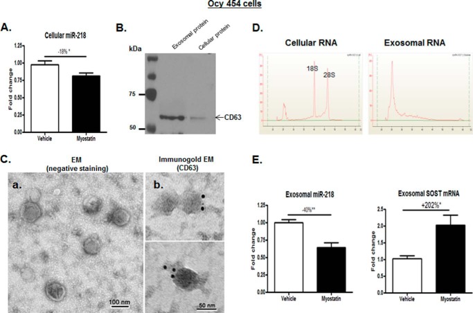
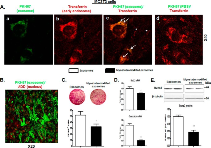
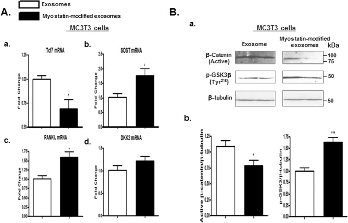
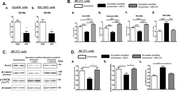
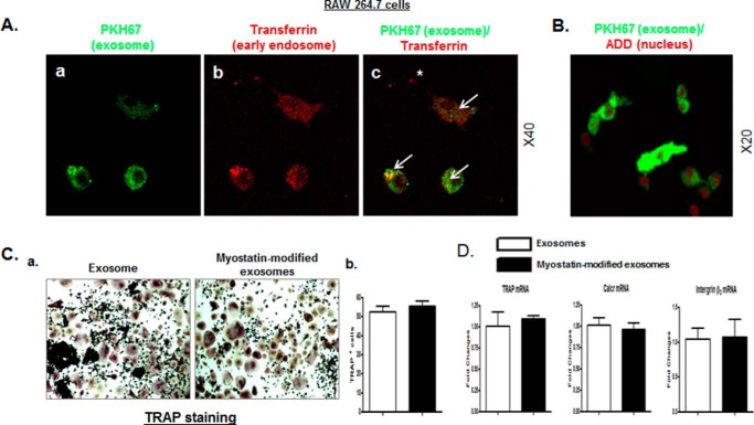
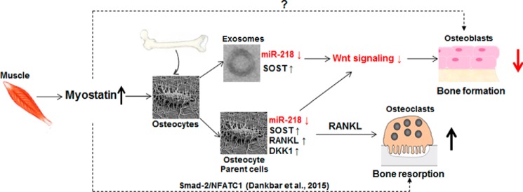
Muscle and bone are closely associated in both anatomy and function, but the mechanisms that coordinate their synergistic action remain poorly defined. Myostatin, a myokine secreted by muscles, has been shown to inhibit muscle growth, and the disruption of the myostatin gene has been reported to cause muscle hypertrophy and increase bone mass. Extracellular vesicle-exosomes that carry microRNA (miRNA), mRNA, and proteins are known to perform an important role in cell-cell communication. We hypothesized that myostatin may play a crucial role in muscle-bone interactions and may promote direct effects on osteocytes and on osteocyte-derived exosomal miRNAs, thereby indirectly influencing the function of other bone cells. We report herein that myostatin promotes expression of several bone regulators such as sclerostin (SOST), DKK1, and RANKL in cultured osteocytic (Ocy454) cells, concomitant with the suppression of miR-218 in both parent Ocy454 cells and derived exosomes. Exosomes produced by Ocy454 cells that had been pretreated with myostatin could be taken up by osteoblastic MC3T3 cells, resulting in a marked reduction of Runx2, a key regulator of osteoblastic differentiation, and in decreased osteoblastic differentiation via the down-regulation of the Wnt signaling pathway. Importantly, the inhibitory effect of myostatin-modified osteocytic exosomes on osteoblast differentiation is completely reversed by expression of exogenous miR-218, through a mechanism involving miR-218-mediated inhibition of SOST. Together, our findings indicate that myostatin directly influences osteocyte function and thereby inhibits osteoblastic differentiation, at least in part, through the suppression of osteocyte-derived exosomal miR-218, suggesting a novel mechanism in muscle-bone communication.

Keywords: Wnt signaling; bone; exosome (vesicle); microRNA (miRNA); myostatin; osteoblast; osteocyte; skeletal muscle metabolism.


Conflict of interest statement

The authors declare that they have no conflicts of interest with the contents of this article

Figures

Figure 1.
Figure 1.
Effects of myostatin on SOST, DKK1, RANKL, and OPG mRNA and/or protein expression in Ocy454 cells. A, a schematic diagram illustrating experimental procedures employed. B and C, Ocy454 cells were differentiated for 12 days and then treated with 100 ng/ml myostatin or vehicle for 48 h. B, levels of SOST, DKK1, RANKL, and OPG mRNA were determined by real-time PCR. C, levels of SOST protein were determined by Western blotting. Images shown in C (a) are representative of Western blot analysis. Data shown are the mean values ± S.E. for three separate determinations. **, p < 0.01; ***, p < 0.001.
Figure 2.
Figure 2.
Myostatin inhibited miR-218 expression in Ocy454 parent cells and their exosomes. As shown in Fig. 1A, Ocy454 cells were differentiated for 12 days and then treated with 100 ng/ml myostatin or vehicle for 48 h followed by extraction of total RNA from Ocy454 parent cells or the isolation of exosomes released form Cyc454 cells. A, levels of cellular miR-218 in Ocy454 parent cells were determined by real-time PCR. B, the Western blot showed that CD63 proteins are enriched in isolated exosome compared with that in cellular protein lysate. C, electron microscopy analysis of exosomes; a, exosomes were stained with uranyl acetate (EM (negative staining)); b, exosomes were labeled with 10-nm immunogold using antibody against exosomal membrane marker CD63 and stained with uranyl acetate (Immunogold EM (CD63)). Exosome morphology was then visualized using electron microscope Hitachi H7000. D, isolated cellular or exosomal RNA was analyzed using a Bioanalyzer. The results showed the absence of the ribosomal RNA peaks (18S and 28S rRNA) in exosomal RNA compared with that of cellular RNA. E, levels of miR-218 and SOST mRNA in exosomes produced by Ocy454 cells were determined by real-time PCR. Data shown are mean values ± S.E. for three separate determinations. *, p < 0.05 and **, p < 0.01.
Figure 3.
Figure 3.
The myostatin-modified osteocytic exosomes were internalized into MC3T3 cells, inhibiting osteoblastic differentiation. As shown in Fig. 1A, Ocy454 cells were differentiated for 12 days and were treated with 100 ng/ml myostatin or vehicle for 48 h followed by the isolation of exosomes that are released form Cyc454 cells. MC3T3 cells were then co-cultured with the myostatin-modified osteocytic exosomes for 48 h. A and B, uptake of osteocytic exosomes by MC3T3 cells. 10 μg of the PKH67-labeled osteocytic exosomes or a PKH67-PBS control were added into per 75,000 MC3T3 cells and incubated at 37 °C for 30 min. The uptake of the fluorescently labeled exosomes by MC3T3 cells was detected with confocal fluorescence microscopy. PKH67 (green) was used to label the exosomes. Texas Red-transferrin (red, in A) or 7-AAD (red, in B) was used to detect the early endosomes and the nucleus (red) of the MC3T3 (red), respectively. PKH67 exosomes (green) were rapidly internalized into early endosomes labeled with Texas Red-transferring (yellow is indicative of co-localization of green and red). The arrows indicate co-localization; the asterisks indicate no co-localization. C–E, co-culturing myostatin-modified osteocytic exosomes with MC3T3 cells inhibits osteoblastic differentiation. C, representative images showing alkaline phosphatase staining (CFU-F) of cultures of MC3T3 cells and cell counts of alkaline phosphatase-positive cells. D, changes in mRNA levels of Runx2 and osteocalcin in MC3T3 cells were determined by real-time PCR. E, quantification of Runx2 protein content in cell lysate from MC3T3 cells assessed by immunoblot analysis (inset). Data are expressed as the mean ± S.E. for three separate determinations. *, p < 0.05; ***, p < 0.001 versus indicated group.
Figure 4.
Figure 4.
Co-culturing MC3T3 cells with the myostatin-modified osteocyte-derived exosomes inactivated Wnt signaling. As shown in Fig. 1A, Ocy454 cells were differentiated for 12 days and were treated with 100 ng/ml myostatin or vehicle for 48 h followed by the isolation of exosomes released form Ocy454 cells. MC3T3 cells were then co-cultured with the myostatin-modified osteocytic exosomes for 48 h. A, changes in mRNA levels of Wnt signaling-related genes Tcf7, SOST, RANKL, and DKK2 in MC3T3 cells were determined by real-time PCR. B, Western blotting was performed on total proteins from MC3T3 cells. Images shown in (Ba) were representative Western blotting. Bb, blots in (Ba) were quantified by scanning densitometry and normalized relative to β-tubulin. Data are expressed as the mean ± S.E. for three separate determinations. *, p < 0.05; ***, p < 0.001 versus the indicated group.
Figure 5.
Figure 5.
Expression of exogenous miR-218 reversed the inhibitory effects of myostatin-modified osteocyte-derived exosomes on osteoblastic differentiation. As shown in Fig. 1A, Ocy454 cells were differentiated for 12 days and were treated with 100 ng/ml myostatin or vehicle for 48 h followed by the isolation of exosomes released form Cyc454 cells. MC3T3 cells were then co-cultured with the myostatin-modified osteocytic exosomes in the presence of lentivirus-mediated exogenous expression of miR-218 or scramble control plasmid (miR-C) for 48 h. Total RNA or protein was isolated on day 3 and subjected to real-time PCR or Western blot analysis, respectively. A, efficacy of exogenous expression of miR-218 on inhibiting SOST mRNA expression in Ocy454 cells and IDG-SW3 cells. B, changes in mRNA levels of RunX2, osteocalcin, and OPG in MC3T3 cells were determined by real-time PCR. C, Western blotting was performed on total proteins from MC3T3 cells. Images shown were representative Western blottings. D, blots in C were quantified by scanning densitometry and normalized relative to β-tubulin. Data are expressed as the mean ± S.E. for three separate determinations. *, p < 0.05; **, p < 0.01, ***, p < 0.001 versus indicated group.
Figure 6.
Figure 6.
The myostatin-modified osteocyte-derived exosomes have no direct effect on osteoclastic differentiation. As shown in Fig. 1A, Ocy454 cells were differentiated for 12 days and were treated with 100 ng/ml myostatin or vehicle for 48 h followed by the isolation of exosomes released form Cyc454 cells. RAW 264.7 cells were then co-cultured with the myostatin-modified osteocytic exosomes for 48 h. A and B, uptake of osteocytic exosomes by RAW 264.7 cells was examined by using the approaches as described in Fig. 3, A and B. The arrows indicate co-localization; the asterisk indicates no co-localization. C and D, co-culture of myostatin-modified osteocytic exosomes with RAW 264.7 cells had no effect on osteoclastic differentiation. C, representative images showing TRAP staining of cultures of RAW 264.7 cells and cell counts of TRAP-positive cells. D, changes in mRNA levels of TRAP, calcitonin receptor (Calcr), and integrin β3 in RAW 264.7 cells were determined by real-time PCR. Data are expressed as the mean ± S.E. for three separate determinations.
Figure 7.
Figure 7.
A schema of the putative mechanisms by which myostatin may influence the function of bone cells. Myostatin influences osteoblastic differentiation through the osteocyte and its release of exosomal miR-218 in a mechanism that involves the activation of SOST and its inhibition of the Wnt signaling pathway. Additionally, myostatin promotes RANKL production in osteocytes and, in turn, has the potential to increase osteoclast differentiation, activity, and viability. We note that myostatin has been recently reported to directly accelerate RANKL-mediated osteoclast formation by a mechanism through transcription factor Smad2-dependent regulation of NFATC1 (nuclear factor of activated T-cells; Ref. 61). Whether there is a direct effect of myostatin on osteoblasts remains to be determined.

References

    1. Schoenau E. (2005) From mechanostat theory to development of the “Functional Muscle-Bone-Unit.” J. Musculoskelet. Neuronal Interact. 5, 232–238 - PubMed
    1. Hamrick M. W. (2012) The skeletal muscle secretome: an emerging player in muscle-bone crosstalk. Bonekey Rep. 1, 60. - PMC - PubMed
    1. Bloomfield S. A. (2010) Disuse osteopenia. Curr. Osteoporos. Rep. 8, 91–97 - PubMed
    1. Karinkanta S., Piirtola M., Sievänen H., Uusi-Rasi K., and Kannus P. (2010) Physical therapy approaches to reduce fall and fracture risk among older adults. Nat. Rev. Endocrinol. 6, 396–407 - PubMed
    1. Rittweger J., Beller G., Ehrig J., Jung C., Koch U., Ramolla J., Schmidt F., Newitt D., Majumdar S., Schiessl H., and Felsenberg D. (2000) Bone-muscle strength indices for the human lower leg. Bone 27, 319–326 - PubMed

MeSH terms

LinkOut - more resources