. 2017 Jun 5;214(6):1753-1767.
doi: 10.1084/jem.20161645. Epub 2017 May 2.

Inflammatory signals from photoreceptor modulate pathological retinal angiogenesis via c-Fos

Affiliations

Inflammatory signals from photoreceptor modulate pathological retinal angiogenesis via c-Fos

Ye Sun et al. J Exp Med. .

Abstract

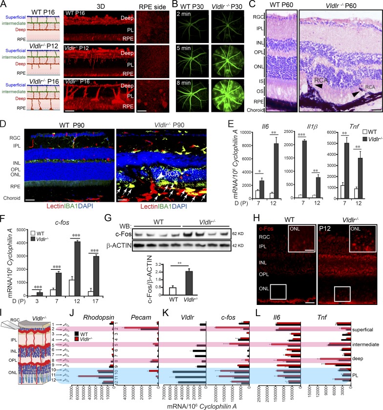
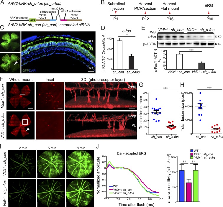
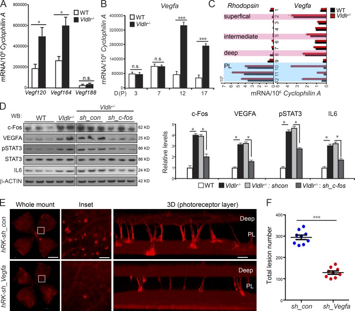
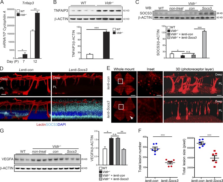
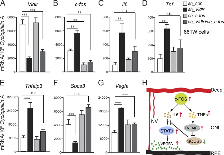
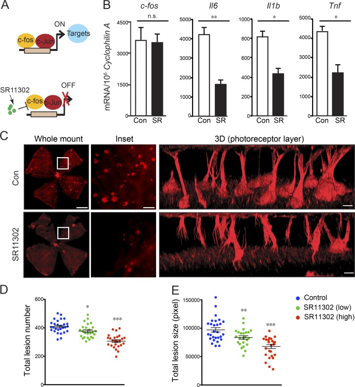
Pathological neovessels growing into the normally avascular photoreceptors cause vision loss in many eye diseases, such as age-related macular degeneration and macular telangiectasia. Ocular neovascularization is strongly associated with inflammation, but the source of inflammatory signals and the mechanisms by which these signals regulate the disruption of avascular privilege in photoreceptors are unknown. In this study, we found that c-Fos, a master inflammatory regulator, was increased in photoreceptors in a model of pathological blood vessels invading photoreceptors: the very low-density lipoprotein receptor-deficient (Vldlr-/- ) mouse. Increased c-Fos induced inflammatory cytokines interleukin 6 (IL-6) and tumor necrosis factor (TNF), leading to activation of signal transducer and activator of transcription 3 (STAT3) and increased TNFα-induced protein 3 (TNFAIP3) in Vldlr-/- photoreceptors. IL-6 activated the STAT3/vascular endothelial growth factor A (VEGFA) pathway directly, and elevated TNFAIP3 suppressed SOCS3 (suppressor of cytokine signaling 3)-activated STAT3/VEGFA indirectly. Inhibition of c-Fos using photoreceptor-specific AAV (adeno-associated virus)-hRK (human rhodopsin kinase)-sh_c-fos or a chemical inhibitor substantially reduced the pathological neovascularization and rescued visual function in Vldlr-/- mice. These findings suggested that the photoreceptor c-Fos controls blood vessel growth into the normally avascular photoreceptor layer through the inflammatory signal-induced STAT3/VEGFA pathway.


Figures

Figure 1.
Figure 1.
c-Fos was induced in Vldlr−/− retinas. (A) Vldlr deficiency led to neovascularization in the normally avascular photoreceptor layer shown by 3D reconstruction of representative confocal images. n = 6. (B) Vldlr deficiency led to retinal vascular leakage at 2, 5, and 8 min after intraperitoneal injection of fluorescent dye, as shown by FFA images from P30 WT and Vldlr−/− mice. n = 6. (C) H&E staining showed retinal layer disorganization and retinal-choroidal vascular anastomoses in P60 Vldlr−/− retinas. Black arrowheads indicate neovascularization. n = 6. (D) Macrophage marker IBA1 (green) costained with the endothelial cell marker isolectin (red) and nuclear marker DAPI (blue) in 3-mo Vldlr−/− retinas with retinal-choroidal vascular anastomoses (arrowhead); macrophages were seen in the subretinal space between the ONL and RPE (arrows). (E) Cytokine expression, including Il6, Il1β, and Tnf, was increased during development in Vldlr−/− retinas. n = 6. (F–L) c-fos mRNA and protein expression were markedly increased (F and G) in Vldlr−/− retinas during development (F), mainly in the P12 ONL (K) and colocalized with the expression of Vldlr in P12 WT (H–K) and its target genes including Il6 and Tnf (L). n = 6. (H) IHC staining showed increased c-Fos expression in the ONL of P12 Vldlr−/− retinas. INL, inner nuclear layer; IPL, inner plexiform layer; IS, photoreceptor inner segment; OPL, outer plexiform layer; OS, photoreceptor outer segment; PL, photoreceptor layer; RCA, retinal-choroidal anastomosis; RGC, retinal ganglion cell; WB, Western blot. Bars: (A, 3D) 100 µm; (A, RPE) 250 µm; (C, D, and H) 50 µm; (H, inset) 25 µm. All data are representative of at least three independent experiments. *, P < 0.05; **, P < 0.01; ***, P < 0.001. Results are presented as mean ± SEM.
Figure 2.
Figure 2.
Attenuation of c-Fos in the photoreceptor layer extending to the subretinal space reduced neovascularization. (A) Schematic diagram illustrating AAV2 carrying sh_c-fos within a mir30 cassette under the control of an hRK promoter (hRK-sh_c-fos). sh_con, sh_control. (B) Time line for AAV subretinal injection and retina collection. (C) The infection of AAV2 in the subretinal space (SS) and photoreceptor layer was confirmed by AAV2-hRK-eGFP. n = 6. INL, inner nuclear layer; RGC, retinal ganglion cell. The white arrowhead indicates the retinal areas were not affected by AAV2-hRK-eGFP because of the limitation of subretinal injection. (D and E) The knockdown efficiency of AAV2-hRKsh_c-fos via subretinal injection into Vldlr−/− retinas was confirmed by comparison with AAV2-hRK-sh_control–injected retinas at both mRNA (D) and protein (E) levels. (D) *, P < 0.05. n = 6. (E) ***, P < 0.001. n = 4. WB, Western blot. (F) Knocking down c-fos in the photoreceptor layer (PL) extending to the subretinal space using AAV2-hRKsh_c-fos–inhibited neovascularization shown by whole-mounted images of AAV2-hRK-sh_control– or AAV2-hRKsh_c-fos–treated Vldlr−/− retinas stained with isolectin IB4 to label endothelial cells and representative 3D reconstruction images for neovascularization in photoreceptor layers, including the retinal areas, which may not be completely affected by AAV2-hRKsh_c-fos (about 25% not transfected) as shown in C (white arrowhead in C and F). (G and H) Both total lesion number and total lesion size (pixels) were reduced in AAV-hRKsh_c-fos–treated compared with AAV2-hRK-sh_control–treated retinas. ***, P < 0.001. n = 8–13. (I) AAV2-hRKsh_c-fos injected in the subretinal space at P1 rescued Vldlr deficiency–induced retinal vascular leakage shown by FFA images at P60 in Vldlr−/−-treated mice. n = 6–8. (J) Representative dark-adapted (scotopic) ERG graph (left) and quantification of photoreceptor a-wave sensitivity (right) showed that photoreceptor function was attenuated in 3-mo-old Vldlr−/− retinas and was particularly rescued by AAV-hRKsh_c-fos (one-time treatment on P1) in 3-mo-old Vldlr−/− retinas. *, P < 0.05; **, P < 0.01; ***, P < 0.001. Bars: (C and F, whole-mount images) 1,000 µm; (C, cross section) 50 µm; (F, inset) 250 µm; (F, 3D) 100 µm. All data are representative of at least three independent experiments. Results are presented as mean ± SEM.
Figure 3.
Figure 3.
c-Fos promoted retinal angiogenesis via VEGFA signaling modulated by neuronal IL-6/STAT3. (A) Vegf120 and Vegf164 but not Vegf188 expression was increased in P12 Vldlr−/− retinas. n = 6. (B and C) Vegfa expression was markedly increased in Vldlr−/− whole retinas at P12 and P17 (B) as well as in the photoreceptor layer (PL) of P12 Vldlr−/− retinas (C). n = 6. (D) The protein levels of c-Fos, pSTAT3, total STAT3, IL-6, and VEGFA in P12 WT, Vldlr−/−, and Vldlr−/− treated with AAV2-hRKsh_c-fos or AAV2-hRKsh_control (sh_con). n = 4–6. WB, Western blot. (E and F) Representative P16 whole mount images stained with isolectin IB4 and quantification showed that knocking down VEGFA in Vldlr−/− retinas using AAV2-hRK-sh_Vegfa reduced total lesion number by 55% compared with AAV2-hRK-sh_control–treated Vldlr−/− retinas. (E) Bars: (whole-mount images) 1,000 µm; (inset) 250 µm; (3D) 100 µm. PL, photoreceptor layer. All data are representative of at least three independent experiments. *, P < 0.05; ***, P < 0.001. Results are presented as mean ± SEM.
Figure 4.
Figure 4.
c-Fos promoted retinal angiogenesis via TNF/TNFAIP3/SOCS3. (A and B) Tnfaip3 mRNA (A; n = 6) and protein levels (B; n = 4) were increased in the developing Vldlr−/− retinas compared with WT controls. (C) Lenti-Socs3 was injected subretinally into Vldlr−/− eyes, and SOCS3 protein levels were confirmed by Western blotting (WB). n = 3. con, control; non-treat, nontreated. (D) The infection of lenti-Socs3 in the photoreceptor layer (PL) was confirmed by anti-SOCS3 antibody staining (cyan). 3D reconstructed representative images show that SOCS3 was mainly expressed in the photoreceptor layer after subretinal injection. n = 4. The asterisk in the lenti-control image indicates nonspecific staining. (E and F) Overexpression of Socs3 using lenti-Socs3 inhibited neovascularization, including the retinal areas, which may not be completely affected by lenti-Socs3 (∼25% not transfected; white arrowhead). (E) The representative whole-mount images of lenti-control– or lenti-Socs3–treated Vldlr−/− retinas stained with isolectin IB4 to label endothelial cells. 3D reconstruction images show neovascularization in the photoreceptor layer. n = 6. (F) Quantification shows a decrease in both total lesion number and total lesion size in lenti-Socs3–treated Vldlr−/− retinas compared with lenti-control–treated Vldlr−/− retinas. n = 6. (G) Increased protein level of VEGFA in Vldlr−/− retinas was suppressed by lenti-Socs3 treatment. n = 3. Bars: (E, whole-mount images) 1,000 µm; (E, inset) 250 µm; (D and E, 3D) 100 µm. All data are representative of at least three independent experiments. *, P < 0.05; **, P < 0.01; ***, P < 0.001. Results are presented as mean ± SEM.
Figure 5.
Figure 5.
c-Fosregulated Vegfa via TNF/TNFAIP3/SOCS3 in photoreceptor cells. (A–G) The mRNA levels of Vldlr, c-fos, Il6, Tnf, Tnfaip3, Socs3, and Vegfa in 661W photoreceptor cells treated with AAV2-hRK-sh_Vldlr and/or AAV2-hRKsh_c-fos or AAV2-hRK-sh_Control (sh_con). **, P < 0.01; ***, P < 0.001. n = 6. (H) A schematic diagram illustrates pathological neovascularization (NV) in the normal avascular photoreceptor layer controlled by transcriptional factor c-FOS through both IL-6/STAT3/VEGFA and TNF/TNFAIP3/SOCS3/STAT3/VEGFA pathways. All data are representative of at least three independent experiments. Results are presented as mean ± SEM.
Figure 6.
Figure 6.
c-Fos inhibitor SR11302 prevented retinal neovascularization. (A) Schematic diagram illustrating how c-Fos inhibitor SR11302 may block c-Fos binding to its target promoters to inhibit target gene expression. (B) The mRNA levels of c-fos and its target genes (Il6, Il1β, and Tnf) in SR11302 (SR)-treated and control (con) Vldlr−/− retinas confirmed the efficiency of the c-Fos inhibitor. n = 6. (C) SR11302 inhibited neovascularization. Representative whole-mount images of control or SR11302-treated Vldlr−/− retinas stained with isolectin IB4 to label endothelial cells. Enlarged images show reduced total lesion number and lesion size in the SR11302-treated retinas. 3D reconstruction images show neovascularization in the photoreceptor cell layer. Bars: (whole-mount images) 1000 µm; (inset) 250 µm; (3D) 100 µm. (D and E) Quantification shows reduced total lesion number and lesion size (pixels) in SR11302-treated retinas compared with controls in a dose-dependent manner. n = 22–29. All data are representative of at least three independent experiments. *, P < 0.05; **, P < 0.01; ***, P < 0.001. Results are presented as mean ± SEM.

Similar articles

Cited by

References

    1. Aikawa Y., Morimoto K., Yamamoto T., Chaki H., Hashiramoto A., Narita H., Hirono S., and Shiozawa S.. 2008. Treatment of arthritis with a selective inhibitor of c-Fos/activator protein-1. Nat. Biotechnol. 26:817–823. 10.1038/nbt1412 - DOI - PubMed
    1. al-Ubaidi M.R., Font R.L., Quiambao A.B., Keener M.J., Liou G.I., Overbeek P.A., and Baehr W.. 1992. Bilateral retinal and brain tumors in transgenic mice expressing simian virus 40 large T antigen under control of the human interphotoreceptor retinoid-binding protein promoter. J. Cell Biol. 119:1681–1687. 10.1083/jcb.119.6.1681 - DOI - PMC - PubMed
    1. Ambati J., Atkinson J.P., and Gelfand B.D.. 2013. Immunology of age-related macular degeneration. Nat. Rev. Immunol. 13:438–451. 10.1038/nri3459 - DOI - PMC - PubMed
    1. Ames A. III, Li Y.Y., Heher E.C., and Kimble C.R.. 1992. Energy metabolism of rabbit retina as related to function: high cost of Na+ transport. J. Neurosci. 12:840–853. - PMC - PubMed
    1. Angel P., and Karin M.. 1991. The role of Jun, Fos and the AP-1 complex in cell-proliferation and transformation. Biochim. Biophys. Acta. 1072:129–157. - PubMed

Publication types

MeSH terms