

Indoleamine 2,3-dioxygenase 1 deficiency attenuates CCl4-induced fibrosis through Th17 cells down-regulation and tryptophan 2,3-dioxygenase compensation
- PMID: 28465467
- PMCID: PMC5522192
- DOI: 10.18632/oncotarget.17119
Indoleamine 2,3-dioxygenase 1 deficiency attenuates CCl4-induced fibrosis through Th17 cells down-regulation and tryptophan 2,3-dioxygenase compensation
Abstract
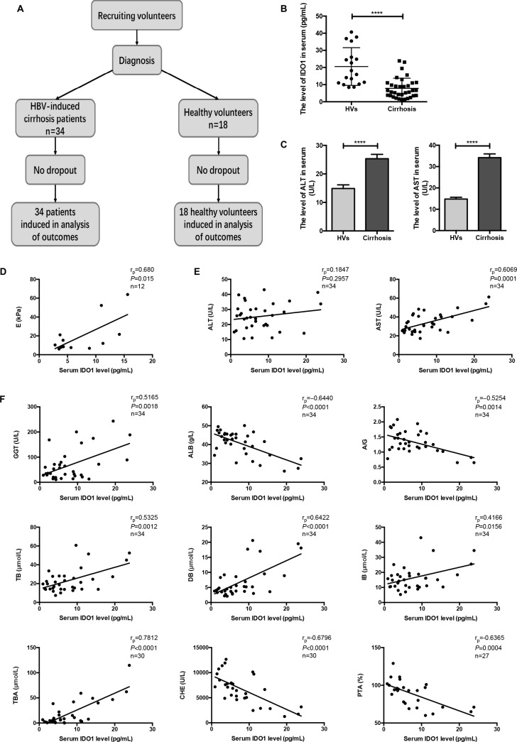
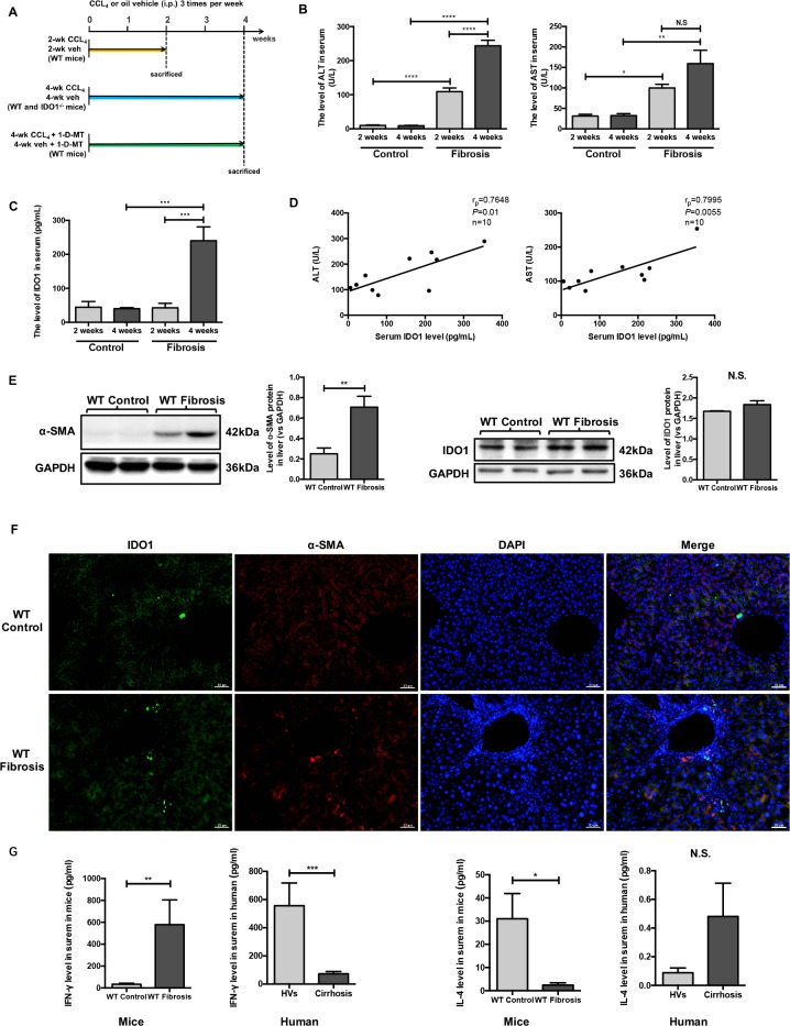
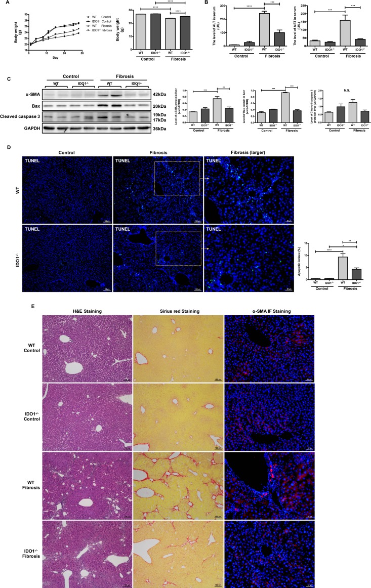
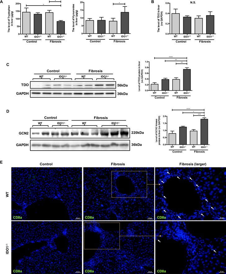
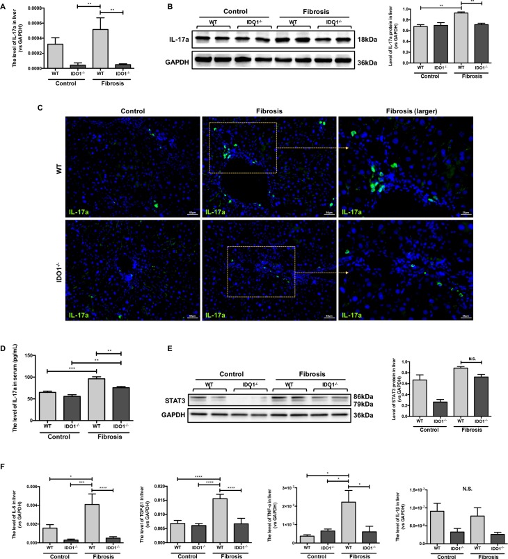
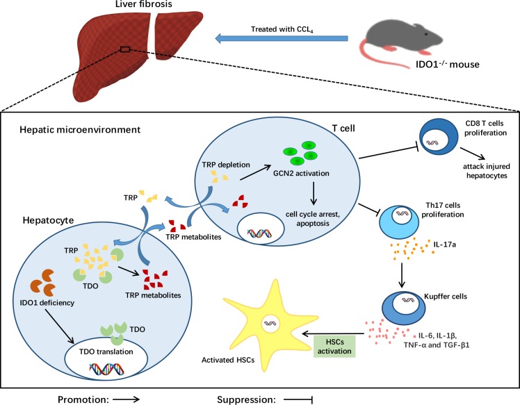
Indoleamine 2,3-dioxygenase 1 (IDO1) is an intracellular rate-limiting enzyme in the metabolism of tryptophan along the kynurenine pathway, subsequently mediating the immune response; however, the role of IDO1 in liver fibrosis and cirrhosis is still unclear. In this study, we investigated the role of IDO1 in the development of hepatic fibrosis and cirrhosis. Patients with hepatitis B virus-induced cirrhosis and healthy volunteers were enrolled. For animals, carbon tetrachloride (CCl4) was used to establish liver fibrosis in wild-type and IDO1 knockout mice. Additionally, an IDO1 inhibitor (1-methyl-D-tryptophan) was administered to WT fibrosis mice. Liver lesions were positively correlated with serum IDO1 levels in both the clinical subjects and hepatic fibrosis mice. A positive correlation between serum IDO1 levels and liver stiffness values was found in the cirrhosis patients. Notably, IDO1 knockout mice were protected from CCl4-induced liver fibrosis, as reflected by unchanged serum alanine transaminase and aspartate transaminase levels and lower collagen deposition, α-smooth muscle actin expression and apoptotic cell death rates. On the other hand, tryptophan 2,3-dioxygenase (TDO), another systemic tryptophan metabolism enzyme, exhibited a compensatory increase as a result of IDO1 deficiency. Moreover, hepatic interleukin-17a, a characteristic cytokine of T helper 17 (Th17) cells, and downstream cytokines' mRNA levels showed lower expression in the IDO1-/- model mice. IDO1 appears to be a potential hallmark of liver lesions, and its deficiency protects mice from CCl4-induced fibrosis mediated by Th17 cells down-regulation and TDO compensation.
Keywords: 1-methyl-D-tryptophan; T helper 17 cells; indoleamine 2,3-dioxygenase 1; liver fibrosis; tryptophan 2,3-dioxygenase.
Conflict of interest statement
The authors do not have any disclosures to report.
Figures
Similar articles
-
Kynurenine produced by indoleamine 2,3-dioxygenase 2 exacerbates acute liver injury by carbon tetrachloride in mice.Toxicology. 2020 May 30;438:152458. doi: 10.1016/j.tox.2020.152458. Epub 2020 Apr 11. Toxicology. 2020. PMID: 32289347
-
The Deficiency of Indoleamine 2,3-Dioxygenase Aggravates the CCl4-Induced Liver Fibrosis in Mice.PLoS One. 2016 Sep 6;11(9):e0162183. doi: 10.1371/journal.pone.0162183. eCollection 2016. PLoS One. 2016. PMID: 27598994 Free PMC article.
-
Tryptophan 2,3-Dioxygenase Expression Identified in Murine Decidual Stromal Cells Is Not Essential for Feto-Maternal Tolerance.Front Immunol. 2020 Dec 8;11:601759. doi: 10.3389/fimmu.2020.601759. eCollection 2020. Front Immunol. 2020. PMID: 33363543 Free PMC article.
-
Tryptophan: A Rheostat of Cancer Immune Escape Mediated by Immunosuppressive Enzymes IDO1 and TDO.Front Immunol. 2021 Feb 23;12:636081. doi: 10.3389/fimmu.2021.636081. eCollection 2021. Front Immunol. 2021. PMID: 33708223 Free PMC article. Review.
-
Targeting Indoleamine Dioxygenase and Tryptophan Dioxygenase in Cancer Immunotherapy: Clinical Progress and Challenges.Drug Des Devel Ther. 2022 Aug 8;16:2639-2657. doi: 10.2147/DDDT.S373780. eCollection 2022. Drug Des Devel Ther. 2022. PMID: 35965963 Free PMC article. Review.
Cited by
-
The Tryptophan and Kynurenine Pathway Involved in the Development of Immune-Related Diseases.Int J Mol Sci. 2023 Mar 17;24(6):5742. doi: 10.3390/ijms24065742. Int J Mol Sci. 2023. PMID: 36982811 Free PMC article. Review.
-
A narrative review of the roles of indoleamine 2,3-dioxygenase and tryptophan-2,3-dioxygenase in liver diseases.Ann Transl Med. 2021 Jan;9(2):174. doi: 10.21037/atm-20-3594. Ann Transl Med. 2021. PMID: 33569476 Free PMC article. Review.
-
The Complex World of Kynurenic Acid: Reflections on Biological Issues and Therapeutic Strategy.Int J Mol Sci. 2024 Aug 20;25(16):9040. doi: 10.3390/ijms25169040. Int J Mol Sci. 2024. PMID: 39201726 Free PMC article. Review.
-
Metabolic Signature of Hepatic Fibrosis: From Individual Pathways to Systems Biology.Cells. 2019 Nov 12;8(11):1423. doi: 10.3390/cells8111423. Cells. 2019. PMID: 31726658 Free PMC article. Review.
-
Inhibition of Rho-Kinase Downregulates Th17 Cells and Ameliorates Hepatic Fibrosis by Schistosoma japonicum Infection.Cells. 2019 Oct 16;8(10):1262. doi: 10.3390/cells8101262. Cells. 2019. PMID: 31623153 Free PMC article.
References
-
- Sun J, Xie Q, Tan D, Ning Q, Niu J, Bai X, Fan R, Chen S, Cheng J, Yu Y, Wang H, Xu M, Shi G, et al. The 104-week efficacy and safety of telbivudine-based optimization strategy in chronic hepatitis B patients: a randomized, controlled study. Hepatology. 2014;59:1283–1292. - PubMed
-
- Tsochatzis EA, Bosch J, Burroughs AK. Liver cirrhosis. Lancet. 2014;383:1749–1761. - PubMed
-
- Mallat A, Lotersztajn S. Cellular mechanisms of tissue fibrosis. 5. Novel insights into liver fibrosis. Am J Physiol. 2013;305:C789–799. - PubMed
MeSH terms
Substances
LinkOut - more resources
Full Text Sources
Other Literature Sources
Medical
Molecular Biology Databases
Research Materials
