. 2017 May 2;7(1):1315.
doi: 10.1038/s41598-017-01501-0.

The role of hydrophobicity in tuberculosis evolution and pathogenicity

Affiliations

The role of hydrophobicity in tuberculosis evolution and pathogenicity

Monika Jankute et al. Sci Rep. .

Abstract

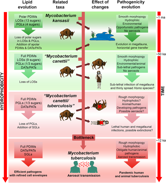
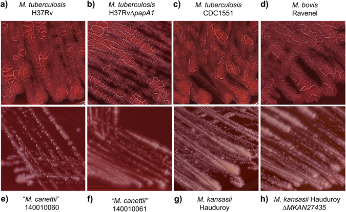
The evolution of tubercle bacilli parallels a route from environmental Mycobacterium kansasii, through intermediate "Mycobacterium canettii", to the modern Mycobacterium tuberculosis complex. Cell envelope outer membrane lipids change systematically from hydrophilic lipooligosaccharides and phenolic glycolipids to hydrophobic phthiocerol dimycocerosates, di- and pentaacyl trehaloses and sulfoglycolipids. Such lipid changes point to a hydrophobic phenotype for M. tuberculosis sensu stricto. Using Congo Red staining and hexadecane-aqueous buffer partitioning, the hydrophobicity of rough morphology M. tuberculosis and Mycobacterium bovis strains was greater than smooth "M. canettii" and M. kansasii. Killed mycobacteria maintained differential hydrophobicity but defatted cells were similar, indicating that outer membrane lipids govern overall hydrophobicity. A rough M. tuberculosis H37Rv ΔpapA1 sulfoglycolipid-deficient mutant had significantly diminished Congo Red uptake though hexadecane-aqueous buffer partitioning was similar to H37Rv. An M. kansasii, ΔMKAN27435 partially lipooligosaccharide-deficient mutant absorbed marginally more Congo Red dye than the parent strain but was comparable in partition experiments. In evolving from ancestral mycobacteria, related to "M. canettii" and M. kansasii, modern M. tuberculosis probably became more hydrophobic by increasing the proportion of less polar lipids in the outer membrane. Importantly, such a change would enhance the capability for aerosol transmission, affecting virulence and pathogenicity.


Conflict of interest statement

The authors declare that they have no competing interests.

Figures

Figure 1
Figure 1
The importance of lipid composition and hydrophobicity in the evolution of tubercle bacilli. Abbreviations: PDIMs, phthiocerol dimycocerosates; LOSs, lipooligosaccharides, PGLs, glycosyl phenolphthiocerol dimycocerosates (phenolic glycolipids); DATs & PATs, di- & pentaacyl trehaloses; SGLs, sulfoglycolipids. Graded green and red backgrounds indicate environmental and mammalian pathogen associations, respectively. Evolution cannot take place directly from modern M. kansasii and “M. canettii”, which are shown as related taxa. The hypothetical transitional taxon, labelled “Mycobacterium canettii/tuberculosis”, possibly corresponds to the well-characterised So93R rough variant of “M. canettii”. Increasing hydrophobicity, with time, applies to both lipid composition and whole cells.
Figure 2
Figure 2
Staining of test strains with Congo Red dye on solid media. (a) M. tuberculosis H37Rv; (b) M. tuberculosis H37Rv ΔpapA1; (c) M. tuberculosis CDC1551; (d) M. bovis Ravenel; (e)“M. canettii” 140010060; (f) “M. canettii” 140010061; (g) M. kansasii Hauduroy; (h) M. kansasii Hauduroy ΔMKAN27435.
Figure 3
Figure 3
Quantitative assessment of relative mycobacterial hydrophobicity. (a) Quantification of Congo Red extracted from solid media growth. Hexadecane/aqueous buffer partitioning of: (b) Live cells, (c) Killed cells, (d) Defatted killed cells. Results are expressed as mean ± standard deviation of at least three independent experiments. Statistical significance was determined by Student’s t-test (p < 0.01).

References

    1. Veyrier FJ, Dufort A, Behr MA. The rise and fall of the Mycobacterium tuberculosis genome. Trends Microbiol. 2011;19:156–161. doi: 10.1016/j.tim.2010.12.008. - DOI - PubMed
    1. Minnikin DE, et al. Ancient mycobacterial lipids: Key reference biomarkers in charting the evolution of tuberculosis. Tuberculosis. 2015;95(Suppl 1):S133–139. doi: 10.1016/j.tube.2015.02.009. - DOI - PubMed
    1. Minnikin, D. E. et al. Pathophysiological implications of cell envelope structure in Mycobacterium tuberculosis and related taxa. In Tuberculosis - Expanding Knowledge (ed. Ribón, W.) 145–175 (InTech Open Access Publisher, Rijeka, 2015).
    1. Wang J, et al. Insights on the emergence of Mycobacterium tuberculosis from the analysis of Mycobacterium kansasii. Genome Biol. Evol. 2015;7:856–870. doi: 10.1093/gbe/evv035. - DOI - PMC - PubMed
    1. Fabre M, et al. Molecular characteristics of ‘Mycobacterium canettii’ the smooth Mycobacterium tuberculosis bacilli. Infect. Genet. Evol. 2010;10:1165–1173. doi: 10.1016/j.meegid.2010.07.016. - DOI - PubMed

Publication types

MeSH terms

Substances