. 2017 May 2;7(1):1424.
doi: 10.1038/s41598-017-01542-5.

The Rational Design of Therapeutic Peptides for Aminopeptidase N using a Substrate-Based Approach

Affiliations

The Rational Design of Therapeutic Peptides for Aminopeptidase N using a Substrate-Based Approach

Shilvi Joshi et al. Sci Rep. .

Abstract

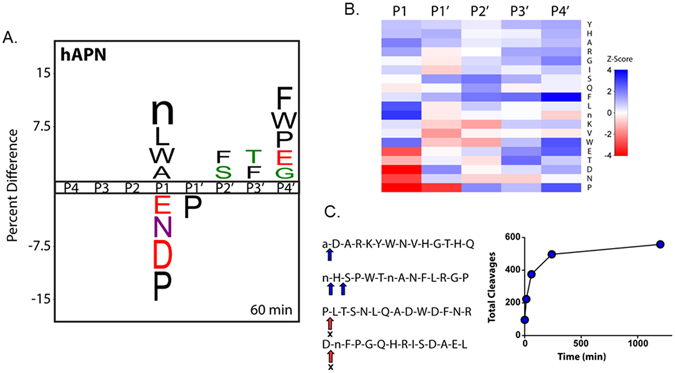
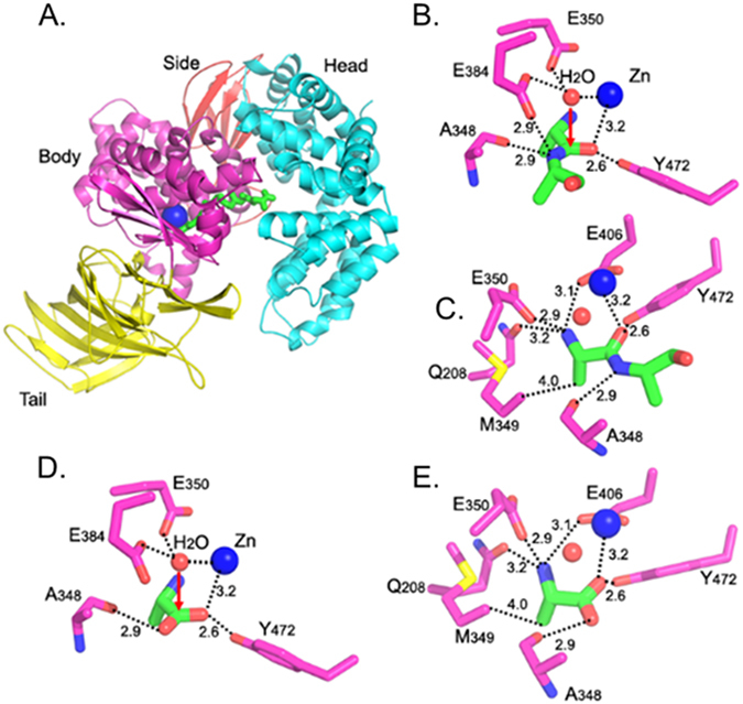
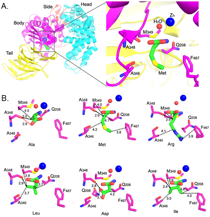
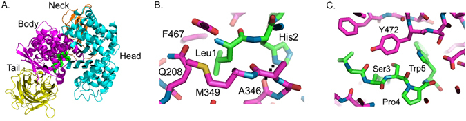
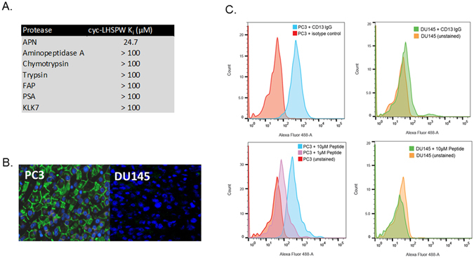
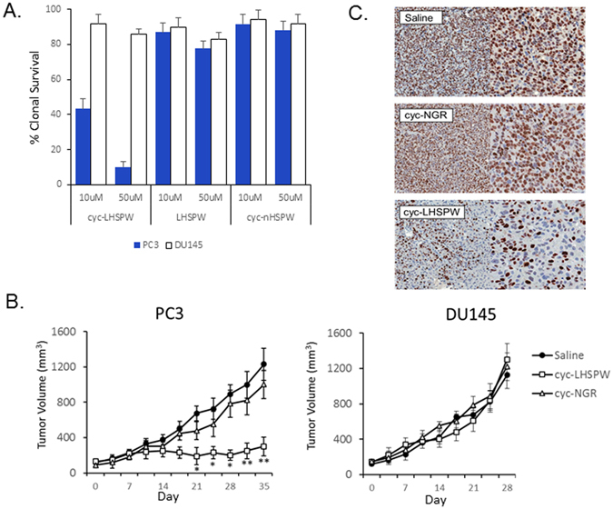
The M1 family of metalloproteases represents a large number of exopeptidases that cleave single amino acid residues from the N-terminus of peptide substrates. One member of this family that has been well studied is aminopeptidase N (APN), a multifunctional protease known to cleave biologically active peptides and aide in coronavirus entry. The proteolytic activity of APN promotes cancer angiogenesis and metastasis making it an important target for cancer therapy. To understand the substrate specificity of APN for the development of targeted inhibitors, we used a global substrate profiling method to determine the P1-P4' amino acid preferences. The key structural features of the APN pharmacophore required for substrate recognition were elucidated by x-ray crystallography. By combining these substrate profiling and structural data, we were able to design a selective peptide inhibitor of APN that was an effective therapeutic both in vitro and in vivo against APN-expressing prostate cancer models.


Conflict of interest statement

The authors declare that they have no competing interests.

Figures

Figure 1
Figure 1
Global identification of human aminopeptidase N (hAPN) substrate specificity with the MSP-MS assay. (A) IceLogo representation of P1–P4′ specificity at the 60 min assay time point (P ≤ 0.05 for all residues shown; “n” is norleucine). Residues with a positive percent difference are considered favorable at a given position; residues with a negative percent difference are considered disfavorable. (B) Heat map representation of hAPN P1–P4′ specificity at the 60 min assay time point calculated using Z-scores at each position. Favored residues are colored blue (Z-score > 0) and disfavored residues are colored red (Z-score < 0). iceLogo representations and heat maps for the 15, 240, and 1200 min assay time points are provided (Supplementary Figure 1). (C) Example 14-mer peptides from the MSP-MS library are shown with primary and secondary cleavages indicated with a blue arrow. “X” indicates that no cleavage was detected at the indicated position. A progress curve is provided depicting the total cleavages observed at each assay time point.
Figure 2
Figure 2
Catalytic mechanism of pAPN. (A) Overall structure of pAPN complexed with a peptide substrate (PDB 4FKF). pAPN contains four domains: head (in cyan), side (in brown), body (in magenta), and tail (in yellow). Zinc is shown as a blue ball, and the peptide substrate is in green. (B) Interactions between catalytic residues of pAPN (in magenta) and the scissile peptide bond of the peptide substrate (in green). Catalytic water is shown as a red ball. (C) Another view of the structure in panel (B) to show all of the interactions between pAPN and the N-terminal residue of the peptide substrate. (D) Interactions between catalytic residues of pAPN (in magenta) and product of APN catalysis - free alanine (in green) (PDB 4FKH). (E) Another view of the structure in panel (D) to show all the interactions between pAPN and free alanine.
Figure 3
Figure 3
Crystal structures of pAPN complexed with free amino acids. (A) The amino acid-binding pocket in pAPN. Left: the overall structure of pAPN complexed with methionine (in green). Right: an enlarged view of the amino acid-binding pocket in the pAPN body domain. The orientation of the view on the right is derived by rotating the view on the left 90° clockwise along a vertical axis. (B) Crystal structures of pAPN complexed with amino acids that are favored (Ala, Met, Arg and Leu) and disfavored (Ile and Asp) P1 residues for APN. pAPN residues are in magenta, and amino acids are in green. The catalytically critical hydrogen bond between the carbonyl oxygen of Ala348 and the C-terminal carboxyl group of amino acids is shown as a red dashed line in the structure with Ala, but is omitted in other panels for clarity. The interactions between the carbonyl oxygen of Ala348 and amino acid side chains are shown as black dashed lines. The distance between the carbonyl oxygen of Ala348 and the nearest atom on amino acid side chains is shown as a bidirectional arrow.
Figure 4
Figure 4
Molecular modeling of the peptide LHSPW in the active site of pAPN. (A) Structure of pAPN complexed with the modeled LHSPW peptide (green) depicting the pAPN ectodomain and its four components: head (cyan), neck (brown), body (in magenta), and tail (in yellow). For the modeling of peptide LHSPW in the active site of APN, two crystal structures were used: (B) the first three residues (Leu1-His2-Ser3) were modeled from the structure of pAPN complexed with polyalanines (PDB ID: 4NAQ), and (C) last two residues (Pro4-Trp5) were from the structure of pAPN complexed with substance P, which also contains a proline at the 4th position (PDB ID: 4HOM).
Figure 5
Figure 5
Determining the specificity of cyc-LHSPW for APN. (A) The cyc-LHSPW peptide was specific inhibitor of APN when compared to a panel of proteases. (B) Fluorescence microscopy of frozen PC3 (left) and DU145 (right) xenograft sections with cyc-LHSPW-(Gly)4-FITC. Sections were incubated with 250 nM of cyc-LHSPW-(Gly)4-FITC overnight and then visualized. The merged fluoresence channels are cyc-LHSPW-(Gly)4-FITC (green) and nuclei (DAPI, blue). (C) Analysis of APN inhibitor binding to PC3 (left) and DU145 cells (right) by flow cytometry. The peptide (cyc-LHSPW-(Gly)4-FITC) selectively labeled the APN-expressing PC3 cells over the APN-null DU145 cells. The cells surface expression of APN was by confirmed by staining both cells with a FITC conjugated anti-CD13 (APN) antibody from Miltentyi.
Figure 6
Figure 6
The therapeutic evaluation of cyc-LHSPW in vivo and in vitro. (A) The effect of the three peptides cyc-LHSPW, LHSPW and cyc-nHSPW on the clonogenic survival of APN-expressing PC3 cells and APN-null DU145 cells. (B) Tumor growth of PC3 and DU145 xenograft mice treated with 40 mg/kg of cyc-LHSPW, cyc-NGR or saline control three times a week for four weeks. Mice were first injected with drug via tail vein starting at day 0 of the study and continued until tumor volumes >1000 mm3 were observed as dictated by our animal protocol. Each treatment group consisted of n = 9 mice/xenograft. Statistical significance of cyc-LHSPW compared with control is denoted by: *P < 0.05; and **P < 0.01, as determined by the Student t test. (C) At the end of the study, PC3 tumors were removed from the treated arms and stained for the proliferation maker Ki67. Decreased Ki67 staining is evident in the cyc-LHSPW treated arm.

Similar articles

Cited by

References

    1. Mina-Osorio P. The moonlighting enzyme CD13: old and new functions to target. Trends Mol Med. 2008;14:361–371. doi: 10.1016/j.molmed.2008.06.003. - DOI - PMC - PubMed
    1. Luan Y, Ma C, Wang Y, Fang H, Xu W. The characteristics, functions and inhibitors of three aminopeptidases belonging to the m1 family. Curr Protein Pept Sci. 2012;13:490–500. doi: 10.2174/138920312802430554. - DOI - PubMed
    1. Tsujimoto M, Goto Y, Maruyama M, Hattori A. Biochemical and enzymatic properties of the M1 family of aminopeptidases involved in the regulation of blood pressure. Heart Fail Rev. 2008;13:285–291. doi: 10.1007/s10741-007-9064-8. - DOI - PubMed
    1. Liu C, Yang Y, Chen L, Lin YL, Li F. A unified mechanism for aminopeptidase N-based tumor cell motility and tumor-homing therapy. J Biol Chem. 2014;289:34520–34529. doi: 10.1074/jbc.M114.566802. - DOI - PMC - PubMed
    1. Drag M, Bogyo M, Ellman JA, Salvesen GS. Aminopeptidase fingerprints, an integrated approach for identification of good substrates and optimal inhibitors. J Biol Chem. 2010;285:3310–3318. doi: 10.1074/jbc.M109.060418. - DOI - PMC - PubMed

Publication types

LinkOut - more resources