Review
. 2017 Apr 28:23:26.
doi: 10.1186/s40409-017-0117-8. eCollection 2017.

Strategies in 'snake venomics' aiming at an integrative view of compositional, functional, and immunological characteristics of venoms

Affiliations
Review

Strategies in 'snake venomics' aiming at an integrative view of compositional, functional, and immunological characteristics of venoms

Bruno Lomonte et al. J Venom Anim Toxins Incl Trop Dis. .

Abstract

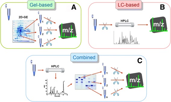
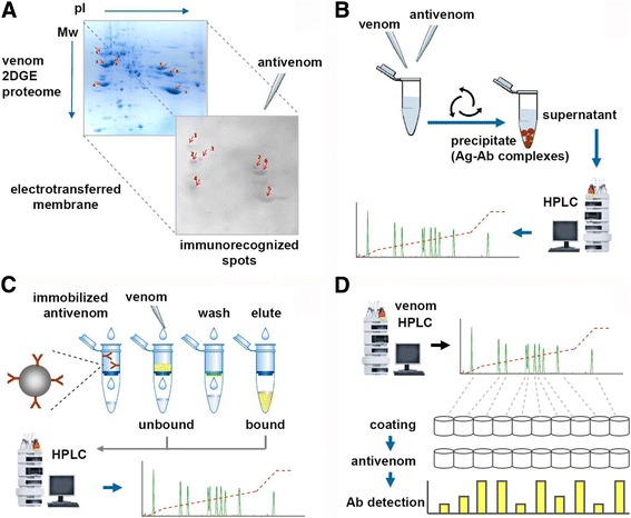
This work offers a general overview on the evolving strategies for the proteomic analysis of snake venoms, and discusses how these may be combined through diverse experimental approaches with the goal of achieving a more comprehensive knowledge on the compositional, toxic, and immunological characteristics of venoms. Some recent developments in this field are summarized, highlighting how strategies have evolved from the mere cataloguing of venom components (proteomics/venomics), to a broader exploration of their immunological (antivenomics) and functional (toxicovenomics) characteristics. Altogether, the combination of these complementary strategies is helping to build a wider, more integrative view of the life-threatening protein cocktails produced by venomous snakes, responsible for thousands of deaths every year.

Keywords: Antivenomics; Proteomics; Snake venoms; Toxicovenomics; Venomics.


Figures

Fig. 1
Fig. 1
General types of analytical bottom-up strategies employed in the proteomic profiling of snake venoms. a Gel-based strategies involve the separation of the venom proteins by two-dimensional gel electrophoresis (2DE) followed by staining and spot picking. Protein spots are then in-gel digested (usually with trypsin, scissors icon) and the resulting proteolytic peptides submitted to tandem mass spectrometry (MS/MS) analysis. b Liquid-chromatography (LC)-based strategies (shotgun proteomics) digest the whole venom with trypsin and separate the resulting peptides usually by multidimensional nano-flow HPLC, hyphenated to MS/MS analysis. c The combined strategy of ‘snake venomics’ takes advantage of the opportunity of performing the fractionation and the quantification of the venom components in the same reversed-phase chromatography step. A second step of separation and quantification is performed by SDS-PAGE followed by gel densitometry. Protein bands are excised, in-gel digested with trypsin, and submitted to MS/MS analysis
Fig. 2
Fig. 2
Scheme for RP-HPLC fractionation of snake venoms. A considerable number of snake venomic studies have used the chromatographic conditions indicated in the diagram. The venom proteins are separated using an analytical (4.6 × 250 mm, particle diameter of 5 μm) reverse-phase C18 column, eluted at a flow rate of 1 mL/min by a linear gradient of water containing 0.1% of trifluoroacetic acid (TFA) (solution A) and 70% acetonitrile (CNCH3) containing 0.1% TFAa, and the eluate monitored at 215 nm. The timetable for the mixing of these solutions (A, B), and the shape of the gradient (dashed line) are indicated. As an example, the approximate elution regions for some of the common protein components of snake venoms are indicated by colored boxes. This procedure has been applied to venoms of a number of viperid and elapid snakes, helping in the standardization and comparability of results between different laboratories. 3FTx: three-finger toxin; Kunitz: Kunitz-type serine protease inhibitor; PLA2: phospholipase A2; CTL: C-type lectin; SP: serine protease; CRiSP: cystein-rich secretory protein; NGF: nerve-growth factor; VEGF: vascular endothelium growth factor; MP: metalloproteinase; LAAO: L-amino acid oxidase; PDE: phosphodiesterase; 5′-NU: 5′-nucleotidase; HYA: hyaluronidase; PLB: phospholipase B
Fig. 3
Fig. 3
Antivenomic analytical strategies. A schematic representation of immunological approaches that have been combined with proteomic analysis of snake venoms, aiming to assess the immunorecognition of venom components by antibodies present in a given antivenom. a Immunoblotting, performed on electrotransferred membranes from two-dimensional gel electrophoresis (2DE) venom separations, identifies spots that are immunorecognized by the antivenom, in an essentially qualitative way. Immunoblotting can also be performed on membranes from the electrophoresis step (second dimension separation by SDS-PAGE) of the snake venomics strategy (see text and Fig. 1c). b ‘First generation’ antivenomics evaluates the immunodepletion of venom components after addition of antivenom and removal of precipitated immunocomplexes. The remaining supernatant is analyzed by HPLC and its profile is compared to that of a control venom aliquot. Differences in the chromatographic peaks between the antivenom-treated venom and the control venom can be quantified by integration of their peak areas, representing the immunodepletion of recognized components. c ‘Second generation’ antivenomics evaluates the venom components that are captured by an antivenom that has been covalently linked to beads, following the principles of immunoaffinity chromatography. Whole venom is incubated with this matrix and the unbound components are collected. After washing out the non-binding venom components, a change in pH elutes the bound venom fraction. Both samples are finally analyzed by HPLC, and their profiles are compared to that of a control sample of venom. Quantitative estimations of the degree of immunorecognition of components are performed as described for panel b by integration of chromatographic peak areas [58]. d HPLC/ELISA-based assessment of immunorecognition of venom components by an antivenom, or HPLC/ELISA-based immunoprofiling, is performed by coating microwell plates with a normalized amount of venom fractions obtained from the HPLC profile of the venom. Then, antivenom is added to each well and the bound antibodies (Ab) are detected by conventional ELISA
Fig. 4
Fig. 4
Evolution of analytical strategies in the characterization of snake venoms by proteomic tools, used in combination with appended methodologies. Initial proteomic studies on venoms essentially focused on the qualitative cataloguing of components. The introduction of the snake venomics strategy led to a valuable increase in the informative value of these analyses, by providing an estimation of the abundances of venom components. In combination with antivenomics, the immunogenicity of venom components can be inferred by evaluating their recognition by antibodies present in a given antivenom. A third dimension in the characterization of venoms is provided by a combination with toxicovenomics, which evaluates the toxic activities of components. Altogether, these combined strategies increase the informative value of studies characterizing venoms by disclosing their composition (venomics), immunorecognition (antivenomics) and toxicity (toxicovenomics)

References

    1. Garfield E. Research and immunotherapy are taking the bite out of venom. Curr Contents. 1988;5(11):29–38.
    1. Mackessy SP, (editor). Handbook of Venoms and Toxins of Reptiles. CRC Press, Taylor & Francis Group, Boca Raton, FL, USA; 2009. ISBN 978-0849391651.
    1. Fry B, editor. Venomous Reptiles and Their Toxins: Evolution, Pathophysiology and Biodiscovery. Oxford: Oxford University Press; 2015.
    1. Williams DJ, Gutiérrez JM, Calvete JJ, Wüster W, Ratanabanangkoon K, Paiva O, et al. Ending the drought: new strategies for improving the flow of affordable, effective antivenoms in Asia and Africa. J Proteomics. 2011;74(9):1735–1767. doi: 10.1016/j.jprot.2011.05.027. - DOI - PubMed
    1. Fry BG, Sunagar K, Casewell NR, Kochva E, Roelants K, Jackson TNW, et al. The origin and evolution of the Toxicofera reptile venom system. In: Brian F, editor. Venomous Reptiles and their Toxins. Oxford: Oxford University Press; 2015. p. 1-31.