. 2017:2017:8325049.
doi: 10.1155/2017/8325049. Epub 2017 Mar 29.

Extract from Periostracum cicadae Inhibits Oxidative Stress and Inflammation Induced by Ultraviolet B Irradiation on HaCaT Keratinocytes

Affiliations

Extract from Periostracum cicadae Inhibits Oxidative Stress and Inflammation Induced by Ultraviolet B Irradiation on HaCaT Keratinocytes

Tsong-Min Chang et al. Evid Based Complement Alternat Med. 2017.

Abstract

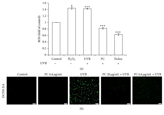
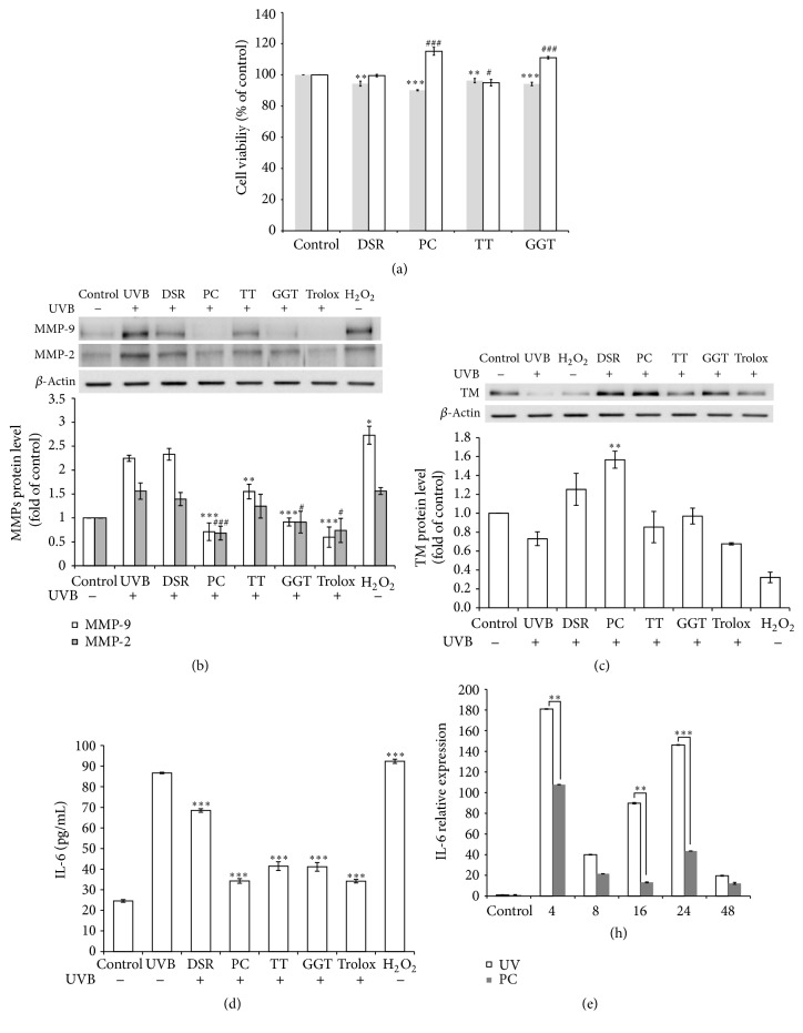
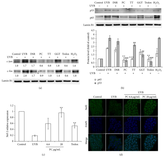
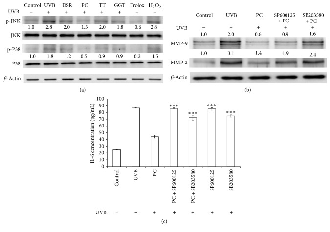
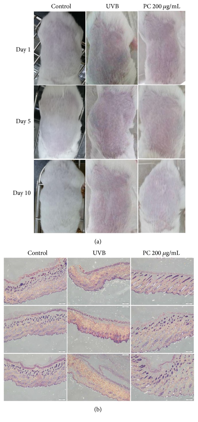
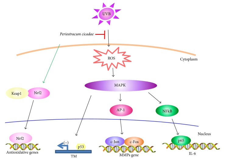
Periostracum cicadae is widely used for the treatment of skin diseases such as eczema, pruritus, and itching. The current study sought to evaluate the effect of P. cicadae extract on ultraviolet B (UVB) irradiation and identify the mechanisms involved. Photodamage-protective activity of P. cicadae extracts against oxidative challenge was screened using HaCaT keratinocytes. P. cicadae extracts did not affect cell viability but decreased reactive oxygen species (ROS) production. The extract attenuates the expression of interleukin-6 (IL-6), matrix metalloproteinase-2 (MMP-2), and MMP-9 in UVB-treated HaCaT cells. Also, P. cicadae abrogated UVB-induced activation of NF-κB, p53, and activator protein-1 (AP-1). The downmodulation of IL-6 by P. cicadae was inhibited by the p38 inhibitor (SB203580) or JNK inhibitor (SP600125). Moreover, the extract attenuated the expression of NF-κB and induced thrombomodulin in keratinocytes and thereby effectively downregulated inflammatory responses in the skin. The nuclear accumulation and expression of NF-E2-related factor (Nrf2) were increased by P. cicadae treatment. Furthermore, treatment with P. cicadae remarkably ameliorated the skin's structural damage induced by irradiation. This study demonstrates that P. cicadae may protect skin cells against oxidative insult by modulating ROS concentration, IL-6, MMPs generation, antioxidant enzymes activity, and cell signaling pathways.


Figures

Figure 1
Figure 1
P. cicadae reduced the ROS production of HaCaT cells after UVB exposure. HaCaT cells were treated with 20 μg/mL of P. cicadae for 4 hours, followed by exposure to 12 mJ/cm2 of UVB. The levels of ROS were measured by the DCFH-DA assay. (a) Fluorescence intensity of untreated control cells was set as 1 and results were expressed in X-fold. Trolox was used as an antioxidant control. H2O2 were used to generate hydroxyl radicals. P. cicadae effectively prevented UVB-produced ROS in HaCaT cells. (b) Representative immunofluorescent microscopy images, 40x magnification; scale bar = 500 μm. p < 0.05 was considered as a significant difference compared with the only UVB-irradiated group. ∗∗∗p < 0.001 was considered as a highly significant difference compared with the UVB-irradiated only group.
Figure 2
Figure 2
Effects of P. cicadae and herb extracts on MMP-9 and MMP-2 production in UVB-stimulated HaCaT cells. (a) Cell viability. HaCaT cells were exposed to extracts (gray bar) or added after UVB treated (white bar). For gray bar, stands for p < 0.05 versus control. For white bar, # stands for p < 0.05 versus control, ## stands for p < 0.01 versus control, and ### stands for p < 0.001 versus control. After 2 days of exposure, cell viability of the treated cells was examined by means of MTS assay (n = 5, mean ± SD). (b) The MMP-9 and MMP-2 levels were determined by Western blotting and β-actin was used as the loading control. The relative expression levels of MMP-9 (white bar) and MMP-2 (gray bar) were compared to control cells and presented as the mean ± SD for three independent experiments performed in triplicate. (c) The relative expression levels of TM were compared to control cells and presented as the mean ± SD for three independent experiments performed in triplicate. (d) IL-6 was quantified by ELISA in cell culture supernatants 24 hours following UVB and herb extracts (10 μg/mL). Results are expressed as mean ± SEM of five independent experiments. (e) Relative IL-6 mRNA levels were measured by Q-PCR to 52 hours following UVB (20 μg/mL). Histograms represent mean ± SEM of relative mRNA levels after normalization with 18S (n = 4-5 independent experiments). Trolox was used as an antioxidant control. H2O2 were used to generate hydroxyl radicals. DSR, Divaricate saposhnikovia root; TT, Tribulus ter; PC, P. cicadae; GGT, Ge Gen Tang. p < 0.05 or #p < 0.05, ∗∗p < 0.01 or ##p < 0.01, and  ∗∗∗p < 0.001 or ###p < 0.001 were considered as a significant difference compared to control group (p < 0.05 versus control).
Figure 3
Figure 3
Effects of P. cicadae and herb extracts on UVB-stimulated transcription factors activated in HaCaT cells. (a) The p53 and p65 levels in cell nuclei were determined by Western blotting and lamin B1 was used as a loading control. The protein expression levels of p53 (gray bar) or p65 (white bar) were presented. For gray bar (p53), stands for p < 0.05 versus control. For white bar (p65), # stands for p < 0.05 versus control, ## stands for p < 0.01 versus control, and ### stands for p < 0.001 versus control. The relative expression levels of p53 (gray bar) and p65 (white bar) were compared to control cells and presented as the mean ± SD for three independent experiments performed in triplicate. (b) Relative Nrf-2 mRNA levels were measured by Q-PCRs following UVB and PC (20 μg/mL). Histograms represent mean ± SEM of relative mRNA levels after normalization with 18S (n = 4-5 independent experiments). (c) Nrf-2 was revealed by green fluorescence and DAPI was revealed by blue fluorescence. Confocal microscope analysis was used to demonstrate the Nrf-2 accumulation in the nuclear fraction in HaCaT cells. (d) The c-jun and c-fos levels in cell nuclei were determined by Western blotting and lamin B1 was used as a loading control. The relative expression levels were compared to control cells and presented as the mean ± SD for three independent experiments performed in triplicate. Trolox was used as an antioxidant control. H2O2 were used to generate hydroxyl radicals. DSR, Divaricate saposhnikovia root; TT, Tribulus ter; PC, P. cicadae; GGT, Ge Gen Tang. p < 0.05 or #p < 0.05,  ∗∗p < 0.01 or ##p < 0.01, and ∗∗∗p < 0.001 or ###p < 0.001 were considered as a significant difference compared to control group (p < 0.05 versus control).
Figure 4
Figure 4
P. cicadae inhibits UVB-induced MMPs and IL-6 via suppression of JNK MAPK in HaCaT cells. (a) The JNK and p38 levels in cell nucleus were determined by Western blotting and β-actin was used as loading controls. The relative expression levels of JNK and p38 were compared to control cells and presented as the mean ± SD for three independent experiments performed in triplicate. (b) The expression of MMPS in the presence of SP600125 and SB203580, respectively. MMPs levels in cell were determined by Western blotting and β-actin was used as a loading control. The relative expression levels of MMP-9 and MMP-2 were compared to control cells and presented as the mean ± SD for three independent experiments performed in triplicate. (c) Effects of P. cicadae on the IL-6 in the presence of SP600125 and SB203580, respectively. The UVB-incubated HaCaT cells pretreated with signal inhibitors were incubated with P. cicadae for 24 hours. The production of IL-6 was analyzed by ELISA. The data represent at least three independent experiments. p < 0.05,  ∗∗p < 0.01, and  ∗∗∗p < 0.001 were considered as a significant difference compared to control group (p < 0.05 versus control).
Figure 5
Figure 5
Effect of topical treatment of mouse skin with P. cicadae on UVB-induced damage. (a) ICR mice skin was treated with either 200 μg/mL P. cicadae or PBS and was exposed to UVB every 5 days, as described in Methods. Mouse skin treated topically with P. cicadae before UVB exposure reduced UVB-induced signs of sunburn tissue damage. (b) Skin biopsies were processed by H&E staining by a routine procedure. Micrographs show representative findings of skin in response to UVB, sampled after UVB exposure, and a representative section of H&E staining from three independent experiments is shown. Pretreatment of P. cicadae inhibited UVB-induced extensive structural damage in both epidermis and dermis (magnification, 40x).
Figure 6
Figure 6
Proposed mechanism by which P. cicadae abolished UVB-stimulated inflammation. Cartoon shows that P. cicadae decreases expression of MMPs, TM, and IL-6 in HaCaT cells. The suppressive effect of P. cicadae on photodamage was induced through JNK and p38 activation. P. cicadae suppression of the JNK or p38 pathway could diminish the UVB-induced AP-1, p53, and p65 activation. Moreover, P. cicadae-induced Nrf-2 activation and translocation from the cytosol to the nucleus.

References

    1. Lin J.-F., Liu P.-H., Huang T.-P., et al. Characteristics and prescription patterns of traditional chinese medicine in atopic dermatitis patients: ten-year experiences at a medical center in taiwan. Complementary Therapies in Medicine. 2014;22(1):141–147. doi: 10.1016/j.ctim.2013.12.003. - DOI - PubMed
    1. Lee S.-H., Cekanova M., Baek S. J. Multiple mechanisms are involved in 6-gingerol-induced cell growth arrest and apoptosis in human colorectal cancer cells. Molecular Carcinogenesis. 2008;47(3):197–208. doi: 10.1002/mc.20374. - DOI - PMC - PubMed
    1. Dugasani S., Pichika M. R., Nadarajah V. D., Balijepalli M. K., Tandra S., Korlakunta J. N. Comparative antioxidant and anti-inflammatory effects of [6]-gingerol, [8]-gingerol, [10]-gingerol and [6]-shogaol. Journal of Ethnopharmacology. 2010;127(2):515–520. doi: 10.1016/j.jep.2009.10.004. - DOI - PubMed
    1. Xu M.-Z., Lee W. S., Han J.-M., et al. Antioxidant and anti-inflammatory activities of N-acetyldopamine dimers from Periostracum Cicadae. Bioorganic and Medicinal Chemistry. 2006;14(23):7826–7834. doi: 10.1016/j.bmc.2006.07.063. - DOI - PubMed
    1. M.-T. Hsieh. Studies on the anticonvulsive, sedative and hypothermic effects of Periostracum Cicadae extracts. Journal of Ethnopharmacology. 1991;35(1):83–90. doi: 10.1016/0378-8741(91)90136-2. - DOI - PubMed

LinkOut - more resources