. 2016 Mar;11(1):5-25.

2016 WHO Clinical Molecular and Pathological Criteria for Classification and Staging of Myeloproliferative Neoplasms (MPN) Caused by MPN Driver Mutations in the JAK2, MPL and CALR Genes in the Context of New 2016 WHO Classification: Prognostic and Therapeutic Implications

Affiliations

2016 WHO Clinical Molecular and Pathological Criteria for Classification and Staging of Myeloproliferative Neoplasms (MPN) Caused by MPN Driver Mutations in the JAK2, MPL and CALR Genes in the Context of New 2016 WHO Classification: Prognostic and Therapeutic Implications

Jan Jacques Michiels et al. Maedica (Bucur). 2016 Mar.

Abstract

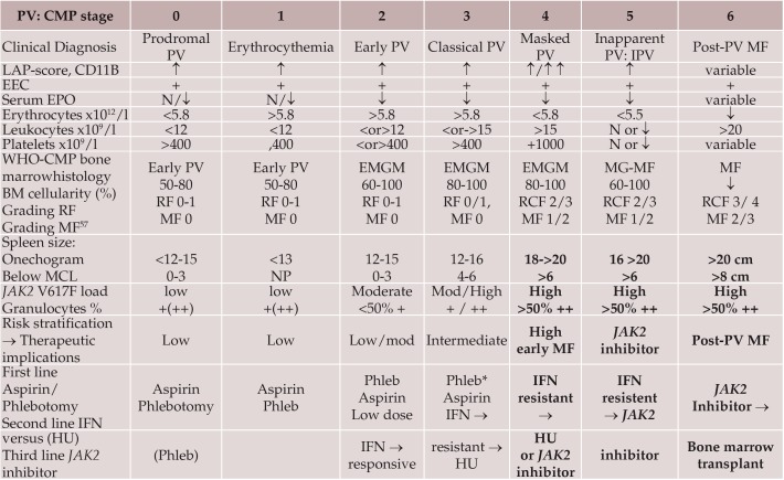
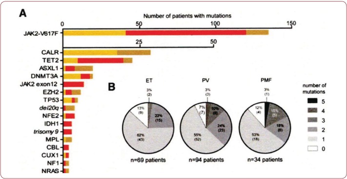
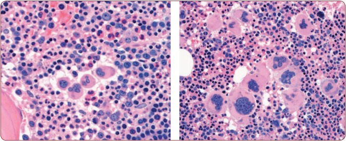
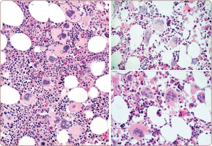
The 2016 WHO-CMP classification proposal defines a broad spectrum of JAK2 V617F mutated MPN phenotypes: normocellular ET, hypercellular ET due to increased erythropoiesis (prodromal PV), hypercellular ET with megakaryocytic-granulocytic myeloproliferation and splenomegaly (EMGM or masked PV), erythrocythemic PV, early and overt classical PV, advanced PV with MF and post-PV MF. ET heterozygous for the JAK2 V617F mutation is associated with low JAK2 mutation load and normal life expectance. PV patients are hetero-homozygous versus homozygous for the JAK2 V617F mutation in their early versus advanced stages with increasing JAK2 mutation load from less than 50% to 100% and increase of MPN disease burden during life long follow-up in terms of symptomatic splenomegaly, constitutional symptoms, bone marrow hypercellularity and secondary MF. Pretreatment bone marrow biopsy in prefibrotic MPNs is of diagnostic and prognostic importance. JAK2 exon 12 mutated MPN is a distinct benign early stage PV. CALR mutated hypercellular thrombocythemia show distinct PMGM bone marrow characteristics of clustered larged immature dysmorphic megakaryocytes with bulky (bulbous) hyperchromatic nuclei, which are not seen in JAK2 mutated ET and PV. MPL mutated normocellular thrombocythemia is featured by clustered giant megakaryocytes with hyperlobulated stag-horn-like nuclei without features of PV in blood and bone marrow. Myeloproliferative disease burden in each of the JAK2, CALR and MPL MPNs is best reflected by the degree of anemia, splenomegaly, mutation allele burden, bone marrow cellularity and myelofibrosis.


Figures

Figure 1.
Figure 1.
Figure 1. The 1950 Dameshek one cause hypothesis of trilinear PV (2) has been confirmed by Vainchenker’s discovery in 2005 (13,14) of heterozygous and homozygous JAK2 V617F mutations as the driver cause of the trilinear erythroctic, megakaryocytic granulocytic myeloproliferation (EMGM) myeloproliferative neoplasms polycythemia vera (PV, essential thrombocythemia (ET) and secondary myelofibrosis (MF). In the editorial on speculations of myeloproliferative syndromes in 1951 (5), Dameshek recognized megakaryocytic leukemia (ML) as a distinct entity without features of PV at diagnosis and follow-up. This ML entity has recently be recognized as MPL or calreticulin (CALR) mutated thrombocythemia without features of PV (8,9).
Figure 2.
Figure 2.
Figure 2. Translation of the 2008 WHO clinical criteria (11,12) into 2016 WHO-CMP criteria (6-9) for the classification of JAK2 V617F mutated ET, PV and EMGM or masked PV (red), versus CALR mutated primary megakaryocytic granulocytic myeloproliferation (PMGM, blue) and MPL mutated normocellular ET (black). All molecular variants of MPN disease burden is reflected by the degree of anemia, myeloid neoplasia of the spleen (splenomegaly), bone marrow cellularity and secondary myelofibrosis.
Table 1.
Table 1.
Table 1. 2016 WHO Clinical Molecular and Pathobiological (2016 WHO-CMP) criteria for diagnosis of JAK2 V617F mutated essential throbocythemia (ET) (6-9).
Table 2.
Table 2.
Table 2. 2016 WHO Clinical Molecular andPathological (2016 WHO-CMP) criteria for the diagnosis of prodromal, masked and classical JAK2 mutated polycythemia vera (PV) versus primary or secondary erythrocytoses (6-9). 2014 WHO-CMP criteria for staging of prodromal, erythrocythemic, and advanced PV A2 + B1 + P1 establish early PV (mimicking ET) prodromal PV CMP stage 0 A1 + A2 + A3 + P1and none of B establish idiopathic erythrocythemia (IE) or stage 1 PV A1 + A2 + A3 + P1 and one or more of B establish classic stages of PV stage 2 and 3 A2 + B3 + P1 detect masked cases of PV with splenomegaly and hypersplenism to be labelled as Inapparent PV (IPV) frequently seen Budd-Chiari syndrome or splanchnic vein thrombosis
Figure 3.
Figure 3.
Figure 3. Erythrocyte counts x1012/L vertical axis versus red cell mass (RCM) horizontal axis according to Michiels et al. (6-9,52,53). At erythrocyte values below 5.8 x10¹²/L the red cell mass (RCM) values are between 25 and 30 ml/kg in essential thrombocythemia (ET). At erythrocyte values above 5.7x10¹²/L all values of RCM area bove 30/kg in all polycythemia vera (PV) cases indicating that the erythrocyte cut-off level of 5.7 x10¹²/L dicriminates between ET and PV The numbers in the blue line are erythrocyte counts x1012/l.
Table 3.
Table 3.
Table 3. The relation between RCM, erythrocyte count and bone marrow histology findings at time of diagnosis in 26 MPN patients: 10 ET and 14 PV and in 2 ET cases at time of evolution into PV as compared to the 2008 WHO cut-of levels of hemoglobin (Hb) and hematocrit (Ht) for PV: Hb >18.5 g/dl and Ht >0.60 in men and Hb >16.5 and Ht >0.56 in women for the diagnosis of PV. At RCM above 30 ml/kg (Red) the erythrocytes are above 5.7 x10¹²/L = PV (Red). Of 10 ET cases 7 had ET and 3 had PV with erythrocytes above 5.7x10¹²/L (Bold) At erythrocytes above 5.7 x10¹²/L Hb ranges from 15.0 to 20.9 and are below WHO criteria in 3 females and 2 males (Blue) At erythrocytes above 5.7 x10¹²/L the Ht ranges from 0.46 to 0.72 and are below WHO criteria in 7 females and 1 male (Blue) For further interpretation of these data see figure 1.
Figure 4.
Figure 4.
Figure 4. Clinical course in a casewith idiopathic erythrocythemia (IE) or erythrocythemic polycythemia vera treated with venesections (arrows). The development of microcytic hypochromic erythrocytes due to iron deficiency was associated with persistent increased red cell count (>6x10¹²/L), which is diagnostic for PV. Phlebotomy on top of low dose aspirin induces iron deficiency with microcytic erythrocytes (MCV around 65 fL), normal values for haemoglobin (Hb) and hematocrit (Ht) and relief of hypervolumic symptoms.
Table 4.
Table 4.
Table 4. Staging of JAK2 V617F positive prodromal PV, erythrocythemic PV, classical PV, early MF, inapparent PV, spent phase PV and post-PV myelofibrosis (MF) according to 2016 WHO-CMP criteria related to therapy (6-9,52,53). *↑ = increased, ↓ = decreased, N = normal, + = present or heterozygous; ++ = homozygous
Figure 5.
Figure 5.
Figure 5. WHO bone marrow features in JAK2 V617F positive ET cases 2, 3, 4, 5 and 6, and PV case 7. Case 2. Clinically JAK2 V617F positive ET 2 (low serum EPO) and a normocellular ET (WHO-ET) bone marrow wit pleomorph small and large megakaryocytes and reticulin fibers: RF grade 1. Case 3 and 4. Clinically JAK2 V617F positive ET 2 with a trilinear hypercellular PV bone marrow and RF 0 in case 2 and increased cellularity due to increased erythropoiesis in case 4, and no increase of reticulin fibers: RF grade 0. Case 5. Clinically JAK2 V617F positive ET with slight to moderate splenomegaly and a hypercellular megakaryocytic granulocytic myeloproliferation (EMGM = ET 3), with dysmorphic megakaryocytes (not cloud-like) and no increase of reticulin fibers: RF grade 0 in the bone marrow. Case 6. Clinically JAK2 V617F positive ET 2 with a trilinear hypercellular PV bone marrow picture and no oncrease of reiculinefibers: RF 0. Case 7. Clinically JAK2 V617F positive PV with a 65% hypercellular ET/PV bone marrow picture in between “normocellular ET” (WHO-ET) and trilinear hypercellular (100%) PV picture.
Table 6.
Table 6.
Table 6. Grading of bone marrow biopsy content of secondary reticulin fibrosis (RF), and WHO grading of myelofibrosis (MF) (6-9,29,50,51).
Figure 6.
Figure 6.
Figure 6. Relationship of splenomegaly and MPN disease duration to the JAK2 mutation status of burst forming uniterythropiesis (BFU-E) clonal genotypes in 6 ET, 14 PV and 6 MF patients (21). Progression of heterozygous JAK2 V617F mutation load from about 25% to 100% is seen in ET, PV and MF during longterm follow-up of 10 to 20 years. The mutation load of combined hetero/homozygous JAK2 V617 muated PV in 10 cases of PV was around 25% to 50% in early stage PV and increases to 80% to 100% in advanced myeloproliferative stages (splenomegaly) of PV and post-PV myelofibrosis (21).
Table 7.
Table 7.
Table 7. 2016 WHO-CMP criteria for hypercellular ET associated with primary megakaryocytic, granulocytic myeloproliferation (PMGM) caused by calreticulin (CALR) mutations (8,9). The combination of A1 + A2 and P1 establishes CALR ET and various clinical stages (C1, C2,C3) related to the degree of myelofibosis (MF).
Figure 7.
Figure 7.
Figure 7. Proportions of JAK2 genotypes in BFU-Es from 59 patients with JAK2-mutated essential thrombocythemia (ET) and polycythemia vera (PV) (30). Each vertical bar represents 1 patient, divided according to the proportion of wild-type, heterozygous and homozygous-mutant colonies obtained, with the absolute colony numbers shown above: (wild type white), heterozygous (purple) homozygous (red). Results EEC colony genotypes are presented for 29 JAK2 V617F -positive ET patients (total 2277 colonies; mean 79 per patient) and for 30 JAK2 V617F -positive PV patients (total 2287 colonies; mean 76 colonies per patient) (30). All 29 JAK2 V617F positive ET patients have heterozygous JAK2 mutated EEC colonies and a low percentage less than 10% homozyous colonies in 9 and 20% in 1 of them. Out of 30 JAK2 V617F positive PV patients 8 have heterozygous JAK2 mutated EEC, 13 have homozygous EEC colonies of more than 50% and 7 of less than 50%.
Figure 8.
Figure 8.
Figure 8. Distribution of somatic mutations in 197 MPN patients from the study of Lundberg et al. (35). None of 94 (%) PV patients, 17 of 69 (25%) ET patients and 11 of 34 (32%) MF patients carried mutations in the caleticulin (CALR) gene. After JAK2 V617F and CALR, the most frequently observed mutations affected genes implicated in epigenetic regulation (TET2, ASXL1, DNMT3A, EZH2, and IDH1. Rare epigenetic mutations include NF1, NFE2, and CUX1. Recurrent somatic mutations were observed in the genes TP53, CBL, MPL, and NRAS. On top of the JAK2 or CALR mutation one, two or more somatic mutations were found in 65 of 197 (33%) patients, which appeared to be of impaired prognostic significance (35).
Table 8.
Table 8.
Table 8. 2016 WHO Clinical Molecular andPathological (2016 WHO-CMP) criteria for the diagnosis of normocelular ET carrying one of the MPL mutations (8,9).
Figure 9.
Figure 9.
Figure 9. Bone marrow histology in PV patients with JAK2 exon 12 mutations meeting the criteria for idiopathic erythrocythemia (IE) and classical PV, showed prominent erythroid hyperplasia and clustered atypical small to medium-sized large megakaryocytes with various degrees of monolobation to hyperlobation and abnormal chromatin distribution (Lakey et al. [40]).
Figure 10.
Figure 10.
Figure 10. Hemorrhagic thrombocythemia in case of JAK2 wild type hypercellular ET associated with prefibrotic primary megakaryocytic and granulocytic myeloproliferation (PMGM, colour picture) in the bone marrow by dual myeloproliferation of granulopoiesis and dense clustered enlarged immature dysmorphic megakaryocytes with bulky (bulbous) hyperchromatic nuclei (arrows), which are never seen in JAK2 wild type MPL mutated ET and also not in the prefibrotic JAK2 V617F mutated ET. During long-term follow-up, reduction of platelet count to normal or near normal by treatment with hydroxyurea in 1994 followed by anagrelide from 1995 to 1998 the bleeding manifestations did not recur. After discontinuation of anagrelide in 1998 the patient remained asymptomatic, the platelet counts were between 600 and 800x109/L, which normalized after 8 years of follow-up. From 2001 to 2005 haemoglobin and hematocrit reached completely normal values.
Figure 11.
Figure 11.
Figure 11. Clinical case of calcireticulin (CALR) positive ET who present with aspirin responsive platelet thrombophilia, normal vlaues for hemogobin, hematocrit and erythrocytes, platelet count of 1352x109/L and slight splenomegaly (16 cm lenght diameter on echogram). Bone marrow histology is hypercellular with relative decrease of erythropoisis, dense cluster of immature megakaryocytes with hypolobulated nu clei consistent, and no increase of reticulin fibrosis consistent with a typical PMGM bone marrow (Table 7).
Figure 12.
Figure 12.
Figure 12. Clinical case of CALR positive myelofibrosis (MF): hemoglobin 11.2 g/dL, hematocrit 0.33, leukocytes 9.2x10⁹/L, platelets 347x109/L, LDH 1393 U/l, and the presence of tear drop erythrocytes, poikolocytosis and polychromasie in a peripheral bloodsmear, and hypercellular bone marrow with relative decrease of erythropoisis, dense cluster of immature megakaryocytes with hypolobulated nuclei consistent, and reticulin fibrosis grade 2 consistent with a PMGM bonemarrow (Table 7), clearly distinct from JAK2 V617F mutated ET and PV, and distinct from MPL mutated ET (Figure 13).
Figure 13.
Figure 13.
Figure 13. Comparison of JAK2 V617F mutated ET versus MPL mutated normocellular ET: 65 years old man with RBC 5.37x10¹²/L, HB 15.8 g/dL, MCV 89, WBC 11.95 x10⁹/L, Plts 517x109/L, LDH 600 UI/L and JAK2 V617F mutation (allele burden: 20% on peripheral blood). Diagnosis JAK2 V617F mutated prodromal PV according WHO-ECMP (left). 55 years old woman with MPL-positive normocellular ET with normal values for hemoglobin, hematocrit, leukocytes and platelet around 1000x109/L associatedwith small medium, large and giant megakaryocytes with staghorn-like hyperlobulated nuclei (right).
Figure 14.
Figure 14.
Figure 14. World Health Organisation (WHO) clinical, molecular and pathological (2016 WHO-CMP) translational states of Myeloproliferative Neoplasms (MPN) Classification of at least four distinct clonal JAK2 V617F, JAK2 exon 12, CALR, MPL mutated MPNs.

Similar articles

Cited by

References

    1. Dameshek W, Henstell HH - The diagnosis of polycythemia. Ann Intern Med. 1940;13:1360–1387.
    1. Dameshek W - Physiopathology and corse of polycythemia vera as related to therapy. JAMA. 1950;142:790–797. - PubMed
    1. Michiels JJ - Physiopathology, etiologic factors, diagnosis and course of polycythemia vera as related to therapy according to William Dameshek 1940-1950. Turkish J Hematol. 2013;30:102–110. - PMC - PubMed
    1. Dameshek W - The treatment of Polycythemia. Blood. 1946;1:256.
    1. Dameshek W - Some speculations on the myeloproliferative syndromes. Blood. 1951;6:372–375. - PubMed

LinkOut - more resources