

Review
doi: 10.4103/2211-4122.158415.
Strain Echocardiography and Myocardial Mechanics: From Basics to Clinical Applications
Affiliations
- PMID: 28465921
- PMCID: PMC5353453
- DOI: 10.4103/2211-4122.158415
Item in Clipboard
Review
Strain Echocardiography and Myocardial Mechanics: From Basics to Clinical Applications
J Cardiovasc Echogr.
2015 Jan-Mar.
Abstract
The aim of this review is to summarize the recent developments in strain imaging, an evolving technique - from tissue Doppler to 3D echocardiography - for resolving the complex left ventricular mechanics. Following a brief overview of the different used technique to extract myocardial deformation data, the authors summarize the role of the technique in the assessment of cardiac mechanics and its role in the clinical arena.
Keywords: Cardiac mechanics; clinical application; speckle tracking; strain echocardiography.
Conflict of interest statement
Conflict of Interest: None declared.
Figures
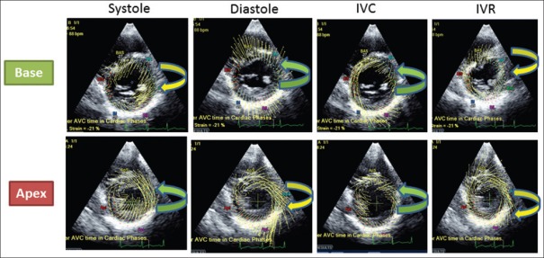
Rotation of LV base and apex during the different phases of the cardiac cycle. Systole: Counterclockwise apical rotation and the smaller clockwise basal rotation. Diastole: The apex moves clockwise and the base counterclockwise. IVC (Isovolumic contraction period): Both the base and apex rotate in a counterclockwise direction. IVR (Isovolumic relaxation period): Apex and base rotate in a clockwise direction
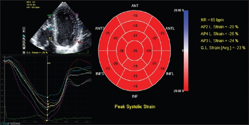
From apical views, longitudinal myocardial strain can be calculated. In most of the commercially available systems, a bull's eye showing the peak systolic longitudinal strain is displayed
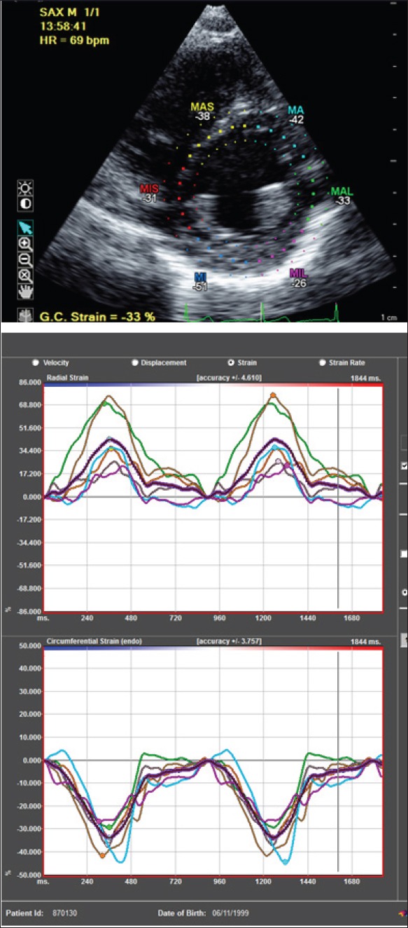
From short axis view, radial strain (upper panel) and circumferential strain (lower panel) can be calculated
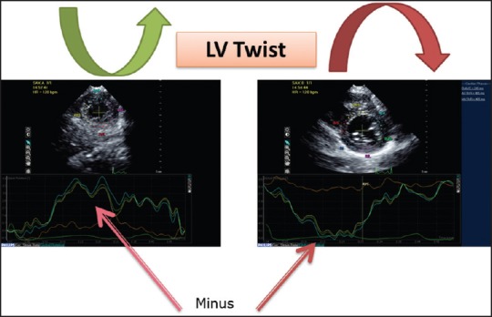
The apical mechanics is the result of the interaction between the subepicardial (ascendant) and the subendocardial (descendant) segments. Because of its larger radius, the ascendant segment overpowers the descent segment leading to a counterclockwise apical rotation and the smaller clockwise basal rotation. Practically, LV twist is calculated as the difference between the maximal rotation (positive) during systole of the apex and the maximal (negative) systolic rotation of the base
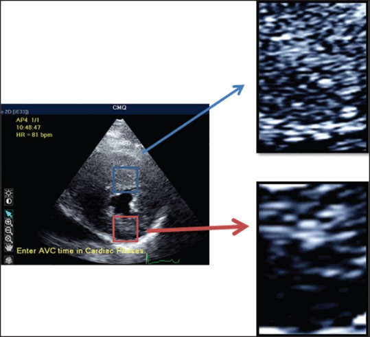
STE is based on the identification and on the tracking of speckles (spots generated by the interaction between the ultrasound beam and myocardial fibers) on routine 2-dimensional ultrasonic derived images. These spots, temporally stable, are used as natural acoustic markers, able to identify, as fingerprint, that specific myocardial segment and moving together with the tissue. Thus, the analysis of the spatial dislocation (tracking) of speckle represents the movement of that segment of tissue. From this, natural strain can be obtained and strain rate can be calculated as temporal derivative

Hypertrophic cardiomyopathy. During systole, the normal myocardial shortening is color-coded in yellow (as shown in the top panel corresponding to the apical segment). Conversely, the mid panel shows during systole abnormal myocardial deformation, suggestive of fibrosis and myocardial disarray, as demonstrated by the abnormal red and blue lines. Minor abnormalities showed in the basal panel (corresponding to the basal septum) suggesting some minor degree of fibrosis
References
-
- Sutherland GR, Di Salvo G, Claus P, D’hooge J, Bijnens B. Strain and strain rate imaging: A new clinical approach to quantifying regional myocardial function. J Am Soc Echocardiogr. 2004;17:788–802. - PubMed
-
- Buckberg G, Hoffman JI, Nanda NC, Coghlan C, Saleh S, Athanasuleas C. Ventricular torsion and untwisting: Further insights into mechanics and timing interdependence: A viewpoint. Echocardiography. 2011;28:782–804. - PubMed
-
- Buckberg G, Hoffman JI, Mahajan A, Saleh S, Coghlan C. Cardiac mechanics revisited: The relationship of cardiac architecture to ventricular function. Circulation. 2008;118:2571–87. - PubMed
-
- Biswas M, Sudhakar S, Nanda NC, Buckberg G, Pradhan M, Roomi AU, et al. Two- and three-dimensional speckle tracking echocardiography: Clinical applications and future directions. Echocardiography. 2013;30:88–105. - PubMed
Publication types
LinkOut - more resources
Full Text Sources
Other Literature Sources