

Transcranial Doppler Ultrasound: Physical Principles and Principal Applications in Neurocritical Care Unit
- PMID: 28465958
- PMCID: PMC5224659
- DOI: 10.4103/2211-4122.183746
Transcranial Doppler Ultrasound: Physical Principles and Principal Applications in Neurocritical Care Unit
Abstract
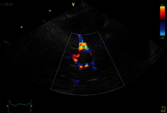
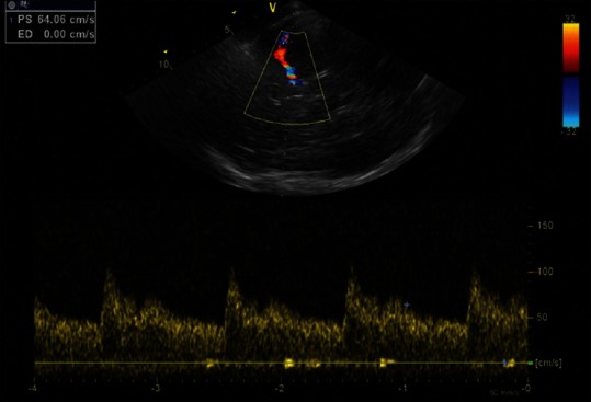
Transcranial Doppler (TCD) ultrasonography is a noninvasive ultrasound study, which has been extensively applied on both outpatient and inpatient settings. It involves the use of a low-frequency (≤2 MHz) transducer, placed on the scalp, to insonate the basal cerebral arteries through relatively thin bone windows and to measure the cerebral blood flow velocity and its alteration in many different conditions. In neurointensive care setting, TCD is useful for both adults and children for day-to-day bedside assessment of critical conditions including vasospasm in subarachnoid hemorrhage, traumatic brain injury, acute ischemic stroke, and brain stem death. It also allows to investigate the cerebrovascular autoregulation in setting of carotid disease and syncope. In this review, we will describe physical principles underlying TCD, flow indices most frequently used in clinical practice and critical care applications in Neurocritical Unit care.
Keywords: Brain stem death; Neurocritical Unit Care; cryptogenic stroke; mean cerebral brain flow; paradoxical embolism; patent foramen ovale; subarachnoid hemorrhage; transcranial Doppler ultrasonography; traumatic brain injury; vasospasm.
Conflict of interest statement
There are no conflicts of interest.
Figures

References
-
- Aaslid R, Markwalder TM, Nornes H. Noninvasive transcranial Doppler ultrasound recording of flow velocity in basal cerebral arteries. J Neurosurg. 1982;57:769–74. - PubMed
-
- Saqqur M, Zygun D, Demchuk A. Role of transcranial Doppler in neurocritical care. Crit Care Med. 2007;35(5 Suppl):S216–23. - PubMed
-
- Rigamonti A, Ackery A, Baker AJ. Transcranial Doppler monitoring in subarachnoid hemorrhage: A critical tool in critical care. Can J Anaesth. 2008;55:112–23. - PubMed
-
- Arenillas JF, Molina CA, Montaner J, Abilleira S, González-Sánchez MA, Alvarez-Sabín J. Progression and clinical recurrence of symptomatic middle cerebral artery stenosis: A long-term follow-up transcranial Doppler ultrasound study. Stroke. 2001;32:2898–904. - PubMed
-
- Christou I, Felberg RA, Demchuk AM, Grotta JC, Burgin WS, Malkoff M, et al. A broad diagnostic battery for bedside transcranial Doppler to detect flow changes with internal carotid artery stenosis or occlusion. J Neuroimaging. 2001;11:236–42. - PubMed
Publication types
LinkOut - more resources
Full Text Sources
Other Literature Sources