

Three-dimensional Transesophageal Echocardiography Demonstration of Left Atrial Appendage Echocontrast Regression after 6 Months Therapy with Dabigatran and not with Warfarin
- PMID: 28465961
- PMCID: PMC5224660
- DOI: 10.4103/2211-4122.183751
Three-dimensional Transesophageal Echocardiography Demonstration of Left Atrial Appendage Echocontrast Regression after 6 Months Therapy with Dabigatran and not with Warfarin
Abstract
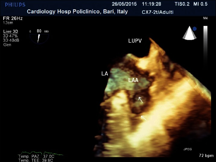
While warfarin therapy is efficacious in treating left atrial (LA) thrombus formation in patients with nonvalvular atrial fibrillation (AF), it does not affect red cell aggregation in vitro or LA spontaneous echo-contrast in patients. In this patient with left ventricular (LV) dysfunction secondary to AF, we observed the disappearance of dense echo-contrast in the atrial appendage after therapy with dabigatran and not with well-controlled warfarin. This allowed us to analyze accurately the appendage with three-dimensional (3D) transesophageal echocardiography (TEE) excluding thrombi and to perform electrical cardioversion to obtain a significant improvement of LV function. This case demonstrates the capability of dabigatran and not of warfarin in reducing the intense spontaneous echocontrast in atrial appendage and the ability of 3D TEE in analyzing accurately the appendage, excluding thrombi to safely perform cardioversion.
Keywords: Atrial appendage; atrial fibrillation; cardioversion; dabigatran etexilate; three-dimensional echocardiography; transesophageal echocardiography.
Conflict of interest statement
There are no conflicts of interest.
Figures


Similar articles
-
Use of transesophageal contrast echocardiography for excluding left atrial appendage thrombi in patients with atrial fibrillation before cardioversion.J Am Soc Echocardiogr. 2002 Oct;15(10 Pt 2):1256-61. doi: 10.1067/mje.2002.123961. J Am Soc Echocardiogr. 2002. PMID: 12411914
-
[Effect of non-vitamin K antagonist oral anticoagulants on left atrial or atrial appendage thrombi in patients with nonvalvular atrial fibrillation].Zhonghua Xin Xue Guan Bing Za Zhi. 2018 Aug 24;46(8):606-610. doi: 10.3760/cma.j.issn.0253-3758.2018.08.006. Zhonghua Xin Xue Guan Bing Za Zhi. 2018. PMID: 30139010 Chinese.
-
[Transesophageal echocardiography in patients with atrial fibrillation, candidates for cardioversion: usefulness and limitations].G Ital Cardiol. 1995 May;25(5):543-52. G Ital Cardiol. 1995. PMID: 7642059 Italian.
-
Transesophageal echocardiography-guided approach to cardioversion of atrial fibrillation.Prog Cardiovasc Dis. 1996 Jul-Aug;39(1):21-32. doi: 10.1016/s0033-0620(96)80038-7. Prog Cardiovasc Dis. 1996. PMID: 8693093 Review.
-
Diagnosis and Management of Left Atrium Appendage Thrombosis in Atrial Fibrillation Patients Undergoing Cardioversion.Medicina (Kaunas). 2019 Aug 21;55(9):511. doi: 10.3390/medicina55090511. Medicina (Kaunas). 2019. PMID: 31438560 Free PMC article. Review.
Cited by
-
Morphological and functional assessment of the left atrial appendage in daily practice: a comprehensive approach using basic and advanced echocardiography with practical tips.J Cardiovasc Imaging. 2024 Jul 29;32(1):12. doi: 10.1186/s44348-024-00017-2. J Cardiovasc Imaging. 2024. PMID: 39069633 Free PMC article. Review.
-
3D transesophageal echocardiography assists in evaluating the morphology, function, and presence of thrombi of left atrial appendage in patients with atrial fibrillation.Ann Transl Med. 2021 May;9(10):876. doi: 10.21037/atm-21-1981. Ann Transl Med. 2021. PMID: 34164510 Free PMC article.
-
Evaluation of left atrial appendage function and thrombi in patients with atrial fibrillation: from transthoracic to real time 3D transesophageal echocardiography.Int J Cardiovasc Imaging. 2017 Apr;33(4):491-498. doi: 10.1007/s10554-016-1026-6. Epub 2016 Nov 17. Int J Cardiovasc Imaging. 2017. PMID: 27853971
-
Anticoagulation efficacy of dabigatran etexilate for left atrial appendage thrombus in patients with atrial fibrillation by transthoracic and transesophageal echocardiography.Medicine (Baltimore). 2018 Jun;97(26):e11117. doi: 10.1097/MD.0000000000011117. Medicine (Baltimore). 2018. PMID: 29952953 Free PMC article.
References
-
- Zabalgoitia M, Halperin JL, Pearce LA, Blackshear JL, Asinger RW, Hart RG. Transesophageal echocardiographic correlates of clinical risk of thromboembolism in nonvalvular atrial fibrillation. Stroke Prevention in Atrial Fibrillation III Investigators. J Am Coll Cardiol. 1998;31:1622–6. - PubMed
-
- Bernhardt P, Schmidt H, Hammerstingl C, Lüderitz B, Omran H. Patients with atrial fibrillation and dense spontaneous echo contrast at high risk a prospective and serial follow-up over 12 months with transesophageal echocardiography and cerebral magnetic resonance imaging. J Am Coll Cardiol. 2005;45:1807–12. - PubMed
-
- Ito T, Suwa M, Nakamura T, Miyazaki S, Hirota Y, Kawamura K. Influence of warfarin therapy on left atrial spontaneous echo contrast in nonvalvular atrial fibrillation. Am J Cardiol. 1999;84:857–9. - PubMed
-
- Yuan YW, Shung KK. Ultrasonic backscatter from flowing whole blood. I: Dependence on shear rate and hematocrit. J Acoust Soc Am. 1988;84:52–8. - PubMed
Publication types
LinkOut - more resources
Full Text Sources
Other Literature Sources