

The Diagnostic Challenge of Dipyridamole-atropine Stress Echocardiography in a Patient with Myocardial Bridge
- PMID: 28465977
- PMCID: PMC5224666
- DOI: 10.4103/2211-4122.192175
The Diagnostic Challenge of Dipyridamole-atropine Stress Echocardiography in a Patient with Myocardial Bridge
Abstract
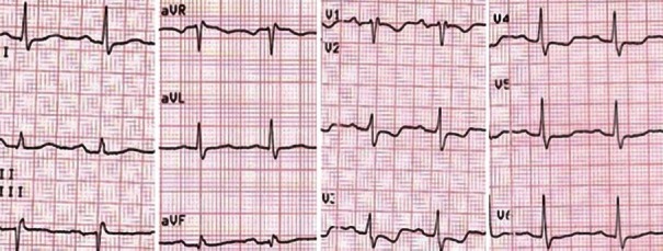
A 60-year-old male patient was submitted to dipyridamole-atropine stress echocardiography (DSE) for chest pain during exertion. At rest, no electrocardiographic (ECG) and transthoracic echocardiographic (TTE) abnormalities were observed. After dipyridamole infusion, the patient complained a mild chest discomfort, without ECG changes and TTE wall-motion abnormalities. Subsequently, worsening of the anginal symptoms combined with descending ST-depression and T-negative waves occurred after atropine and unexpectedly, aminophylline administration. Coronary angiography was performed showing a myocardial bridge (MB) of the left anterior descending artery. The occurrence, during DSE, of worsening ischemic abnormalities after atropine and aminophylline administration may be a particular diagnostic feature of MB.
Keywords: Dipyridamole stress echocardiography; left anterior descending artery; longitudinal strain; myocardial bridge.
Conflict of interest statement
There are no conflicts of interest.
Figures


Similar articles
-
Regional distribution and timing of wall motion abnormalities during echo-dipyridamole stress test in patients with stable angina: the elusive link between coronary stenoses and myocardial ischemia.Ital Heart J. 2000 Jan;1(1):33-8. Ital Heart J. 2000. PMID: 10868920
-
[Diagnosis of ischemic heart disease by dipyridamole-stress two-dimensional echocardiography].J Cardiol. 1994 Jan-Feb;24(1):9-16. J Cardiol. 1994. PMID: 8158534 Japanese.
-
[Problem of false positives in dipyridamole-echocardiography test. Description of a case and review of the literature].Minerva Cardioangiol. 1994 Nov;42(11):517-22. Minerva Cardioangiol. 1994. PMID: 7700541 Italian.
-
Simultaneous assessment of wall motion and coronary flow velocity in the left anterior descending coronary artery during dipyridamole stress echocardiography.J Am Soc Echocardiogr. 2003 May;16(5):457-63. doi: 10.1016/s0894-7317(03)00101-9. J Am Soc Echocardiogr. 2003. PMID: 12724655
-
Italian Society of Cardiovascular Echography (SIEC) Consensus Conference on the state of the art of contrast echocardiography.Ital Heart J. 2004 Apr;5(4):309-34. Ital Heart J. 2004. PMID: 15185894 Review.
Cited by
-
Myocardial Bridge: If You Wait Longer You Can Find It!J Cardiovasc Echogr. 2017 Apr-Jun;27(2):78-79. doi: 10.4103/jcecho.jcecho_15_17. J Cardiovasc Echogr. 2017. PMID: 28466001 Free PMC article. No abstract available.
-
A Case of Symptomatic Myocardial Bridge Treated with Calcium Channel Blocker.Int Med Case Rep J. 2022 May 31;15:259-262. doi: 10.2147/IMCRJ.S360819. eCollection 2022. Int Med Case Rep J. 2022. PMID: 35669125 Free PMC article.
References
-
- Andò G, Morabito G, de Gregorio C, Trio O, Saporito F, Oreto G. The ACEF score as predictor of acute kidney injury in patients undergoing primary percutaneous coronary intervention. Int J Cardiol. 2013;168:4386–7. - PubMed
-
- Andò G, Capodanno D. Radial versus femoral access in invasively managed patients with acute coronary syndrome: A Systematic Review and Meta-analysis. Ann Intern Med. 2015;163:932–40. - PubMed
-
- Sakuma M, Kamishirado H, Inoue T, Ichihara M, Takayanagi K, Hayashi T, et al. Acute myocardial infarction associated with myocardial bridge and coronary artery vasospasm. Int J Clin Pract. 2002;56:721–2. - PubMed
-
- Zoghi M, Duygu H, Nalbantgil S, Kirilmaz B, Turk U, Ozerkan F, et al. Impaired endothelial function in patients with myocardial bridge. Echocardiography. 2006;23:577–81. - PubMed
-
- Herrmann J, Higano ST, Lenon RJ, Rihal CS, Lerman A. Myocardial bridging is associated with alteration in coronary vasoreactivity. Eur Heart J. 2004;25:2134–42. - PubMed
Publication types
LinkOut - more resources
Full Text Sources
Other Literature Sources
Miscellaneous