Review
. 2017 Oct;23(5):454-465.
doi: 10.1177/1073858417691030. Epub 2017 Feb 2.

The Glymphatic Pathway: Waste Removal from the CNS via Cerebrospinal Fluid Transport

Affiliations
Review

The Glymphatic Pathway: Waste Removal from the CNS via Cerebrospinal Fluid Transport

Helene Benveniste et al. Neuroscientist. 2017 Oct.

Abstract

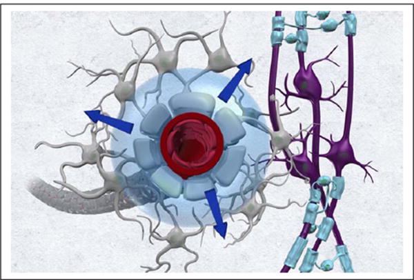
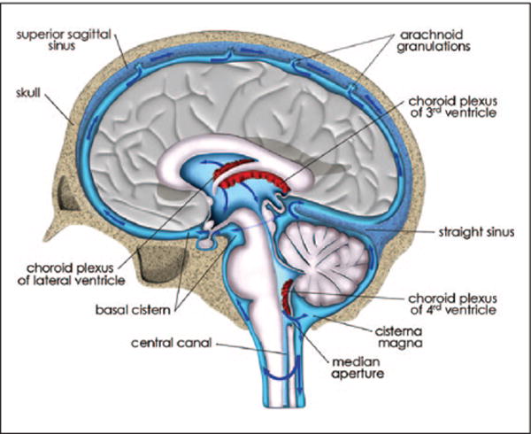
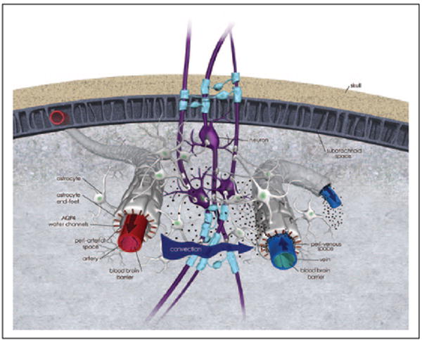
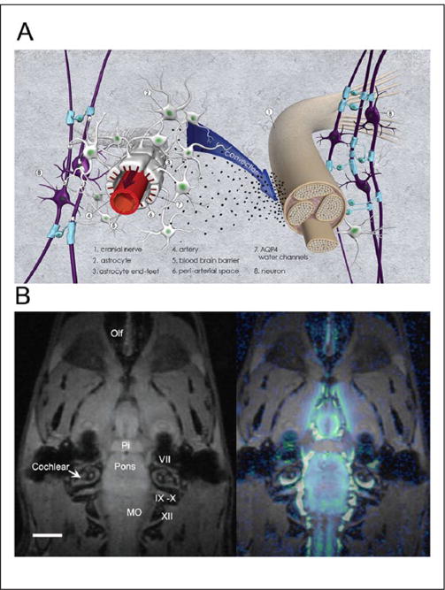
The overall premise of this review is that cerebrospinal fluid (CSF) is transported within a dedicated peri-vascular network facilitating metabolic waste clearance from the central nervous system while we sleep. The anatomical profile of the network is complex and has been defined as a peri-arterial CSF influx pathway and peri-venous clearance routes, which are functionally coupled by interstitial bulk flow supported by astrocytic aquaporin 4 water channels. The role of the newly discovered system in the brain is equivalent to the lymphatic system present in other body organs and has been termed the "glymphatic pathway" or "(g)lymphatics" because of its dependence on glial cells. We will discuss and review the general anatomy and physiology of CSF from the perspective of the glymphatic pathway, a discovery which has greatly improved our understanding of key factors that control removal of metabolic waste products from the central nervous system in health and disease and identifies an additional purpose for sleep. A brief historical and factual description of CSF production and transport will precede the ensuing discussion of the glymphatic system along with a discussion of its clinical implications.

Keywords: brain; cerebrospinal fluid; glymphatic pathway; microcirculation; transport.


Conflict of interest statement

Declaration of Conflicting Interests

The author(s) declared no potential conflicts of interest with respect to the research, authorship, and/or publication of this article.

Figures

Figure 1
Figure 1
The brain microcirculation contributes to cerebrospinal fluid (CSF) production. It has been proposed that the capillary unit secretes fluid which enters the interstitial space and communicates with ventricular and subarachnoid CSF via bulk flow. The figure illustrates this concept from the perspective of the capillary and astrocytic end-foot.
Figure 2
Figure 2
Cerebrospinal fluid (CSF) transport and reabsorption from ventricles and subarachnoid space: CSF transport from the site of formation in the choroid plexuses in the lateral, 3rd and 4th ventricle and out into the subarachnoid space via the median aperture is illustrated in an anatomical drawing of the human brain at the level of the superior sagittal sinus. The blue arrows in the subarachnoid space indicate CSF transport above the hemispheric convexities toward the arachnoid granulations; from where CSF exits into the dural sinuses. There is also CSF transport from the subarachnoid space and down toward the spine.
Figure 3
Figure 3
Cerebrospinal fluid (CSF) transport of metabolically inert tracer in human brain The distribution pattern of 111In-labeled diethylenetriamine pentaacetic acid (111In-DTPA) in brain and cervical spine in three orthogonal planes from a 63-year-old female, 24 hrs. after lumbar intrathecal injection of the radiotracer. The images were acquired using combined SPECT/CT. The radiotracer in CSF was transported from the lumbar intrathecal space to the cranial subarachnoid space and into brain parenchyma over 24 hrs. There is evidence of the radiotracer over the hemispheric convexities as well as in parenchyma. The uptake in the brain in inhomogeneous with excessive uptake in the cerebellum, frontoorbital cortex (CTX) and pons. There was no evidence of dural leak in this patient and the brain 111In-DTPA uptake pattern was read as normal. Data courtesy: Professor Dinko Franceschi, MD, Department of Nuclear Medicine, Radiology Stony Brook University.
Figure 4
Figure 4
Glymphatic pathway function conceptualized. The principle of cerebrospinal fluid (CSF) transport and waste solute removal via the glymphatic pathway is illustrated on this 3-dimensional anatomical drawing. First, CSF enters along the peri-arterial space; the outer perimeter of the peri-arterial space is made up by astrocytic end-feet on which AQP4 water channels are strategically positioned to facilitate rapid water exchange across the capillary-endfeet complex. Second, CSF is transported from the peri-arterial space into brain parenchyma; a process driven by diffusion, convection (arrow indicate convective force) and local mixing whereby CSF exchanges with interstitial fluid (ISF). The CSF-ISF mixing facilitates waste removal. The CSF-ISF and waste leaves the brain parenchyma along peri-venous channels (large central veins). The CSF and solute transport system illustrated has been designed “glymphatic” pathway or (g)lymphatics due to its dependence on glial cells.
Figure 5
Figure 5
Cerebrospinal fluid (CSF) and waste drainage along cranial and peripheral nerves A: Anatomical drawing (3-dimensional perspective) of CSF and waste drainage along cranial or peripheral nerves. Contrast agents administered into CSF can exit along cranial and peripheral nerves. However, it is currently unknown how CSF and waste solutes exit from the subarachnoid space around the nerve roots. Studies from the olfactory nerves show that CSF and large contrast molecules are transported along the nerve (i.e., the space defined by the epineurium and perineurium) and then drain into veins and/or lymph vessels outside of the nerve. There is also some evidence in the literature (optic nerve studies) indicating that CSF and waste solutes can access the nerve itself. B: In vivo MRI study of rat after injection of paramagnetic contrast (Gd-DTPA) into the CSF via the cisterna magna demonstrating drainage along the cranial nerve. The MRI on the left shows the anatomical template from the rat at the level of the pituitary gland and cranial nerves exiting from pons and medulla oblongata (MO). Pi = pituitary gland; MO = medulla oblongata; Olf = olfactory nerves. Scale bar = 3 mm. The MRI on the right shows a color-coded map overlaid on the anatomical template, which represents distribution of Gd-DTPA 1 hour after administration into the cisterna magna. It is evident that CSF tagged with contrast has exited along the cranial nerves. The spatial resolution of the MRI (0.0013 mm3) is insufficient to document contrast within the nerve sheaths.
Figure 6
Figure 6
Cerebrospinal fluid (CSF) transport of 18F in nonhuman primate visualized by positron emission tomography (PET). Static PET whole body image of anesthetized female non-human primate (baboon/Papio) 4.5 hours after intrathecal administration of 18F via the L3/L4 lumbar level. The baboon was intubated but allowed to breathe spontaneously during the study. Normal physiological parameters where maintained throughout the study. The 18F distribution pattern is shown in the whole baboon body, and visualized as a volume rendered, color-coded map where red and blue colors represent high and low tracer uptake, respectively. The area associated with the spine has the highest uptake of 18F. The 18F tracer has reached the cranium but penetration into the brain parenchyma is not apparent. There is also 18F uptake in bony structures, including jaw, skull, and vertebrae. Color scale: Arbitrary units. Data courtesy: This study was performed by Helene Benveniste, Joanna Fowler, and Nora Volkow on May 3, 2013 at Brookhaven National Laboratory, Upton, New York; after approval by the Institutional Animal Use and Care Committee (unpublished data). After the conclusion of the study anesthesia was discontinued and the baboon recovered without problems and was returned to the non-human primate housing colony.
Figure 7
Figure 7
Wide field confocal images showing AQP4 immunoreactivity from postmortem cortical tissue of a young, cognitively intact subject (A) and of a subject with Alzheimer’s disease (B). In the young subject, there is uniform AQP4 expression (green) throughout; whereas in the Alzheimer’s disease (AD) subject the distribution is inhomogeneous within the cortex. The inhomogeneous AQP4 expression pattern in the AD post-mortem tissue is associated with amyloid beta plaques (red) and reactive astrocytes. Data courtesy: Dr. Jeffrey Iliff, PhD, Oregon Health & Science University. For more details, see (Zeppenfeld and others 2017).

References

    1. Abbott NJ, Bundgaard M, Cserr HF. Tightness of the blood-brain barrier and evidence for brain interstitial fluid flow in the cuttlefish, Sepia officinalis. J Physiol. 1985;368:213–26. - PMC - PubMed
    1. Algin O, Turkbey B. Intrathecal gadolinium-enhanced MR cisternography: a comprehensive review. AJNR Am J Neuroradiol. 2013;34:14–22. - PMC - PubMed
    1. Asgari M, de Zelicourt D, Kurtcuoglu V. Glymphatic solute transport does not require bulk flow. Sci Rep. 2016;6:38635. - PMC - PubMed
    1. Aston-Jones G, Bloom FE. Activity of norepinephrine-containing locus coeruleus neurons in behaving rats anticipates fluctuations in the sleep-waking cycle. J Neurosci. 1981;1:876–86. - PMC - PubMed
    1. Bakker EN, Bacskai BJ, Arbel-Ornath M, Aldea R, Bedussi B, Morris AW, et al. Lymphatic clearance of the brain: perivascular, paravascular and significance for neurodegenerative diseases. Cell Mol Neurobiol. 2016;36:181–94. - PMC - PubMed

Publication types

MeSH terms