. 2017 Nov 1;34(21):3012-3026.
doi: 10.1089/neu.2016.4844. Epub 2017 Jun 30.

Spinal Transection Alters External Urethral Sphincter Activity during Spontaneous Voiding in Freely Moving Rats

Affiliations

Spinal Transection Alters External Urethral Sphincter Activity during Spontaneous Voiding in Freely Moving Rats

Brandon K LaPallo et al. J Neurotrauma. .

Abstract

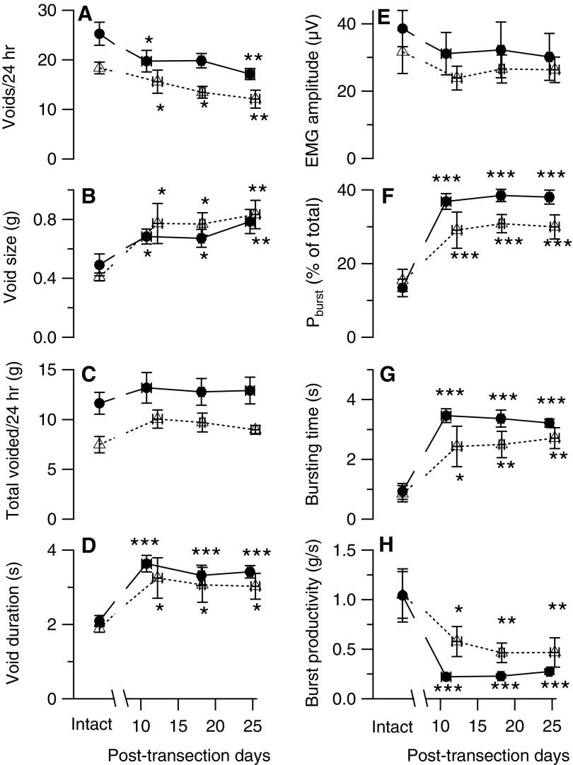
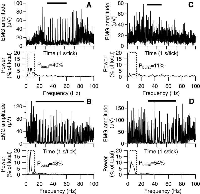
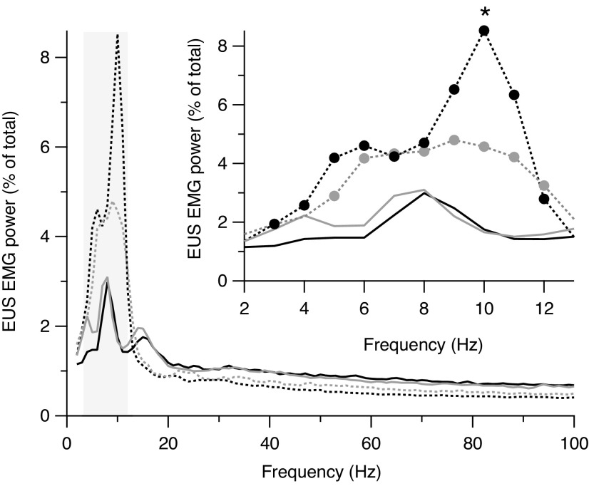
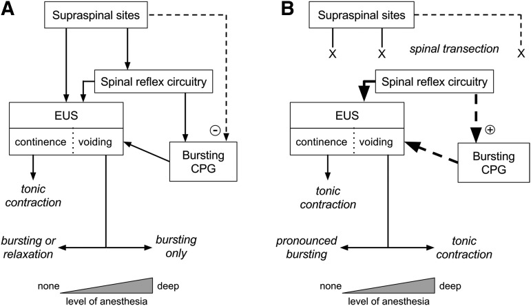
The rat is a commonly used model for the study of lower urinary tract function before and after spinal cord injury. We have previously reported that in unanesthetized freely moving rats, although phasic external urethral sphincter (EUS) activity (bursting) is most common during micturition, productive voiding can occur in the absence of bursting, which differs from results seen in anesthetized or unanesthetized restrained animals. The purpose of the present study was to characterize EUS behavior in unanesthetized, freely moving rats before and after mid-thoracic (T8) or thoraco-lumbar (T13-L1) spinal transection to determine how EUS behavior after spinal cord injury differs from that seen in anesthetized or unanesthetized restrained rats. Several abnormalities became evident that were comparable after transection at either level, including the following: repetitive non-voiding EUS contractions; increased prevalence, intensity, and duration of EUS bursting; decreased rate of urine evacuation during bursting; increased void size and decreased number of daily voids; shorter inter-burst silent period and increased frequency of bursting; and loss of the direct linear relationships that are evident in intact animals between void size and bursting silent period. These data suggest that transection-induced delayed initiation of EUS bursting allows co-contraction of the bladder and the EUS that prevents or limits urine evacuation, resulting in a detrusor-sphincter dyssynergia-like phenomenon. In addition, the higher-than-normal frequency at which EUS bursting occurs after transection is associated with shorter silent periods during which urine typically flows, which interferes with voiding by slowing the rate of urine evacuation. That results were comparable after either transection suggests that the central pattern generator responsible for EUS bursting is located caudal to the L1 spinal segment.

Keywords: EMG; external urethral sphincter; implanted electrodes; longitudinal study; spinal cord.


Conflict of interest statement

No competing financial interests exist.

Figures

<b>FIG. 1.</b>
FIG. 1.
Examples of 24-h recordings of voided urine acquired from a rat before (A) and 4 weeks after (B) thoraco-lumbar transection. Time is shown on the abscissa in military format with the black bar denoting the 12-h dark cycle. Step-like increases in weight indicate the occurrence of voiding. Fewer but larger voids tend to occur in the spinal-transected than in the spinal-intact rat.
<b>FIG. 2.</b>
FIG. 2.
Time course of urodynamic and external urethral sphincter electromyography (EUS EMG) variables recorded before and up to 4 weeks after thoraco-lumbar (open symbols with dashed lines) or mid-thoracic (filled symbols with solid lines) transections. Data recorded in intact animals were stable over time, and thus were averaged over all pre-transection recordings. Weekly averages (± standard error) are shown as the number of days after spinal transection. Data are shown for: (A) number of voids per 24 h; (B) void size (urine weight/void/200 g of body weight); (C) total voided/24 h (total urine weight/200 g of body weight per 24 h); (D) void duration (duration of urine weight accumulation in seconds); (E) EMG amplitude (average intra-void rectified EMG amplitude); (F) bursting power (Pburst; average percent of total power in bursting frequency bands normalized to a 7-Hz wide frequency range; see “Data analysis” in the Methods section for details); (G) bursting time (cumulative time during a void when Pburst exceeded the power in the same frequency range of amplitude-matched non-voiding epochs by 2 standard deviations); and (H) bursting productivity (urine weight/void/200 g of body weight divided by the bursting time). Significant difference from intact:*p < 0.05; **p < 0.01; ***p < 0.001 by repeated measures analysis of variance and post hoc contrast.
<b>FIG. 3.</b>
FIG. 3.
Examples of external urethral sphincter electromyography (EUS EMG) activity during voiding in two rats before (A, C) and after (B, D) thoraco-lumbar (left panels) or mid-thoracic (right panels) transection. Within each panel, the upper traces show rectified EUS EMG (1-sec time axis divisions) during single voids (time of urine flow indicated by horizontal bars above each trace), and the lower traces show EUS EMG spectral power (expressed as % of total power) as a function of frequency. The pattern of EUS activity is highly variable in intact rats (upper panels). Some voids exhibit strong phasic activity (i.e., bursting; illustrated in A) associated with peaks in EMG power in frequencies corresponding to the rate of bursting. Within the entire bursting frequency range (indicated by dashed rectangle), the percent of total power associated with bursting (Pburst; shown to right of range) is high. Other voids of intact rats exhibit little or no phasic EUS EMG activity (illustrated in C), having no distinct peaks in the power spectrum and a low value of Pburst. After transection (lower panels), bursting is seen in the vast majority of voids after thoraco-lumbar or mid-thoracic transection and is associated with large values of Pburst.
<b>FIG. 4.</b>
FIG. 4.
Average power spectra during voiding before (solid lines) and after (dashed lines) mid-thoracic (black lines) or thoraco-lumbar (gray lines) spinal transection. Both transections enhance power in frequencies corresponding to the rate of external urethral sphincter (EUS) bursting outlined by the gray rectangle. In the enlargement of this region (inset), the filled circles indicate individual frequencies with statistically significantly differences (p < 0.05) between mid-thoracic–transected (black) or thoraco-lumbar–transected (gray) rats and intact rats. The asterisk indicates that the power at 10 Hz is greater in mid-thoracic– than thoraco-lumbar– transected rats.
<b>FIG. 5.</b>
FIG. 5.
Distribution of power in the bursting frequency range expressed as a percent of the total power (Pburst) during epochs of external urethral sphincter electromyography (EUS EMG) associated with voiding (black lines) or not associated with voiding (gray lines) in two animals before (A, C) and after (B, D) thoraco-lumbar (left panels) mid-thoracic (right panels) spinal transection. The variable contribution of both tonic and phasic voiding patterns of EUS activity during voiding in rats with intact neuraxes (upper panels) results in a broad distribution of bursting power. This distribution is higher than but still overlaps considerably that of bursting power during non-voiding EMG epochs. Spinal transection at either level (lower panels) results in robust EUS bursting during the vast majority of voids, such that the distribution of bursting power is consistently high and minimally overlaps that of non-voiding EMG epochs.
<b>FIG. 6.</b>
FIG. 6.
Regression analysis of relationships between urine weight per void and bursting time (A, D), bursting power (Pburst; B, E), and external urethral sphincter (EUS) bursting silent period (C, F) before (circles with thick black regression line) and after (triangles with thin gray regression line) thoraco-lumbar (left panels) or mid-thoracic (right panels) spinal transection. All values are z-transformed to remove inter-animal differences in mean value and variance. Void size is positively correlated with bursting time and with %Pburst both before and after either spinal transection. Void size is positively correlated with silent period duration in spinal-intact animals, but not in spinal-transected animals.
<b>FIG. 7.</b>
FIG. 7.
Typical example of rhythmic external urethral sphincter (EUS) activation during inter-void intervals in transected rats. (A) EUS activations (lower trace of pair) occurring at regular intervals for extended periods precede voiding of urine (upper trace of pair) in a rat 4 weeks after mid-thoracic spinal transection. (B) Expansion of the area in panel (A) indicated by the gray rectangle illustrates that the roughly uniform contractions increase in amplitude and occur at shorter intervals during the time leading up to the void. (C) Expansion of the area in panel B indicated by the gray rectangle shows that phasic EUS activity (i.e., bursting) begins and continues for >6 sec before urine accumulation begins. The small amount of drift that is apparent in the urine weight trace (A) has a sufficiently slow time course that it does not affect the measurement of the weight that falls during the void.
<b>FIG. 8.</b>
FIG. 8.
Relationship between bladder weight and external urethral sphincter (EUS) bursting silent period in rats after thoraco-lumbar (open circles) or mid-thoracic (filled circles) spinal transection. Linear regression analysis shows a significant inverse relationship between these variables (fit shown by dashed line) for all transections. Inclusion of transection level in this analysis did not alter the relationship between bladder weight and silent period.
<b>FIG. 9.</b>
FIG. 9.
Interaction of anesthesia and spinal transection on external urethral sphincter (EUS) activity patterns in continence and voiding. Spinal and supraspinal elements are shown in text boxes, and EUS activity outcomes are shown in italics. During voiding, outcomes vary over a continuum (indicated by the horizontal two-headed arrows) depending on the depth of anesthesia (indicated by the width of the triangles at the bottom). (A) In rats with intact spinal cords, the EUS is under descending control from spinal and supraspinal sites. It is tonically active during continence to prevent urine flow, while during voiding, it exhibits some form of relaxation that permits urine flow. Under deep anesthesia, the EUS exhibits primarily phasic relaxation (i.e., bursting) that is patterned by intrinsic spinal circuitry (bursting central pattern generator [CPG] reviewed in Fraser). During voiding in unanesthetized, unstressed rats (i.e., no anesthesia, no restraint), the EUS expresses either bursting (∼75% of time) or tonic relaxation (∼25% of time). Voiding during intermediate levels of anesthesia is expected to be associated with an intermediate level of EUS bursting (i.e., between 75 and 100% of voids). Thus, we hypothesize that there is a pathway of supraspinal origin (dashed line) that inhibits (minus sign in circle) the spinal bursting CPG, and that this pathway is itself inhibited by anesthesia or under stressful conditions. (B) In rats with spinal transection, the normal descending control over EUS and bursting CPG function is interrupted, but over time, spontaneous voiding re-emerges as a result of plasticity in spinal reflex pathways (thick solid arrow). The present study has shown that unanesthetized, unrestrained transected rats nearly exclusively exhibit EUS bursting during voiding; anesthetized rats exhibit tonic EUS activity during spontaneous bladder contraction comparable to detrusor-sphincter dyssynergia in humans. Lightly anesthetized rats or unanesthetized rats shortly after bladder implantation surgery exhibit a range of EUS activity patterns that fall between these two extremes (see “EUS behavior before and after spinal transection and the influence of anesthesia” in the Discussion section). Thus, we hypothesize that plasticity in spinal reflex pathways after transection enhances the EUS bursting CPG (down-pointing heavy dashed arrow), which in the absence of anesthesia leads to pronounced bursting (up-pointing heavy dashed arrow) and that anesthesia or stressful conditions suppress this injury-induced enhancement of the bursting CPG, leading to tonic EUS activity during voiding.

Similar articles

Cited by

References

    1. Fowler C.J., Griffiths D., and de Groat W.C. (2008). The neural control of micturition. Nat. Rev. Neurosci. 9, 453–466 - PMC - PubMed
    1. de Groat W.C. and Wickens C. (2013). Organization of the neural switching circuitry underlying reflex micturition. Acta Physiol. (Oxf.) 207, 66–84 - PMC - PubMed
    1. Kruse M.N., Belton A.L., and de Groat W.C. (1993). Changes in bladder and external urethral sphincter function after spinal cord injury in the rat. Am. J. Physiol. 264, R1157–1163 - PubMed
    1. Weld K.J., Graney M.J., and Dmochowski R.R. (2000). Clinical significance of detrusor sphincter dyssynergia type in patients with post-traumatic spinal cord injury. Urology 56, 565–568 - PubMed
    1. Swartz D. (1946). The neurogenic bladder in spinal cord injury. Can. Med. Assoc. J. 54, 333–339 - PubMed

Publication types

LinkOut - more resources