. 2017 Jul 25;8(30):49093-49109.
doi: 10.18632/oncotarget.17095.

HistoneH3 demethylase JMJD2A promotes growth of liver cancer cells through up-regulating miR372

Affiliations

HistoneH3 demethylase JMJD2A promotes growth of liver cancer cells through up-regulating miR372

Jiahui An et al. Oncotarget. .

Abstract

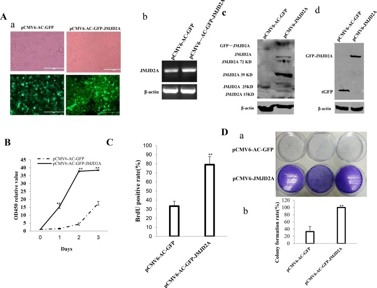
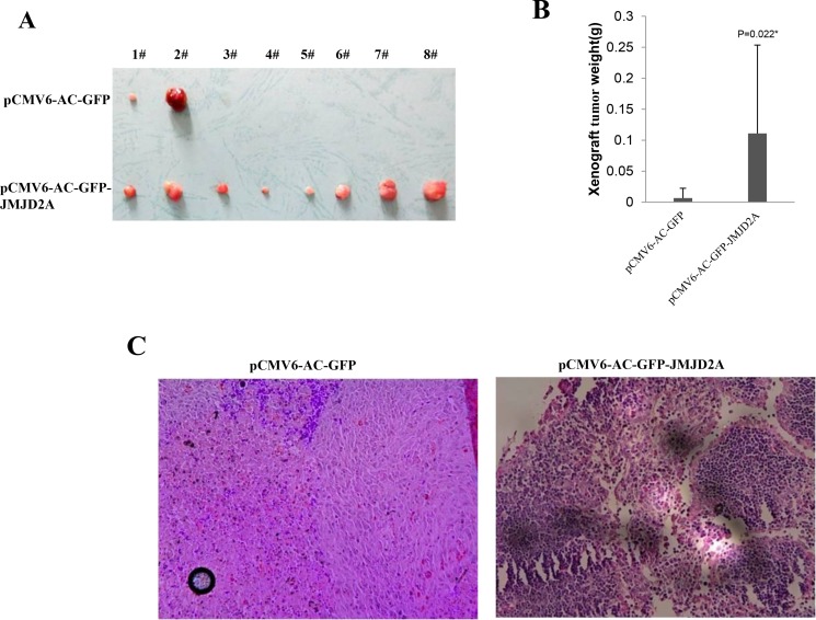
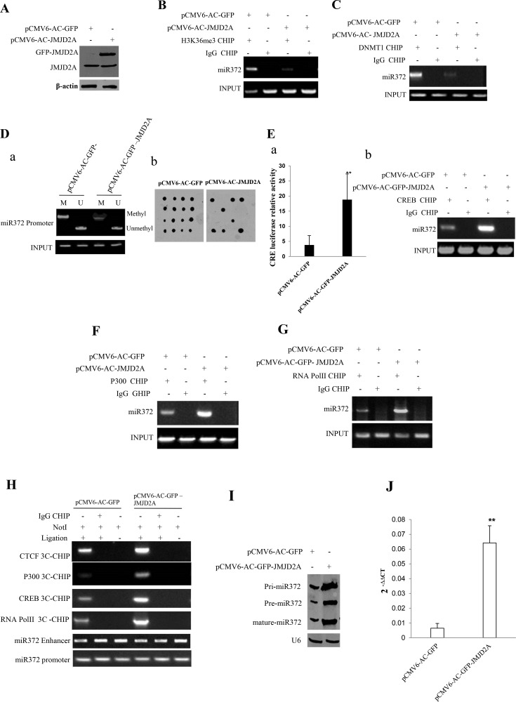
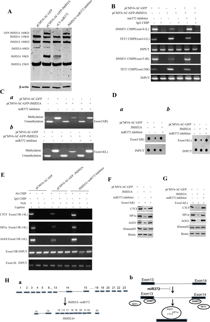
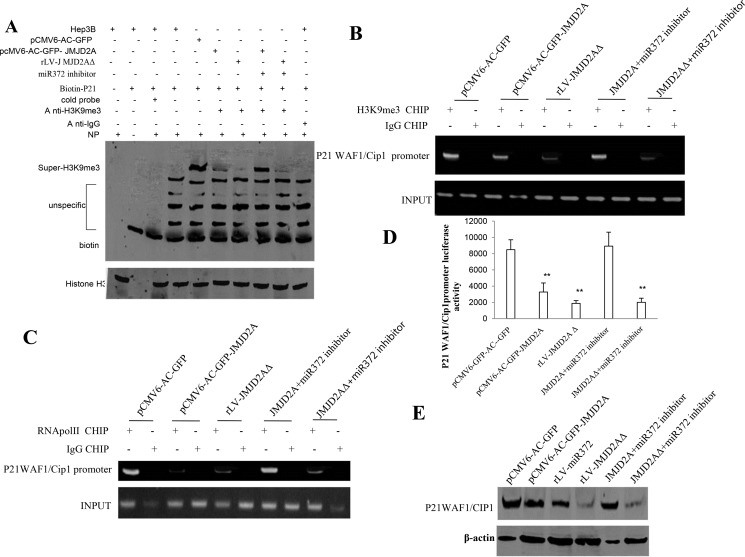
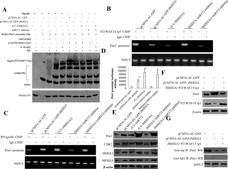
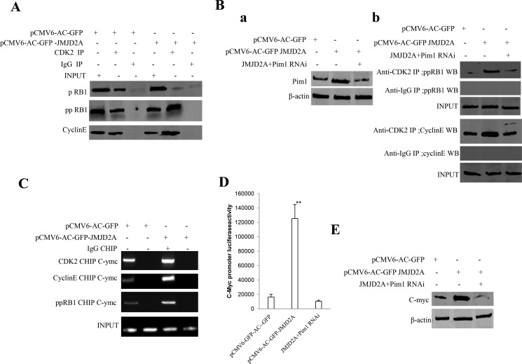
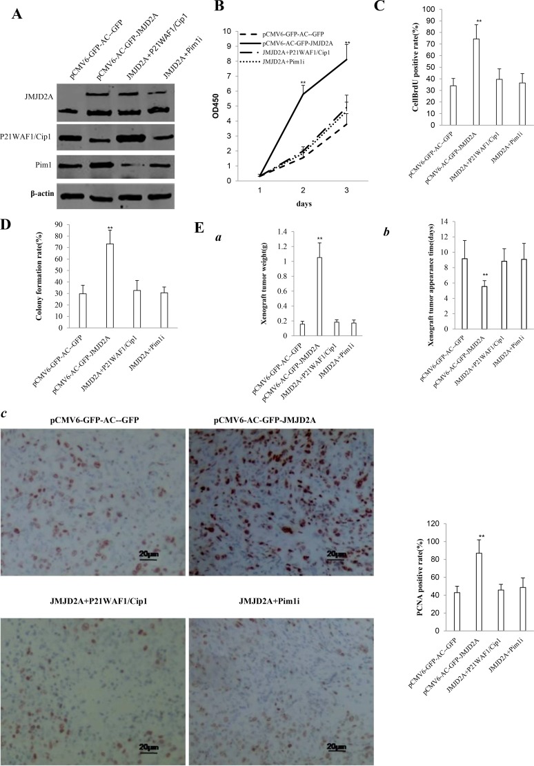
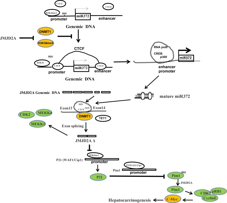
Changes in histone lysine methylation status have been observed during cancer formation. JMJD2A protein is a demethylase that is overexpressed in several tumors. Herein, our results demonstrate that JMJD2A accelerates malignant progression of liver cancer cells in vitro and in vivo. Mechanistically, JMJD2A promoted the expression and mature of pre-miR372 epigenetically. Notably, miR372 blocks the editing of 13th exon-introns-14th exon and forms a novel transcript( JMJD2AΔ) of JMJD2A. In particular, JMJD2A inhibited P21(WAF1/Cip1) expression by decreasing H3K9me3 dependent on JMJD2AΔ. Thereby, JMJD2A could enhance Pim1 transcription by suppressing P21(WAF1/Cip1). Furthermore, through increasing the expression of Pim1, JMJD2A could facilitate the interaction among pRB, CDK2 and CyclinE which prompts the transcription and translation of oncogenic C-myc. Strikingly, JMJD2A may trigger the demethylation of Pim1. On the other hand, Pim1 knockdown and P21(WAF1/Cip1) overexpression fully abrogated the oncogenic function of JMJD2A. Our observations suggest that JMJD2A promotes liver cancer cell cycle progress through JMJD2A-miR372-JMJD2AΔ-P21WAF1/Cip1-Pim1-pRB-CDK2-CyclinE-C-myc axis. This study elucidates a novel mechanism for JMJD2A in liver cancer cells and suggests that JMJD2A can be used as a novel therapeutic targets of liver cancer.

Keywords: HistoneH3 demethylase JMJD2A; P21WAF1/Cip1; Pim1; liver cancer; miR372.


Conflict of interest statement

CONFLICTS OF INTEREST

The authors disclose no conflicts.

Figures

Figure 1
Figure 1. JMJD2A accelerates liver cancer cell growth in vitro
(Aa) The photography of the Hep3B cell lines transfected with pCMV6-AC-GFP or pCMV6-AC-GFP-JMJD2A. (Ab) RT-PCR for JMJD2A cDNA in JMJD2A overexpressed control Hep3B stable cell lines;β-actin as internal control. (Ac) Western blotting with anti- JMJD2A in JMJD2A overexpressed control Hep3B stable cell lines; β-actin as internal control. (Ad) Western blotting with anti-tGFP in JMJD2A overexpressed control Hep3B stable cell lines; β-actin as internal control. (B) Cell proliferation assay was performed in 96-well format using the CCK8 cells proliferation kit to determine the cell viability as described by the manufacturer. Each sample was assayed in triplicates for 3 days consecutively. Cell growth curve was based on the corresponding the relative values of OD450 and each point represents the mean of three independent samples. Data are means of value from three independent experiments, bar ± SEM. **P < 0.01; *P < 0.05. (C) Cell BrdU assay. Data are means of value from three independent experiment, bar ± SEM. **P < 0.01; *P < 0.05. (D) (upper) The photography of colonies from the cell lines indicated in left. (lower) Cell plate colony formation ability assay. Data are means of value from three independent experiment, bar ± SEM. **P < 0.01; *P < 0.05.
Figure 2
Figure 2. JMJD2A promotes liver cancer cell growth in vivo
(A) The photography of xenograft tumors from Balb/C null mouse injected with Hep3B cells transfected with pCMV6-AC-GFP, pCMV6-AC-GFP-JMJD2A subcutaneously at armpit (B) The xenograft tumors weight(gram) in the two groups. Data were means of value from nine Balb/c mice, mean ± SEM, n = 8, *P < 0.05; **P < 0.01. Data were means of value from nine Balb/c mice, mean ± SEM, n = 8, *P < 0.05; **P < 0.01. (C) A portion of each xenograft tumor was fixed in 4% formaldehyde and embedded in paraffin, and the micrometers of sections (4 μm) were made for hematoxylin-eosin (HE) staining (original magnification×100).
Figure 3
Figure 3. JMJD2A enhances miR372 expression epigenetically
(A) Western blotting analysis with anti-JMJD2A in liver cancer cells Hep3B cell lines transfected with pCMV6-AC-GFP, pCMV6-AC-GFP-JMJD2A respectively. β-actin as internal control. (B) Chromatin Immunoprecipitation(CHIP) with anti-H3K9me3 followed by PCR with miR372 promoter primers in liver cancer cells Hep3B cell lines transfected with pCMV6-AC-GFP, pCMV6-AC-GFP-JMJD2A respectively. IgG CHIP as negative control. miR372 promoter as INPUT. (C) Chromatin Immunoprecipitation(CHIP) with anti-DNMT1 followed by PCR with miR372 promoter primers in liver cancer cells Hep3B cell lines transfected with pCMV6-AC-GFP, pCMV6-AC-GFP-JMJD2A respectively. IgG CHIP as negative control. miR372 promoter as INPUT. (Da) Methylation specific PCR (MSP) analysis for miR372 promoter region in Hep3B celllines transfected with pCMV6-AC-GFP, pCMV6-AC-JMJD2A, respectively. pw primer amplification as internal control. methyl DNA fragment is 170 bp.unmethyl DNA fragment is 130 bp. (Db) The dot blot analysis of miR372 promoter DNA methylation using specific biotin-DNA methylation probe. (Ea) CRE binding element luciferase activity assay. (Eb) Chromatin Immunoprecipitation(CHIP) with anti-CREB followed by PCR with miR372 promoter primers in liver cancer cells Hep3B cell lines transfected with pCMV6-AC-GFP, pCMV6-AC-GFP-JMJD2A respectively. IgG CHIP as negative control. miR372 promoter as INPUT. (F) Chromatin Immunoprecipitation(CHIP) with anti-P300 followed by PCR with miR372 promoter primers in liver cancer cells Hep3B cell lines transfected with pCMV6-AC-GFP, pCMV6-AC-GFP-JMJD2A respectively. IgG CHIP as negative control. miR372 promoter as INPUT). (G) Chromatin Immunoprecipitation(CHIP) with anti-RNAPolII followed by PCR with miR372 promoter primers in liver cancer cells Hep3B cell lines transfected with pCMV6-AC-GFP, pCMV6-AC-GFP-JMJD2A respectively. IgG CHIP as negative control. miR372 promoter as INPUT. (H) Chromosome conformation capture (3C)-chromatin immunoprecipitation (ChIP) with anti-P300,anti-RNA polII,anti-CREB in liver cancer cells Hep3B cell lines transfected with pCMV6-AC-GFP, pCMV6-AC-GFP-JMJD2A respectively. The chromatin is cross-linked, digested with restriction enzymes, and ligated under conditions that favor intramolecular ligation. Immediately after ligation, the chromatin is immunoprecipitated using an antibody (anti-P300,anti-RNA polII)against the protein of interest. Thereafter, the cross-links are reversed, and the DNA is purified further. The PCR anlysis is applied for detecting miR372 promoter-enhancer coupling product using miR372 promoter and enhancer primers. The miR372 promoter and enhancer as INPUT. F.Biotin-pre-miR372 pulldown followed by Western blotting with anti-Dicer, anti-ago2 Biotin as INPUT and β-actin as internal control. (I) Northern blotting analysis of miR372 in liver cancer cells transfected with pCMV6-AC-GFP, pCMV6-AC-GFP-JMJD2A. (J) The real-time PCR detection of mature miR372 in liver cancer cells transfected with pCMV6-AC-GFP, pCMV6-AC-GFP-JMJD2A respectively. Each value was presented as mean ± standard error of the mean (SEM). **P < 0.01.
Figure 4
Figure 4. miR372 triggers a novel transcript(JMJD2AΔ) of JMJD2A
(A) Western blotting analysis with anti-JMJD2A in liver cancer cells Hep3B cell lines transfected with pCMV6-AC-GFP, pCMV6-AC-GFP-JMJD2A,rLV-miR372, pCMV6-AC-GFP-JMJD2A plus miR372 inhibitor respectively. β-actin as internal control. (B) Chromatin Immunoprecipitation (CHIP) with anti-DNMT1 and anti-TET followed by PCR with JMJD2A exon13 or 14 primers in liver cancer cells Hep3B cell lines transfected with pCMV6-AC-GFP, pCMV6-AC-GFP-JMJD2A and pCMV6-AC-GFP-JMJD2A plus miR372 inhibitor respectively. IgG CHIP as negative control. JMJD2A exon13 or 14 as INPUT. (C) Methylation specific PCR (MSP) analysis for JMJD2A exon13 right region (a) or JMJD2A exon14 left region (b) in Hep3B cell lines transfected with pCMV6-AC-GFP, pCMV6-AC-JMJD2A and pCMV6-AC-GFP-JMJD2A plus miR372 inhibitor respectively .pw primer amplification as internal control. methyl DNA fragment is 150 bp.unmethyl DNA fragment is 120 bp. (D) The dot blot analysis for JMJD2A exon13 right region (a) or JMJD2A exon14 left region (b) DNA methylation using specific biotin-DNA methylation probe. (E) Chromosome conformation capture (3C)-chromatin immunoprecipitation (ChIP) with anti-CTCF,anti-HP1α,anti-AGOI in liver cancer cells Hep3B cell lines transfected with pCMV6-AC-GFP and pCMV6-AC-GFP-JMJD2A plus miR372 inhibitor respectively. The PCR anlysis is applied for detecting JMJD2A exon13(right)-exon14(left) coupling product using JMJD2A exon13 and exon 14 primers. The JMJD2A exon13 and exon 14 as INPUTI. (F) Biotin-JMJD2A exon13 DNA pulldown followed by Western blotting with anti-CTCF,anti-HP1α,anti-AGOI .Biotin as INPUT and Histone H3 as internal control. (G) Biotin-JMJD2A exon14 DNA pulldown followed by Western blotting with anti-CTCF,anti-HP1α,anti-AGOI .Biotin as INPUT and Histone H3 as internal control. (Ha) Informatics analysis :JMJD2A contains 23 exons(1–23 exon), while JMJD2AΔ contains only contain 7 exons(15–22). (Hb) The schematic illustrates a model of JMJD2A exon-intron editing blocked by miR372.
Figure 5
Figure 5. JMJD2A inhibits P21(WAF1/Cip1) via JMJD2AΔ
(A) Super-EMSA(gel-shift) with biotin-P21WAF1/Cip1 promoter probe and anti-H3K9me3 antibody. The intensity of the band was examined by Western blotting with anti-Bioton.HistoneH3 as internal control. (B) Chromatin Immunoprecipitation(CHIP) with anti-H3K9me3 followed by PCR with P21 WAF1/Cip1 promoter primers in liver cancer cells Hep3B cell lines transfected with pCMV6-AC-GFP, pCMV6-AC-GFP-JMJD2A, rLV-JMJD2AΔ, pCMV6-AC-GFP-JMJD2A plus miR372 inhibitor, rLV-JMJD2AΔ plus miR372 inhibitor, respectively. IgG CHIP as negative control. P21 WAF1/Cip1 promoter as INPUT. (C) Chromatin Immunoprecipitation(CHIP) with anti-RNApolII followed by PCR with P21 WAF1/Cip1 promoter primers in liver cancer cells Hep3B cell lines transfected with pCMV6-AC-GFP, pCMV6-AC-GFP-JMJD2A, rLV-JMJD2AΔ, pCMV6-AC-GFP-JMJD2A plus miR372 inhibitor, rLV-JMJD2AΔ plus miR372 inhibitor, respectively. IgG CHIP as negative control. P21 WAF1/Cip1 promoter as INPUT. (D) The assay of P21 WAF1/Cip1 promoter luciferase activety. Each value was presented as mean ± standard error of the mean (SEM).**P < 0.01. (E) Western blotting analysis with anti- P21WAF1/CIP1 in liver cancer cells Hep3B cell lines transfected with pCMV6-AC-GFP, pCMV6-AC-GFP-JMJD2A, rLV-miR372, rLV-JMJD2AΔ, pCMV6-AC-GFP-JMJD2A plus miR372 inhibitor, rLV-JMJD2AΔ plus miR372 inhibitor, respectively. β-actin as internal control. Antisense-mature miR372 as miR372 inhibitor.
Figure 6
Figure 6. JMJD2A promotes Pim1 through suppressing P21(WAF1/Cip1)
(A) Super-EMSA(gel-shift) with biotin-Pim1 promoter probe and anti- P21(WAF1/Cip1) antibody. The intensity of the band was examined by Western blotting with anti-Biotin.HistoneH3 as internal control. (B) Chromatin Immunoprecipitation(CHIP) with anti- P21(WAF1/Cip1) followed by PCR with Pim1 promoter primers in liver cancer cells Hep3B cell lines transfected with pCMV6-AC-GFP, pCMV6-AC-GFP-JMJD2A, rLV-JMJD2AΔ, pCMV6-AC-GFP-JMJD2A plus miR372 inhibitor, rLV-JMJD2AΔ plus miR372 inhibitor, respectively. IgG CHIP as negative control. Pim1 promoter as INPUT. (C) Chromatin Immunoprecipitation(CHIP) with anti-RNApolII followed by PCR with Pim1 promoter primers in liver cancer cells Hep3B cell lines transfected with pCMV6-AC-GFP, pCMV6-AC-GFP-JMJD2A, rLV-JMJD2AΔ, pCMV6-AC-GFP-JMJD2A plus miR372 inhibitor, rLV-JMJD2AΔ plus miR372 inhibitor, respectively. IgG CHIP as negative control. Pim1 WAF1/Cip1 promoter as INPUT. Antisense-mature miR372 as miR372 inhibitor. (D) The assay of Pim1 promoter luciferase activety. Each value was presented as mean ± standard error of the mean (SEM).**P < 0.01. (E) Western blotting analysis with anti- anti-Pim1, anti-CDK2, anti-MEKK1, anti-MEKK4 in liver cancer cells Hep3B cell lines transfected with pCMV6-AC-GFP, pCMV6-AC-GFP-JMJD2A, rLV-miR372, rLV-JMJD2AΔ, pCMV6-AC-GFP-JMJD2A plus miR372 inhibitor, rLV-JMJD2AΔ plus miR372 inhibitor, respectively. β-actin as internal control. (F) Western blotting analysis with anti-Pim1,anti-P21(WAF1/Cip1) in liver cancer cells Hep3B cell lines transfected with pCMV6-AC-GFP, pCMV6-AC-GFP-JMJD2A, pCMV6-AC-GFP-JMJD2A plus pcDNA3.1-P21(WAF1/Cip1), respectively. β-actin as internal control. (G) The assay of Pim1 methylation through Co-Immunoprecipitation(IP) with anti-me followed by Western blotting with anti-Pim1 in liver cancer cells Hep3B cell lines transfected with pCMV6-AC-GFP, pCMV6-AC-GFP-JMJD2A, pCMV6-AC-GFP-JMJD2A plus pcDNA3.1-P21(WAF1/Cip1), respectively. IgG IP as negative control. INPUT refers to Western blotting with anti-pim1.
Figure 7
Figure 7. JMJD2A promotes C-myc expression by Pim1-CDK2-CyclinE-pRB axis
(A) Co-Immunoprecipitation(IP) with anti-CDK2 followed by Western blotting with anti-CyclinE and anti-pRB1 in the Hep3B cell lines transfected with pCMV6-AC-GFP, pCMV6-AC-JMJD2A. IgG IP as negative control. INPUT refers to Western blotting with anti-pRB and anti-CyclinE. (Ba) Western blotting analysis with anti-Pim1 in liver cancer cells Hep3B cell lines transfected with pCMV6-AC-GFP, pCMV6-AC-GFP-JMJD2A, pCMV6-AC-GFP-JMJD2A plus pGFP-V-RS-Pim1, respectively. β-actin as internal control. (Bb) Co-Immunoprecipitation(IP) with anti-CDK2 followed by Western blotting with anti-CyclinE and anti-pRB1 in the Hep3B cell lines transfected with pCMV6-AC-GFP, pCMV6-AC-JMJD2A, and pCMV6-AC-JMJD2A plus pGFP-V-RS-pim1. IgG IP as negative control. INPUT refers to Western blotting with anti-pRB and anti-CyclinE. (C) Chromatin Immunoprecipitation(CHIP) with anti- CDK2 and anti-CyclinE followed by PCR with C-myc promoter primers in liver cancer cells Hep3B cell lines transfected with pCMV6-AC-GFP, pCMV6-AC-GFP-JMJD2A, respectively. IgG CHIP as negative control. C-myc1 promoter as INPUT. (D) The assay of C-myc promoter luciferase activety. Each value was presented as mean ± standard error of the mean (SEM).**P < 0.01. (E) Western blotting analysis with anti-C-myc in liver cancer cells Hep3B cell lines transfected with pCMV6-AC-GFP, pCMV6-AC-GFP-JMJD2A, pCMV6-AC-GFP-JMJD2A plus pGFP-V-RS-Pim1, respectively. β-actin as internal control.
Figure 8
Figure 8. The rescued experiment of carcinogenesis effect of the JMJD2A in Hep3B cell lines transfected with pCMV6-AC-GFP, pCMV6-AC-JMJD2A (GFP-JMJD2A), pCMV6-AC-GFP-JMJD2A plus pcDNA3.1-P21WAF1/Cip1, pCMV6-AC-JMJD2A puls pGFP-V-RS-Pim1
(A) Western blotting analysis with anti-JMJD2A, and anti-P21WAF1/Cip1 and anti-Pim1. β-actin served as internal control. (B) Cells growth assay using CCK8. Each value was presented as mean ± standard error of the mean (SEM).**P < 0.01. (C) Cell BrdU staining assay. Each value was presented as mean ± standard error of the mean (SEM).**P < 0.01. (D) Cells soft agar colony formation assay. (E) Tumorigenesis test in vivo (a) The wet weight of each tumor was determined for each mouse. Each value was presented as mean ± standard error of the mean (SEM).**P < 0.01. (b) The appearance time of each tumor was determined for each mouse. Each value was presented as mean ± standard error of the mean (SEM).**P < 0.01. (c) A portion of each tumor was fixed in 4% paraformaldehyde and embedded in paraffin for PCNA staining (DAB stainning, original magnification×100).
Figure 9
Figure 9. The schematic illustrates a model of JMJD2A promotes liver cancer cell growth through up-regulating miR372
JMJD2A accelerates malignant progression of liver cancer cells in vitro and in vivo. Mechanistically, JMJD2A promoted the expression and mature of miR372 epigenetically. Thereby, miR372 blocks the editing of 13th exon-introns-14th exon and forms a novel transcript (JMJD2AΔ) of JMJD2A. JMJD2A inhibited P21(WAF1/Cip1) expression by decreasing H3K9me3 dependent on JMJD2AΔ. Moreover, JMJD2A could enhance Pim1 transcription by suppressing P21(WAF1/Cip1). Furthermore, through increasing Pim1, JMJD2A could facilitate the interaction among pRB, CDK2 and CyclinE which prompts the transcription and translation of oncogenic C-myc. Strikingly, JMJD2A may trigger the demethylation of Pim1. Furthermore, Pim1 knockdown and P21(WAF1/Cip1) overexpression fully abrogated the oncogenic function of JMJD2A. JMJD2A promote liver cancer cell cycle progress through miR372-JMJD2AΔ- P21WAF1 /Cip1-Pim1- pRB-CDK2- CyclinE-C-myc axis.

References

    1. Berry WL, Janknecht R. KDM4/JMJD2 histone demethylases: epigenetic regulators in cancer cells. Cancer Res. 2013;73:2936–42. - PMC - PubMed
    1. Kang MA, Kim MS, Kim W, Um JH, Shin YJ, Song JY, Jeong JH. Lanatoside C suppressed colorectal cancer cell growth by inducing mitochondrial dysfunction and increased radiation sensitivity by impairing DNA damage repair. Oncotarget. 2016;7:6074–6087. doi: 10.18632/oncotarget.6832. - DOI - PMC - PubMed
    1. Pfister SX, Ahrabi S, Zalmas LP, Sarkar S, Aymard F, Bachrati CZ, Helleday T, Legube G, La Thangue NB, Porter AC, Humphrey TC. SETD2-dependent histone H3K36 trimethylation is required for homologous recombination repair and genome stability. Cell Rep. 2014;7:2006–18. - PMC - PubMed
    1. Xu W, Jiang K, Shen M, Qian Y, Peng Y. SIRT2 suppresses non-small cell lung cancer growth by targeting JMJD2A. Biol Chem. 2015;396:929–36. - PubMed
    1. Lee HY, Yang EG, Park H. Hypoxia enhances the expression of prostate-specific antigen by modifying the quantity and catalytic activity of Jumonji C domain-containing histone demethylases. Carcinogenesis. 2013;34:2706–15. - PubMed

MeSH terms

Substances